UNIVERSIDADE DE LISBOA
Faculdade de Medicina Veterinária
PAIN REDUCTION IN DAIRY GOAT KIDS’ DISBUDDING
MARIA SARA MARTINS VICENTE PRATAS DOS SANTOS
2018 LISBOA CONSTITUIÇÃO DO JURI
Doutor Miguel Luís Mendes Saraiva Lima Doutor José Manuel Chéu Limão Oliveira Doutor George Thomas Stilwell
ORIENTADOR Doutor George Thomas Stilwell
UNIVERSIDADE DE LISBOA
Faculdade de Medicina Veterinária
PAIN REDUCTION IN DAIRY GOAT KIDS’ DISBUDDING
MARIA SARA MARTINS VICENTE PRATAS DOS SANTOS
DISSERTAÇÃO DE MESTRADO INTEGRADO EM MEDICINA VETERINÁRIA
2018 LISBOA CONSTITUIÇÃO DO JURI
Doutor Miguel Luís Mendes Saraiva Lima Doutor José Manuel Chéu Limão Oliveira Doutor George Thomas Stilwell
ORIENTADOR Doutor George Thomas Stilwell
i
“If we acknowledge that animals have the capacity to experience pain, then we are obligated to minimize its occurrence, through both prevention and treatment”
iii ACKNOWLEDGEMENTS
Aos meus pais. Por desde cedo me terem transmitido o amor e respeito pelos animais e serem os principais responsáveis por este sonho. Pai, por seres diariamente uma inspiração e exemplo de força, por todo o interesse em ouvir histórias de veterinária, por todas as perguntas antes dos exames; fizeste este curso comigo. Mãe, por seres o meu maior apoio, por todo o carinho e palavras de encorajamento sem nunca duvidares das minhas capacidades.
À minha equipa de estágio, com a qual ganhei muito mais do que alguma vez poderia imaginar:
Ao Professor George Stilwell. Por ter acordado a minha paixão pelos ruminantes. Foi um privilégio para mim tê-lo como orientador/mentor, papel que levou à letra. Obrigada por todas as oportunidades, dedicação, apoio e ensinamentos, seja de vida, de vacas ou de pássaros.
À Margarida Ferrador. Pelo apoio incondicional, por me teres dado força e coragem para seguir o caminho que me fazia mais feliz e pela sorte que tive de esse caminho te incluir a ti.
À Joana Domingues. Pela grande companheira de estágio que foste e por todas as partilhas. Às duas pelas horas que pareciam intermináveis a observar cabritos.
Ao Pedro Alves. Por ter permitido a realização deste estudo com os seus animais.
Ao Professor Telmo Nunes. Por, apesar da agenda mais ocupada da história, nunca ter recusado um pedido de ajuda na análise estatística desta dissertação sempre com algum tempo a sobrar para uma boa conversa.
Ao Professor Miguel Saraiva Lima. Pelo entusiasmo na transmissão de conhecimentos e pela constante disponibilidade.
À Margarida Silva. Por termos começado e acabado este percurso juntas. Obrigada pelo alto astral, humor e amizade diários que tornaram esta viagem tão mais divertida e especial. A todos os grandes amigos que tanto a Lusófona como a FMV me deram, nomeadamente a
Teresa, Inês, Sara, Sofia e Madalena. Sou uma sortuda por ter conhecido o melhor dos dois mundos.
iv
Ao Dr. Mulon, Dr. Doré, Dr. Videla, Dr. Sexton, Dr. Lawton, Mary, Dr. Prado, Dr. Sarkisian e Dr. Doherty. Por todo o empenho e amizade com que me ensinaram e toda a confiança que depositaram em mim. Uma página inteira não seria suficiente para agradecer a diferença que fizeram no meu percurso e na minha vida.
À Viktoria, Christin, Simone, Sara e Maria Isabel. Por, debaixo do mesmo teto, terem partilhado comigo uma experiência tão incrível.
Aos alunos da UTCVM. Por me tratarem como se pertencesse à turma desde o início e me proporcionarem uma experiência totalmente americana. Um agradecimento especial à Chloe, Cheryl e Erin.
v ABSTRACT
Cautery disbudding is a routinely done practice in intensive dairy goat farms as hornless animals are easier to handle, cause less damage and have lower space requirements. It is usually carried out within the kids’ first two weeks of life and, in spite of being extremely painful, no anaesthesia or analgesia is generally used. With this work we aimed to assess the efficacy of ketamine, either when used alone or in combination with an NSAID, in reducing pain related behaviours in disbudded kids. The effect of an anaesthetic and antiseptic gel effect on preventing immediate wound inflammation and infection after the procedure was also evaluated.
The behavioural response to ketamine at three different dosages was observed and registered for 1 h. 10mg/kg was considered to be the safest dose after a higher one resulting in a prolonged anaesthetic period and lethargic kids afterwards. Nine healthy goat kids were randomly allocated to one of three treatment groups: K – given ketamine; K+M – given ketamine and meloxicam; S – sham disbudded after being given ketamine. Ketamine was administered intramuscularly to all kids and meloxicam (0.5 mg/kg) subcutaneously to K+M, 2 to 5 minutes and 5 minutes prior to disbudding, respectively. Vocalizations, tail flicking and leg movements were registered during disbudding. After the procedure, the frequency of pain-related behaviours was recorded for 3 h based on a previously developed ethogram. Sixteen healthy goat kids were randomly assigned to one of two groups after disbudding: D – application of an antibacterial spray on each cauterized horn bud site; TS – application of an anaesthetic and antiseptic gel. The same behaviours of the previous trial were registered for 3 h with D group results also being compared with K, K+M and S ones in the statistical analysis. Three days after the procedure, TS and D kids wounds were evaluated based on a 3-point healing scale.
K and K+M kids showed a lower frequency of leg movements during disbudding. Following the procedure, K kids had a higher frequency (p<0.05) of head shaking and head scratching than other groups in the second half-hour and of body shaking in the third hour. No significant differences were found between TS and D groups on wound infection but the use of the gel in detriment of the antibacterial spray may be beneficial in preventing antimicrobial resistance. The use of ketamine either alone or in combination showed to be unsafe, unpractical and not cost-effective in spite of having some effect in reducing pain-related behaviours during disbudding.
vi RESUMO
REDUÇÃO DA DOR NA DESCORNA DE CABRITOS DE EXPLORAÇÕES INTENSIVAS DE LEITE
A descorna de cabritos por termocautério é um procedimento de rotina realizado em explorações intensivas de leite devido à maior facilidade de manipulação dos animais, redução dos danos e menores exigências de espaço. É geralmente efetuada nas duas primeiras semanas de vida do animal e, apesar de extremamente dolorosa, sem recurso anestésicos ou analgésicos. A eficácia da quetamina na redução de comportamentos de dor na descorna de cabritos, quer quando usada isoladamente ou em combinação com um AINE foi avaliada neste estudo, bem como o efeito de um gel anestésico e antisséptico na prevenção de infeções após a descorna. A resposta comportamental à administração três doses diferentes de quetamina foi observada e registada durante 1 h. 10 mg/kg foi considerada a dose mais segura e utilizada nos ensaios subsequentes, após uma dose mais elevada ter resultado num período anestésico prolongado e em letargia. Nove cabritos saudáveis foram aleatoriamente atribuídos a um de três grupos de tratamento: K – administração de quetamina; K+M – administração de quetamina e meloxicam; S – simulação de descorna após administração de quetamina. Foi administrada quetamina a todos os cabritos e meloxicam (0.5 mg/kg) ao grupo K+M, 2 a 5 e 5 minutos antes do procedimento, respetivamente. Vocalizações, movimentos dos membros e abanar da cauda foram comportamentos registados durante a descorna. Após a mesma, a frequência de comportamentos de dor foi registada durante 3 h com base num etograma previamente desenvolvido. Dezasseis cabritos saudáveis foram aleatoriamente atribuídos a um de dois grupos: D – aplicação de um spray antibacteriano no local do botão cornual cauterizado; TS – aplicação de um gel anestésico e antisséptico. Os mesmos comportamentos do ensaio anterior foram registados durante 3 horas e os resultados do grupo D foram comparados com os dos grupos K, K+M e S. Três dias após o procedimento, as feridas resultantes da cauterização foram avaliadas com base numa escala de cicatrização de 3 pontos.
Os grupos K e K+M apresentaram uma menor frequência de movimentos dos membros durante a descorna. Após a mesma, o grupo K demonstrou uma maior frequência de abanar e coçar a cabeça na segunda meia-hora e de abanar o corpo na terceira hora. Não existiram diferenças significativas entre TS e D quanto à presença de infeção, mas o uso do gel em detrimento do spray antibacteriano poderá ser benéfico na prevenção de resistências a antibióticos.
A administração de quetamina demonstrou-se pouco segura ou prática com baixo custo-benefício apesar de algum efeito na redução de comportamentos de dor durante a descorna.
vii INDEX DECLARATION ... i ACKNOWLEDGEMENTS ... iii ABSTRACT ... v RESUMO ... vi INDEX ... vii LIST OF FIGURES ... ix LIST OF TABLES ... ix LIST OF GRAPHS ... x
LIST OF ABBREVIATIONS, INITIALS AND ACRONYMS ... x
PART I - DESCRIPTION OF THE TRAINING PERIOD ... 1
PART II – EXPERIMENTAL STUDY: PAIN REDUCTION IN DAIRY GOAT KIDS’ DISBUDDING ... 3 1. LITERATURE REVIEW ... 3 1.1. DISBUDDING ... 3 1.1.1. Context ... 3 1.1.2. Methods ... 3 1.1.3. Improper disbudding ... 4 1.1.4. Alternatives ... 5 1.2. HORN ANATOMY ... 5
1.2.1. Innervation and nerve blocking ... 6
1.3. PAIN ... 6
1.3.1. Pain physiology ... 7
1.3.1.1. Nociception ... 7
1.3.1.2. Central Nervous System ... 8
1.3.1.3. Hypersensitivity or hyperalgesia ... 9
1.3.2. Pain evaluation – indicators ... 10
1.3.2.1. Behaviour ... 10 1.3.2.1.1. Measures of behaviour ... 11 1.3.2.2. Cortisol ... 12 1.3.2.3. Technological methods ... 13 1.3.3. Pain management ... 13 1.3.3.1. Local anaesthesia ... 14 1.3.3.1.1. Tri-Solfen ... 14 1.3.3.2. General anaesthesia... 15 1.3.3.2.1. Ketamine ... 15 1.3.3.3. Sedatives ... 16
1.3.3.4. Nonsteroidal anti-inflammatory drugs ... 16
1.3.3.4.1. Meloxicam ... 16
1.4. WOUND HEALING ... 17
2. EXPERIMENTAL STUDY ... 19
2.1. OBJECTIVES ... 19
2.2. MATERIAL AND METHODS ... 19
2.2.1. Ethics Statement ... 19
2.2.2. Description of the farm ... 19
2.2.3. Safety study ... 19 2.2.4. Disbudding study ... 20 2.2.5. Healing study ... 22 2.3. STATISTIC ANALYSIS ... 23 3. RESULTS ... 24 3.1.1. Safety study ... 24
viii
3.1.2. Disbudding study ... 24
3.1.2.1. During the procedure ... 24
3.1.2.2. Post disbudding ... 25 3.1.2.2.1. First half-hour ... 25 3.1.2.2.2. Second half-hour ... 26 3.1.2.2.3. Second hour ... 28 3.1.2.2.4. Third hour ... 28 3.1.3. Healing study ... 29 3.2. DISCUSSION ... 32 3.2.1. Safety study ... 32 3.2.2. Disbudding study ... 33
3.2.2.1. During the procedure ... 33
3.2.2.2. Post disbudding ... 33 3.2.2.2.1. First half-hour ... 33 3.2.2.2.2. Second half-hour ... 34 3.2.2.3. Second hour ... 34 3.2.2.4. Third hour ... 35 3.2.3. Healing study ... 35 3.3. CONCLUSIONS ... 36 REFERENCES ... 37 APPENDICES ... 41
ix LIST OF FIGURES
Figure 1 – Goat with horn development after having been disbudded. ... 4
Figure 2 – Polled kid with a single central twirl of hair. (Adapted from Smith & Sherman, 2009) ... 5
Figure 3 – Needle placement for desensitizing the cornual branch of the lacrimal nerve (A) and cornual branch of the infratrochlear nerve (B) in the goat (Adapted from Skarda, 1986). ... 6
Figure 4 – Primary afferent pain transmission. First pain and second pain sensations after a noxious stimulus (A). The first pain sensation is abolished when the A fibers are blocked (B). (Adapted from Ellison, 2017) ... 8
Figure 5 – Pathological pain results from abnormal excitability in the nervous system. This involves both central and peripheral changes and the net result is a low-intensity stimulus can elicit pain. PNS = peripheral nervous system; CNS = central nervous system ... 9
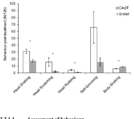
Figure 6 – Back-transformed mean (± exact 95% confidence intervals) frequencies of behaviour post-treatment frequencies. CAUT kids (n=5) were disbudded with a cautery iron and SHAM kids (n=5) were sham handled but not disbudded. * indicates the behaviour that differ between treatments at the 5% significance level. (Adapted from Hempstead et al., 2017) ... 11
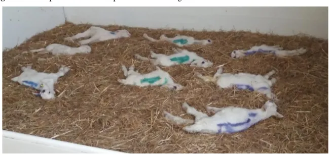
Figure 7 – Kids placed in the same pen after disbudding for behaviour observation. ... 21
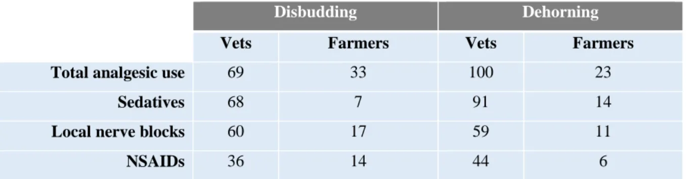
LIST OF TABLES Table 1 – Proportion of veterinarians and goat producers that provide analgesia for any goats during disbudding and dehorning. (Adapted from Valdmanis et al., 2008) ... 13
Table 2 – Treatment description of the experimental groups. ... 20
Table 3 – Intervention and treatment description of the experimental groups. ... 21
Table 4 – Treatment description of the experimental groups. ... 22
Table 5 – Wound evaluation scale description (Photographs taken by the author). ... 23
Table 6 – Mean frequency of tail flicking, leg movements and vocalization performed by each group during disbudding. D (disbudded without anaesthesia), K (administration of ketamine prior to disbudding), K+M (disbudded after being administered ketamine and meloxicam), S (sham disbudded after being administered ketamine). ... 24
Table 7 – Incidence (mean ± S.D) of behaviours in four different groups (D – disbudded without anaesthesia; K – disbudded after being given ketamine; K+M – disbudded after being given ketamine and meloxicam; S – sham disbudded after administration of ketamine) during different observational periods of the disbudding study (first half-hour, second half-hour, second hour and third hour). ... 27
Table 8 – Incidence (mean ± SD) of lesions presented 3 days after disbudding in two different groups, D (application of an antibacterial spray at the horn bud site after the procedure) and TS (application of Tri-Solfen, an anaesthetic and antiseptic gel after disbudding). ... 30
Table 9 – Incidence (mean ± S.D) of behaviours in two different groups (D – application of an antibacterial spray after disbudding; TS – application of anaesthetic and antiseptic gel after disbudding) during different observational periods (first half-hour, second half-hour, second hour and third hour post-disbudding). ... 31
x LIST OF GRAPHS
Graph 1 – Frequency of “leg movements” within and between groups during disbudding. D (disbudded without anaesthesia) differed significantly from K (disbudded after being administered ketamine), K+M (administration of ketamine and meloxicam prior to disbudding) and S (sham disbudded after being administered ketamine)... 25 Graph 2 – Difference in frequency of “vocalization” between groups during disbudding. D (disbudded without anaesthesia) differed significantly from K+M (disbudded after being administered ketamine and meloxicam). K (administration of ketamine prior to disbudding), S (sham disbudded after being administered ketamine). ... 25 Graph 3 – Difference in frequency of “vocalization” between groups in the first half-hour post disbudding. S (sham disbudded after being administered ketamine) kids differed significantly from K (administration of ketamine prior to disbudding), K+M (disbudded after being administered ketamine and meloxicam) and D (disbudded without anaesthesia). ... 26 Graph 4 – Difference in frequency of “head shaking” between groups in the second half-hour of the observation period, with a significant difference between group K (given ketamine prior to disbudding) and groups D (disbudded without any anaesthesia) and K+M (given ketamine and meloxicam prior to disbudding). S (sham disbudded after administration of ketamine) .. 26 Graph 5 – Difference in frequency of “head scratching” between groups in the second half-hour of the observation period, with a significant difference between group K (given ketamine prior to disbudding) and groups D (disbudded without any anaesthesia), K+M (given ketamine and meloxicam prior to disbudding) and S (sham disbudded after administration of ketamine). ... 28 Graph 6 – Difference in frequency of “self-grooming” between groups in the second half-hour of the observation period with a significant difference between S (sham disbudded after administration of ketamine) kids and K+M (ketamine and meloxicam administered prior to disbudding). No significant differences were found between those and D (disbudding without anaesthesia) and K (ketamine given prior to disbudding). ... 28 Graph 7 – Difference in frequency of “body shaking” between groups in the third hour observation period with significant difference between K (ketamine given prior to disbudding) and S (sham disbudded after administration of ketamine), K+M (ketamine and meloxicam administered prior to disbudding) and D (disbudded without anaesthesia). ... 29 Graph 8 – Difference in frequency of “head shaking” between groups in the first half-hour of the observation period with significant differences between group D (antibacterial spray after disbudding) and TS (given and an anaesthetic and antiseptic gel after disbudding – Tri-Solfen®). ... 29 Graph 9 – Difference in frequency of “head scratching” between groups in the first half-hour of the observation period with significant difference between D (antibacterial spray after disbudding) and TS (given and an anaesthetic and antiseptic gel after disbudding – Tri-Solfen®).
... 30
LIST OF ABBREVIATIONS, INITIALS AND ACRONYMS NMDA - N-methil-D-aspartate
NSAID – Non-steroid anti-inflammatory TS – Tri-Solfen
h – hour d – day(s)
1
PART I - DESCRIPTION OF THE TRAINING PERIOD
As part of the 6th year of the Masters in Veterinary Medicine I did my training period in the area of farm animal clinics at Faculdade de Medicina Veterinária, Universidade de Lisboa. From September 2017 to April 2018, under the supervision of Professor George Stilwell, I had the opportunity of accompanying the Farm Animal Clinics ambulatory work which consisted of visits to different farms in the Center-South region of Portugal.
The several farms we visited allowed me to come in contact with an interesting caseload of beef and dairy cattle, goats, sheep and swine. About 70% of the consultations fell into the category of Internal Medicine. For each clinical case, a thorough physical exam was performed and the most likely differential diagnosis and therapeutics were discussed. In a majority of cases, samples were collected for further analysis allowing an accurate diagnosis and follow up of the patients. One particular case resulted in two scientific posters I co-authored, “Caso clínico atípico de besnoitiose (Besnoitia besnoiti) num touro limousine”, which was presented at XIX Jornadas da Associação Portuguesa de Buiatria, in Azores (Appendix I) and “Besnoitiosis as an emerging and exotic disease – new clinical features in breeding bulls”, presented at the 30th World Buiatric Congress in Japan.
The areas of preventive medicine, surgery, reproduction and obstetrics were covered as well in this period in which I was able to develop my skills in pregnancy testing in cattle through rectal palpation and ultrasound and to assist on a few deliveries and two cesarean-sections. In addition to these, I had the opportunity of participating in two different surgical treatments for left displaced abomasum in cows – right flank omentopexy and left flank abomasopexy. There was also the chance to do field necropsies and simulate surgeries on cadavers, both of which played a crucial role in the learning process. Animal welfare was always a present and valued matter. This training period allowed me to reinforce what I had learned in previous curriculum subjects, develop clinical reasoning and apply it on a practical context, communicate with farmers and better understand the farming system, all of which contributed to my improvement as a future veterinarian and human being.
Continuous learning plays a fundamental part in the veterinary medicine profession and for that matter I attended the XIX Jornadas da Associação Portuguesa de Buiatria, V Jornadas Técnico-Veterinárias do Campo Branco and the 8th EFOMV in which a poster I co-authored was presented on “Aplicação tópica de anestésicos locais para controlo da dor durante a aparagem curativa de lesões podais de vacas leiteiras – dados preliminares” (Appendix II). I also got the opportunity of doing an oral presentation about dehorning and disbudding in small ruminants at a Workshop de Descorna em bovinos and of collaborating in Professor George Stilwell’s
2
book “Bem-estar dos Ruminantes” (2017) and in an oral communication on “Topical application of local anaesthesia for pain control during the curative trimming of podal lesions of dairy cows” (Appendix III) at the World Buiatric Congress in Japan.
As an extra training period, I did an externship at the University of Tennessee, College of Veterinary Medicine (UTCVM) from April to July 2018. I rotated through Farm Animal Medicine and Surgery, Theriogenology, Anaesthesiology and a couple of small animal services. The Medicine and Surgery service provides a great amount of advanced patient care, 60% of it being smallholders (goats, sheep, camelids and pet pigs). I participated in all aspects of case management, including receiving, examination, diagnosis, daily management, client communication, discharging and follow-up communication. I was also responsible for daily problem oriented medical records of the cases I was assigned to. Daily rounds were held on inpatient cases and medicine and surgery topics were discussed. This rotation allowed me to improve my diagnostic skills such as abdominal ultrasound and thorough neurological examination, become familiar with a farm animal hospital in an English-speaking setting and, through a hands-on approach of all cases, gain confidence and self-sufficiency. The other rotations allowed me to become comfortable with breeding soundness exam in bulls, breeding soundness exam in mares and cows, artificial insemination in mares, having also mild exposure to cows AI, and anaesthesia mainly focused on pigs and equine.
3
PART II – EXPERIMENTAL STUDY: PAIN REDUCTION IN DAIRY GOAT KIDS’ DISBUDDING
1. LITERATURE REVIEW
1.1. DISBUDDING
1.1.1. Context
With the increase of intensive dairy farms with group housing systems, feed barriers and milking parlours, the prevention of horn growth has become a routine practice. Under the same space conditions, when in comparison to hornless goats, horned animals, especially when low in the hierarchy, have a higher necessity of sharing feeding places which can lead to a reduction in body weight and milk production if they do not have sufficient access to feed (Loretz et al., 2004). Consequently, more space is required at the feed barrier for horned goats in order to prevent high competition for feed and ultimately welfare is compromised. Dehorned animals are considered to be safer to handle, cause less damage to other animals and have less negative impact on their housing environment and social interactions (Alvarez & Gutiérrez, 2010). For all these reasons, disbudding constitutes, at the present moment, an unavoidable management tool for dairy goat farmers.
1.1.2. Methods
Disbudding is achieved by thermal cautery, cutting or chemical methods, most of which have been adopted from cattle. In order to remove the germinal tissue of the horn, disbudding is usually carried out within the goat kids’ first two weeks of life, although further investigation should be done to determine the optimal age for disbudding (Alvarez et al., 2009; Smith & Sherman, 2009).
An online survey (Valdmanis et al., 2008) stated that cauterization is the method of choice for disbudding for 97% of veterinarians and 95% of producers. It consists of an electric dehorning device with a top ring that heats the bud and the surrounding tissue for some seconds until the full thickness of the skin has been destroyed (Stafford & Mellor, 2011). All of the horn corium must be within the ring and the isolated central circle of skin that contains the horn bud can be lifted off (Smith & Sherman, 2009). This type of disbudding causes third degree burns, which can result in a bacterial infection and varying amounts of subcutaneous damage. Because the skull of goat kids is much thinner than calves’ cranium, the safety margin for thermal injury is noticeably reduced, resulting in a higher risk of damage when this method is not used correctly. There have been reports of meningoencephalitis and cerebral infarction following hot-iron
4
disbudding and it is believed that far more kids either die or suffer from these consequences than are brought to the attention of veterinarians (Thompson et al., 2005).
The injection of clove oil essence in the horn bud region has been studied (Molaei et al., 2015) as a new chemical technique for disbudding. It is suggested that the main component of clove oil, eugenol, can inhibit proliferation of the horn germination cells and induce their apoptosis, as well as inducing cell necrosis through its cytotoxic effect on horn bud cells. Because of its anti-inflammatory and analgesic effects, no local anaesthetics and/or sedative drugs were necessary before the injection. A complete necrosis of epidermis and underlying dermis with collagenolysis in horn bud tissues occurred within one week after treatment and stopped horn growth. Hempstead et al. (2018b) suggested that animals treated with clove oil had less tissue damage around the horn buds and appeared to lead to earlier tissue repair with some animals, however, developing scurs. Further studies should be done to assess horn regrowth and stress response to this method.
Chemical burn of underlying tissue is most commonly achieved by caustic chemicals such as sodium hydroxide and potassium hydroxide that burn the underlying tissues. This method should be avoided as it can run to the face and eyes and create skin wounds elsewhere on the kid and other animals in contact with it, as for example the udder of the doe (Smith & Sherman, 2009; Thompson et al., 2005).
Because the hot-iron is the most commonly used technique, it will be this dissertation’s main focus.
1.1.3. Improper disbudding
When goats that have been disbudded still show visible horn tissue (scurs) (Figure 1), they are considered to have been improperly disbudded. Incorrect disbudding can result in scurs pressed against the head or eye and consequently cause lesions and pain (Smith & Sherman, 2009). Scurs getting caught in fences and pen partitions have also been reported (Waiblinger et al., 2011, cited by Battini et al., 2014).
5 1.1.4. Alternatives
It is possible to genetically select for the absence of horns, i.e. polled goats, and this would be the ideal choice for animal welfare. In spite of the polled trait being dominant it is linked to a serious genetic reproductive disorder caused by a recessive gene for infertility. Being homozygous for the polled gene results in a sterile intersex in the female and in a bigger risk for development of sperm granulomas in the head of the epididymis in the male. Nevertheless, breeding pairs with one naturally horned member and another one polled can obtain polled goats with normal reproductive potential and the kid does not require disbudding but, in order to maintain good fertility, horned goats cannot be totally eliminated (Smith & Sherman, 2009). It is simple to identify polled kids as a single central twirl of hair on top of the head is usually presented (Figure 2) instead of one around each horn bud and the part of the skin where the horn buds would be is tightly attached to the underlying bone. However, some producers disbud these animals anyway, cauterizing tissue in the area where horn buds would normally be (Mackenzie, 1975, cited by Smith & Sherman, 2009).
Figure 2 – Polled kid with a single central twirl of hair. (Adapted from Smith & Sherman, 2009)
1.2. HORN ANATOMY
Horns are of continuous and permanent growth; each horn is composed of closely packed tubules produced by corium and germinal epithelium. Its osseous base is provided by the frontal bone’s cornual process and the dermis is firmly adherent to it. The cornual diverticulum of the frontal sinus forms a cavity within the horn, which is consequently opened when an adult goat is dehorned (Dyce, 2010; Smith & Sherman, 2009). The blood to the horn is supplied by the ventral and dorsal cornual arteries, branches of the superficial temporal artery, and blood returns from the horn by two orbital veins, the superficial temporal vein and the angular tributary of the facial vein (Taylor, 2015).
6 1.2.1. Innervation and nerve blocking
Goats differ from cattle in the sense that each horn bud is innervated by the cornual branches of two nerves; lacrimal nerve and infratrochlear nerve. In cattle, horn innervation is achieved only by the lacrimal nerve, which makes nerve blocking and pain mitigation much easier and effective (Hempstead et al., 2017). Because of the dual branching in goats, the cornual nerve blocking requires two injection sites per horn (Figure 3): one being midway between the lateral base of the horn and the lateral canthus of the eye and the other one halfway between the medial base horn and the medial canthus of the eye (Plummer & Schleining, 2013), making the administration of local anaesthesia a challenge.
Figure 3 – Needle placement for desensitizing the cornual branch of the lacrimal nerve (A) and cornual
branch of the infratrochlear nerve (B) in the goat (Adapted from Skarda, 1986).
1.3. PAIN
The absence of pain in routine procedures is one of the criteria for good health and animal welfare (Stilwell, 2017). Disbudding methods are invasive and painful and compromise animal welfare, not only during the procedure but also postoperative. In spite of cautery disbudding being less painful than other methods (Stafford & Mellor, 2011), it has been associated with acute pain and stress in goat kids with high-intensity behaviours and acute cortisol increase. Destruction of the surrounding tissue of the horn bud causes fluid movement from the vascular compartment into that region resulting in inflammation and oedema. The influx of inflammatory mediators and their local actions from the adjacent blood vessels justify the pain experienced during and after disbudding (Hempstead et al., 2017).
7
Pain has negative ethical, biological and economic effects; animals that experience pain show changes in ruminal function with atony or hypotony, decreased immunity as prolonged stress hormonal changes lead to a reduction of the phagocytic capacity, neutrophil mobilization, lymphocytic differentiation and production of antibodies, as well as anorexia and consequentially an increased susceptibility to infectious diseases. Also, pain in mature animals can affect fertility due to the inhibition of reproductive hormones’ production and release and to a nutritional deficit with reduction of normal heat behaviour (Stilwell, 2017).
It is possible to identify and evaluate pain through morphological, physiological, and behavioural measures (Stilwell, 2017).
1.3.1. Pain physiology
Various stimuli are capable of originating a complex pain response. It is important to differentiate between physiological and pathological pain. The first type is part of a normal and useful defence mechanism to noxious stimuli resulting in strategies of avoidance facilitated by a specialized network of primary sensory neurons and nociceptors. Pathological pain is a result of damage to tissue and to nerves (Ellison, 2017) and its prolonged duration has a negative impact on the animal’s homeostasis (Stilwell, 2017).
1.3.1.1. Nociception
Nociception is the physiological element of pain and involves the components of transduction, transmission and modulation of neural signals created by external noxious stimuli (Ellison, 2017). Primary afferent neurons free endings connected to the central nervous system (CNS) intended to detect tissue damage are called nociceptors and are triggered by mechanical, chemical and thermal stimuli (Bourne et al., 2014).
Transduction consists on the nociceptors’ capability of transforming external noxious stimuli into electric activity, generating nerve impulses. These are then transmitted by membrane depolarization of afferent specialized fibers, the primary afferent nociceptive fibers, primarily to the dorsal horn of the medulla and then to the supraspinal structures (Stilwell, 2017). This is achieved through an increase of ion permeability when facing a mechanical stimulus or of membrane permeability due to a chemical one. There are some different types of fibers; the “first pain” is signalized by A-delta fibers (Figure 4). These are lightly myelinated neurons of fast conduction (5 to 30 meters per second) responsible for reflex behaviour related to acute, fast and momentary pain that only lasts as long as the painful stimulus is triggering the nociceptor and is generally associated with mechanical and thermal stimuli. They are easily blocked by local anaesthetics. C fibers are suggested to have a role in monitoring the overall
8
tissue condition as they can be associated with polymodal receptors. They are responsible for the “second” or “slow pain” which is diffuse and prolonged and are of slow conduction (0.5 to 2 meters per second). Inflammation mediators such as substance P, serotonin, potassium, hydrogen, histamine and prostaglandins lead to hyperalgesia with an exaggerated response from these fibers. When tissues are already damaged, further nociceptors are activated (silent fibers). A-beta fibers are responsible for the conduction of non-noxious stimuli, such as tact (Bourne et al., 2014; Stilwell, 2017), but can be involved in pain transmission in some cases.
Modulation consists of pain moderation or inhibition mechanisms, through endogenous opioids and other substances, that act at the level of the CNS and peripherally.
Figure 4 – Primary afferent pain transmission. First pain and second pain sensations after a noxious
stimulus (A). The first pain sensation is abolished when the A fibers are blocked (B). (Adapted from Ellison, 2017)
1.3.1.2. Central Nervous System
The first synapses for the nociceptive fibers are found in the dorsal root ganglia where sensory neuron cell bodies are located and neurotransmitters act. Glutamate is the most common neurotransmitter and induces fast synaptic potentials in the superficial horn neurons, participating in the modulation of noxious stimuli conduction (Ellison, 2017; Stilwell, 2017). Synapses at the dorsal horn of the medulla bind the nociceptors to ascendant neurons that conduct the stimulus to the brain stem. These can be interneurons that produce an instantaneous flight response (e.g. withdrawal reflex) or reduce the conduction of noxious stimuli resulting in an endogenous analgesia and increased pain conduction threshold, propriospinal neurons or projection neurons. The latter are excited only by noxious thermal or mechanical stimuli from C and A-delta fibers and conduct the nerve impulse to supraspinal centres. The fibers ascend through the spinothalamic tract that terminates in the thalamus which is the first part of the brain to receive and redirect noxious stimuli through synapses of these fibers. The impulse is then transmitted to the cerebral cortex where perception of pain occurs (Ellison, 2017)
9
1.3.1.3. Hypersensitivity or hyperalgesia
Due to the accumulation of inflammation mediators and to tissue and nerve damage, the nociceptors’ excitability threshold is decreased, silent fibers are activated and C fibers become more responsive to any stimulus, resulting in peripheral hyperalgesia. It can be primary or secondary. In the first, only the aggression site becomes more sensitive, whereas in the latter it also happens in the surrounding undamaged tissues (Stilwell, 2017).
Central hyperalgesia occurs at the level of the medulla with a reduction of the neuron activation threshold so that they fire at the minimal provocation. After a period of constant C fiber activation in pathological pain, neurotransmitters that are hyperalgesic such as glutamate, substance P, prostaglandins and interleukins accumulate in the spinal cord, an extra number of channels and receptors are created and opened and nociceptive silent fibers and A-beta fibers, which start to act as pain conductors, are activated (Figure 5), resulting in allodynia with any stimuli being felt as painful. Animals with hypersensitivity respond poorly to analgesia or anaesthetic nerve blocks thus prevention plays a major role in pain management (Bourne et al., 2014; Ellison, 2017; Stilwell, 2017).
Figure 5 – Pathological pain results from abnormal excitability in the nervous system. This involves
both central and peripheral changes and the net result is a low-intensity stimulus can elicit pain. PNS = peripheral nervous system; CNS = central nervous system
Pain ain
10 1.3.2. Pain evaluation – indicators
1.3.2.1. Behaviour
Little research exists on the evaluation of pain related behaviours in disbudded kids. Nevertheless, behavioural response constitutes an important and non-invasive measure of pain. The awareness of pain-specific behaviours allows for the study of strategies of pain relief and subsequently improvement of goats’ welfare. Three classes of behaviours can be useful in pain evaluation such as pain-specific behaviours manifested at a higher frequency, a decline in the magnitude or frequency of a certain behaviour and measures of preference based on how the animals perceive different treatments (Weary et al., 2006).
Cautery disbudding increases the presentation of behaviours potentially associated with pain and discomfort. During the procedure, the frequency of high vocalizations, leg movements (kicks, attempts to escape) and tail shaking has been found to be higher in kids that were cautery disbudded in comparison with others in which the procedure was simulated with a cold iron and with other groups given pain relief (Alvarez et al., 2009; Alvarez & Gutiérrez, 2010; Chandrahas et al., 2013). While vocalizations, which are defined as “emission of bleats with open or closed mouth” (Alvarez et al., 2015), are suggested to be a sign of pain in cattle, its evaluation becomes a challenge in goats due a frequent expression of this behaviour in ordinary circumstances and therefore the correlation between the sound’s frequency or amplitude and pain is difficult (Stilwell, 2017). In several studies, a digital sound meter has been used and only vocalisations of >90 dB have been considered as high intensity and indicative of pain ( Alvarez et al., 2009; Alvarez & Gutiérrez, 2010; Alvarez et al., 2015).
Behaviours considered to be associated with pain were described for up to 3 to 6 hours after disbudding, indicating stressful and prolonged experience of pain (Chandrahas et al., 2013). It is not unexpected that behaviours focussed in the top of the head such as head scratching, shaking and rubbing have been found to be the most frequent after disbudding (Figure 6) as a result of the inflammation and damaged tissue on the horn buds (Hempstead et al., 2017). At 5 minutes after disbudding, the frequency of head shakes in disbudded animals is double the one by kids with disbudding simulation (Hempstead et al., 2018a). Scratching and rubbing the burned area may stand as the kids’ effort to relieve the pain and pressure caused by cauterization. Self-grooming was also shown to be performed at higher frequencies following disbudding although individual differences among kids were found. Body shaking was less frequent and the animal’s attention being all diverted to the head was suggested as the main cause for the reduced occurrence of this behaviour. Regardless of being disbudded or not, all kids exhibited these behaviours before and after the procedure indicating that none of the above
11
are pain specific and their simple manifestation is not reliable for measuring pain associated with thermal disbudding in goat kids. Although a few studies on pain mitigation techniques have shown a reduction of some of these behaviours (Chandrahas et al., 2013; Nfor et al., 2016; Hempstead et al., 2018a), further research is required in order to potentially consider them as valid pain indicators.
Figure 6 – Back-transformed mean (± exact 95% confidence intervals) frequencies of behaviour
post-treatment frequencies. CAUT kids (n=5) were disbudded with a cautery iron and SHAM kids (n=5) were sham handled but not disbudded. * indicates the behaviour that differ between treatments at the 5% significance level. (Adapted from Hempstead et al., 2017)
1.3.2.1.1. Assessment of behaviour
In order to assess behaviour either a visual analogue scale or a numerical scale are generally used. The first method scores pain in a subjective manner and normally relies on the presence or absence of several significant behaviours whereas the latter records the frequency of certain pain-related behaviours (Weary et al., 2006). Both have been used for assessing severe pain after caustic paste disbudding in calves and similar results were achieved but, in regards to prolonged pain, the objective method, i.e. numerical scale, was found to be more accurate (Stilwell, 2009). The value of the two methods will depend on the quality of the measures, the pain experienced by the animal and, for the subjective approach, on the observer’s training (Weary et al., 2006); the use of an ethogram that records several behaviours will make pain less likely to be underestimated. A few ethograms have been developed on behaviour patterns of dairy goat kids and used for behaviour analysis following disbudding (Heinrich et al., 2010; Hempstead et al., 2017; Hempstead et al., 2018).
12 1.3.2.2. Cortisol
Plasma cortisol level is usually used as a physiological pain indicator in research with livestock. Stress, be it traumatic, inflammatory or psychological, includes the release of cortisol due to the activation of the hypothalamic-pituitary-adrenocortical (HPA) axis followed by stimulation of the adrenocorticotropic hormone’s (ACTH) release (Smith, 2009). Cortisol acts as a moderator of the inflammatory response and consequently of pain although its levels are not specific for pain and can increase with stress, anxiety or excitement (Stilwell, 2017; Tsigos & Chrousos, 2002). Thus, when using cortisol levels for pain evaluation, the stress involved with handling of the animals for sample collection should be considered so that results are not misperceived. In calves, as long as blood sampling is carried out within one minute of restraining, cortisol levels have been suggested to not be evident (Lane, 2006; Stilwell et al., 2008). As an alternative to plasma cortisol, non-invasive sampling methods such as determination of corticoid in urine, saliva or milk have been explored, as well as faecal samples in chronic stress situations in which cortisol metabolites can be detected through high performance liquid chromatography (HPLC) (Möstl & Palme, 2002). Only about 10% of circulating plasma cortisol is in the free form with biological activity. The level of cortisol in saliva has been confirmed to be a direct reflection of the biologically active cortisol in plasma, with the advantage of less negative impact on animal welfare (Lane, 2006).
Glucocorticoids are known to be produced on a circadian rhythm with physiological variations throughout the day, namely peak levels at pre and post wakening. However, a study on this matter conducted with goats at six different times of the year (Alila-Johansson et al., 2003) showed that no significant daily rhythm in plasma cortisol levels existed at any time of the year, whereas a significant seasonal variance was observed; cortisol concentration was higher in the winter than in any other season, in contrast to low concentrations from early spring to summer. Plasmatic cortisol levels have been determined immediately after kids’ disbudding and shown to significantly increase and last for 2 to 3 hours (Alvarez & Gutiérrez, 2010; Hempstead et al., 2018a) or up to 5 hours (Nfor et al., 2016) with a concentration peak at 15 minutes after the procedure. This shows that not only it is a painful procedure, but that the pain also persists. In calves, elevated cortisol concentrations were found 24 hours after disbudding (Morisse et al., 1995). This variance in time corroborates the suggestion that because the amount of time needed to disbud calves is superior to that of kids and due to the kids’ horn buds being less developed at the time of the procedure, the pain kids experience during disbudding may be lower and relatively short-lived when in comparison with calves (Hempstead et al., 2018a).
13 1.3.2.3. Technological methods
Inflamed lesions and exposure of nerve endings allow to test for the pain tolerance threshold when pressure is applied to an affected area or its surroundings due to the hyperalgesia. Algometers are instruments that measure the sensibility to touch or pressure applied to a certain region (Stilwell, 2017). When the animal tries to move away (withdrawal reflex), a value is registered and it is considered as the first point at which pressure is sensed as pain. Comparing it to healthy regions values aids in quantifying and evaluating pain and possibly tracking healing. To achieve great reliability, the manual force applied should be consistent and perpendicular to the body surface (Kinser et al., 2009). This method has been used to determine pain sensitivity in disbudding wounds in calves (Heinrich et al., 2010; Espinoza et al., 2016). The degree of inflammation can also be assessed by determining the temperature of a specific area and therefore confirm a suspicion of pain (Stilwell, 2017). In this matter, a thermography camera has been used recently to measure and compare skin temperature at the level of the horn buds pre and post different methods of disbudding, including several weeks after treatment to evaluate wound healing (Hempstead et al., 2018b).
1.3.3. Pain management
Disbudding is routinely performed without any anaesthesia or analgesia. A majority (69%) of veterinarians that participated in an online survey (Valdmanis et al., 2008) reported that they include analgesia as part of the disbudding procedures (Table 1) which, along with the reduction of pain related behaviours and physiological parameters modifications in kids given anaesthetics, strongly supports the necessity for pain relief. On the other side, 75% of farmers who do not use analgesic drugs consider it unnecessary, whereas of the 33% of producers that provide analgesia, 91% reported welfare benefits as a reason to do so.
Table 1 – Proportion of veterinarians and goat producers that provide analgesia for any goats during
disbudding and dehorning. (Adapted from Valdmanis et al., 2008)
Disbudding Dehorning
Vets Farmers Vets Farmers
Total analgesic use 69 33 100 23
Sedatives 68 7 91 14
Local nerve blocks 60 17 59 11
14 1.3.3.1. Local anaesthesia
The ionic fluxes generation and conduction that are required for the conduction of nerve impulses are inhibited by local anaesthetics. They interfere with membrane depolarization by blocking Na+ channels and act primarily on mechanical pain, followed by cold, heat and profound pressure. Their action is decreased when necrotic tissue, pus or acid settings are present as they ionize in acid environments because local anaesthetics pKa is normally slightly alkaline (Lomax et al., 2013; Stilwell, 2017).
Local anaesthesia has been shown to be largely ineffective and unsatisfactory at reducing pain-related behaviours in goat kids during and after disbudding. For an effective anaesthesia, both cornual branches of the lacrimal and infratrochlear nerves must be blocked (Galatos, 2011). Physiological and behavioural responses have been studied in cautery disbudded goat kids with and without cornual nerve block with 1 mL of 2% lidocaine per site (Alvarez et al., 2015). This injection did not have a significant effect either on the rising cortisol levels, as they were significantly increased immediately after disbudding and remained high for the following 20 to 30 minutes, or on the behavioural response which did not decrease. Similar findings had been previously achieved (Chandrahas et al., 2013) with the local administration of lidocaine not preventing pain related behaviours after the procedure. In a different study, the administration of lidocaine around each horn bud did not reduce cortisol secretion in response to disbudding. In addition, the raise of cortisol secretion was accelerated due to the puncture required to infiltrate the anaesthetic (Alvarez et al., 2009). The use of a higher dosage could potentially be studied although toxicity needs to be considered as approximately 10mg/kg of lidocaine intramuscularly, i.e. 2 mL of 2% lidocaine for a 4 kg goat kid, cause severe toxicity, resulting in lethargy, unwillingness to feed and convulsions (Galatos, 2011).
1.3.3.1.1. Tri-Solfen
Tri-Solfen® (TS) is a practical topical anaesthetic and antiseptic formulation used in Australia
in painful procedures such as mulesing, castration and tail docking in lambs as well as castration and disbudding in calves. It is only commercially available in Australia. It contains hydrochloride bupivacaine, hydrochloride lidocaine, tartrate adrenaline and cetrimide and comes in the form of a spray-on gel, making it practical and easily applied by farmers immediately after a procedure (Lomax et al., 2008; Windsor et al., 2016).
A rapid onset anaesthesia and prolonged effect is provided with the association of bupivacaine and lidocaine (Lomax et al., 2008) with wounds being desensitised within one minute of the application (Lomax et al., 2013). The presence of a vasoconstrictor (adrenaline) in the preparation also reduces blood loss as haemostasis is rapidly achieved and, through the
15
reduction of systemic absorption, the local effect of the anaesthetics is prolonged (Lomax et al., 2008; Lomax et al., 2010). The fact that it is a gel can itself be advantageous as it can provide a barrier against environmental agents and touch stimulation, offering protection to nerve endings at the surface of the wound. This, along with the antiseptic agent, results in improvement of wound healing (Emflorgo, 1999, cited by Lomax et al., 2008).
A decrease in cortisol secretion and pain-related behaviours in lambs immediately and for up to 24 hours after mulesing supports TS’ effectiveness in pain alleviation and suggests that it inhibits wound nociception for a prolonged time that goes beyond its duration of action (Lomax et al., 2013; Paull et al., 2009). Its use in surgical castration of lambs has also been found to improve wound healing and decrease pain related behaviours with abolishment of primary hyperalgesia. Secondary hyperalgesia has been prevented in animals that were treated with TS post hot-iron tail docking (Lomax et al., 2010).
The management of pain through the administration of this topical anaesthetic immediately after scoop dehorning has also been studied in two-month-old dairy calves with a reduction of pain sensitivity for up to 5 hours following the procedure (Espinoza et al., 2016). However, because the procedure can be associated with considerable haemorrhage, the adherence of TS’ substances to the wound might be inadequate (Espinoza et al., 2013 cited by Windsor et al., 2016).
1.3.3.2. General anaesthesia
There is little research on the efficacy of general anaesthesia on pain management in disbudding of goat kids. The use of isoflurane alone and in combination with meloxicam has been studied (Hempstead et al., 2018a) and a reduction in cortisol secretion in kids after disbudding for up to two hours was achieved but only when used in combination. Behaviours indicative of pain were reduced although the effect of isoflurane alone on behaviour was not assessed and further research should be considered. While it showed to be a good method of anaesthesia and provide good pain relief, it may not be practical in terms of equipment requirements and necessity for trained personnel. The development of an on-farm use portable isoflurane delivery service has been suggested.
1.3.3.2.1. Ketamine
N-methil-D-aspartate (NMDA) is a receptor activated by glutamate that excites post synaptic neurons of the dorsal horn when inflammation or tissue lesion are present and is believed to be a main component in the central sensitization, pain amplification and secondary hyperalgesia’s development (Anderson & Muir, 2005; Stilwell, 2017). Ketamine is a dissociative rapid acting
16
general anaesthetic and NMDA receptor antagonist, responsible for its analgesic properties. It has no depressant effects on the respiratory system, provides stimulation of the sympathetic system, potentially improving cardiovascular function, the peripheral reflexes are maintained and muscle tone is increased (Valverde & Doherty, 2008; Fallis, 2013; Plummer & Schleining, 2013). Regurgitation has occurred in goats following induction with ketamine, so endotracheal intubation should be considered (Prassinos et al., 2005). A good evaluation of the level of anaesthesia should be held as there are reports of animals that do not respond to ketamine (Stilwell, 2017). It should be associated with sedative tranquilizers such as xylazine or diazepam. Nevertheless, in order to avoid prolonged recoveries and an increased risk of regurgitation, sedation is not always performed in ruminants before anaesthesia induction (Prassinos et al., 2005). A combination of ketamine and xylazine given to kids before disbudding has reportedly reduced pain (Ingvast-Larsson et al., 2011) although acute response was not assessed with enough detail.
1.3.3.3. Sedatives
The intramuscular administration of dexmedetomidine hydrochloride in goat kids prior to disbudding (Nfor et al., 2016) both as a single or a combined dose, was found to be successful in reducing the possible long lasting pain and discomfort, as there were no significant increases in baseline cortisol levels and pain-related behaviours had a relatively low incidence. However, because α2 adrenergic agonists provide an intense sedation and muscular relaxation, absence of movement can be misinterpreted as absence of pain (Stilwell, 2017). Further studies are also required to assess the appropriate dosage for kids.
1.3.3.4. Nonsteroidal anti-inflammatory drugs
1.3.3.4.1. Meloxicam
Meloxicam belongs to the class of nonsteroidal anti-inflammatory drugs (NSAID) and its mechanism of action consists in the inhibition of cyclooxygenase, especially COX-2, and prostaglandin synthesis, having anti-inflammatory, antipyretic and analgesic properties (Fallis, 2013). Its elimination rate is faster in goats than in sheep and cattle (Coetzee et al., 2009, cited by Karademir et al., 2016).
The administrations of meloxicam subcutaneously, intramuscularly and orally in healthy goats have been compared in order to better understand each route’s pharmacokinetic parameters. It was found that with subcutaneous administration, plasma concentrations reached a peak faster and a long-term presence of the drug in the goats’ plasma was achieved, making it a preferable route of administration (Karademir et al., 2016).
17
Because of meloxicam’s analgesic qualities, its use has been assessed in disbudded kids. In one recent study (Hempstead et al., 2018a), orally given meloxicam was compared with meloxicam administered subcutaneously to kids prior to disbudding. Contrary to what was expected, oral meloxicam had a better effect on behaviour response. Regardless of the administration route, meloxicam did not seem to have a significant effect on the stress response as the kids had similar cortisol concentrations to those experienced by kids disbudded without any pain relief. These findings are corroborated by former literature, in which no significant differences were observed in cortisol levels of kids treated with and without NSAID (Ingvast-Larsson et al., 2011; Nfor et al., 2016). However, prevalence of pain signs was found to be smaller in kids treated with meloxicam on the first 24 hours after disbudding, even though not subsequently and kids given meloxicam in combination with isoflurane had an inferior physiological and behavioural response to the procedure (Hempstead et al., 2018a) which is consistent with earlier suggestions that either in combination or as a single drug, meloxicam may lessen pain related behaviours, therefore improving animal welfare (Chandrahas et al., 2013).
1.4. WOUND HEALING
A disruption or damage to the normal anatomical structure results in a wound (Velnar et al., 2009). As the disbudding wound generally repairs itself within 30 days, it can be classified as an acute wound (Broughton et al., 2006).
The process of wound healing is normally divided into four phases: coagulation and haemostasis, inflammation, proliferation and remodelling with the formation of scar tissue (Velnar et al., 2009). The first step after wounding is bleeding control (haemostasis) through vasoconstriction of the injured blood vessels and activation of the coagulation cascade, resulting in a clot formed by platelets, collagen, fibronectin and thrombin, all of which release growth factors and cytokines that induce the inflammatory response. Within 24 to 36 hours, a neutrophil response that aims to prevent infection is generated during clot formation and is drawn into the wounded area as surrounding blood vessels dilate. Once neutrophils succeed to remove all contaminating bacteria, its population is eliminated and phagocytosed by former monocytes that transform into macrophages generally 48 to 96 hours following damage. These produce important tissue growth factors such as TGF-β that activate fibroblasts, keratinocytes and endothelial cells that will be crucial for the healing process. Interleukin-1 (IL-1) and immunoglobulin G (IgG) draw lymphocytes to the injury site 72 hours after injury and promote collagenase regulation. On the third day the proliferative phase begins and lasts for about 2 weeks. It is characterized by a profuse formation of granulation tissue, provided by fibroblast
18
migration and deposition of newly produced extracellular matrix supporting cell migration and therefore stimulating re-epithelialization. In the last phase (remodelling), the deposition of collagen in an organized network allows for new epithelium to develop and scar tissue to be formed with an increase in the wound’s strength (Broughton et al., 2006; Velnar et al., 2009). Vasculature and cells are often destroyed in burned tissues as happens in cautery disbudding which may complicate the regular healing response. Neutrophils and macrophages’ access to the injury site is compromised because of insufficient blood flow as well as angiogenesis, essential for re-epithelialization (Rose & Chan, 2016).
Cautery disbudding causes full thickness burns and ceases circulation leading to decreased tissue perfusion and a longer recovery period (Papp, 2012 cited by Hempstead et al., 2018b). Only one study has been conducted on wound healing after thermal disbudding in goat kids, reporting that at 6 weeks post treatment scabs were still displayed (Hempstead et al., 2018b). A study on disbudded dairy calves (Huebner et al., 2017) has been conducted in order to evaluate the efficacy of aluminum-based aerosol bandage spray immediately after the cautery disbudding on wound healing with the suggestion that it is improved at three weeks following disbudding when compared with no treatment at all.
19 2. EXPERIMENTAL STUDY
2.1. OBJECTIVES
With this work we aimed to evaluate the behavioural response of goat kids to disbudding under anaesthesiaso as to suggest to practitioners and farmers a valid and feasible protocol for pain reduction, which is inexistent at the time. Ketamine will be administered intramuscularly to goat kids with dosage, safety and duration of effect being assessed. After the procedure, behaviour will be observed in order to evaluate the efficacy of this drug in decreasing pain related behaviours either when used alone or in combination with an NSAID.
The effect of Tri-Solfen® on preventing wound infection and inflammation after the procedure based on a healing scale will also be assessed.
The conclusions of this study will allow to make progress in pain management and prevention, either by helping to find the best solution or by discarding non-valid ones, therefore contributing to animal welfare.
2.2. MATERIAL AND METHODS
2.2.1. Ethics Statement
All procedures were approved under the ethics committee of University of Lisbon – Faculdade de Medicina Veterinária, Ref. 005/2018, with all recommendations being considered.
2.2.2. Description of the farm
The present study was held between October 2017 and February 2018 at Livretribo leiteira Lda, an intensive dairy goat farm located in the Ribatejo region with 900 lactating Saneen goats with 2% of cross breeds. Kidding seasons are in January, April, June, September and November. Immediately after birth, kids are separated from the does and kept in loose-housing pens with straw bedding. They are fed with milk ad libitum in group buckets until weaning and concentrate is introduced at three weeks of age. Cautery disbudding is routinely done between four and eight days of age.
2.2.3. Safety study
Fourteen healthy goat kids aged between five and seven days were used in this trial, with body weight ranging between 3 and 5.5 kilograms. In order to assess the safety of ketamine, the behavioural response to three different dosages was studied.
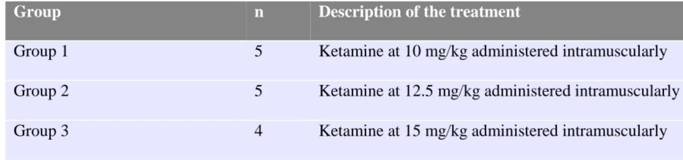
The kids were randomly divided into three groups, two with five animals and one with four, and were given 10, 12.5 and 15 mg/kg of ketamine (100 mg/mL) intramuscularly (Table 2). As
20
to propose a feasible protocol and simulate how a farmer would work in real life, the mean weight was calculated, 4 kg, so that kids were given 0.4 mL of ketamine in Group 1, 0.5 mL in Group 2 and 0.6 mL in Group 3. The kids were individually marked on the back with a coloured spray corresponding to their group to facilitate observation. Behaviours such as vocalization, head scratching, head shaking, tail flicking, allogrooming and self-grooming were visually monitored and registered for one hour every 30 seconds for the first 5 minutes, followed by every 1 minute the next 5 minutes and then every 5 minutes by three observers blind to the dose given (Appendix IV), based on Hempstead et al. (2017) modified ethogram on the behaviour patterns of dairy goat kids (Appendix V). This allowed to determine which may be related to anaesthesia and not to pain, in addition to evaluating the drug’s safety and duration of effect. The latter was considered from the moment the animal was recumbent to it being awake and standing unaided.
All behaviours were recorded using a video camera from the beginning of anaesthesia to one hour after.
Table 2 – Treatment description of the experimental groups.
Group n Description of the treatment
Group 1 5 Ketamine at 10 mg/kg administered intramuscularly
Group 2 5 Ketamine at 12.5 mg/kg administered intramuscularly
Group 3 4 Ketamine at 15 mg/kg administered intramuscularly
2.2.4. Disbudding study
The same direct and video behaviour observations were used in this trial.
Nine healthy goat kids with body weight ranging between 5 and 6.5 kg, aged between 5 and 12 days were randomly allocated to one of three treatment groups (Table 3): (a) given ketamine (K, n = 3), (b) given ketamine and meloxicam (K+M, n = 3) or (c) simulation of disbudding with a cold iron after being given ketamine (S, n = 3). Two to five minutes prior to disbudding, ketamine was administered intramuscularly to all kids at 10 mg/kg. Group K+M additionally received 0.5 mg/kg of meloxicam subcutaneously five minutes before the procedure. For a safer anaesthesia all kids were weighted and each kid was given the exact amount of ketamine for their bodyweight.
The kids were manually restrained, the head being immobilized and the legs free to move, and the procedure was performed by an experienced farmer with a hot-iron while a trained observer evaluated pain based on an ethogram previously developed, registering the number of
21
vocalizations, tail flicking and leg movements per animal (Appendix VI). A Kalfarm HORN’UP C iron was applied one to three times per horn, up to 6 seconds at a time. After disbudding, an antibacterial spray was applied to the cauterized horn bud area. Sham disbudded kids were equally handled but a cold disbudding iron was used with no antibacterial spray being needed. All kids were marked on the back with a coloured spray correspondent to their group to facilitate observation.
Table 3 – Intervention and treatment description of the experimental groups.
Group n Description of the treatment
Ketamine (K) 3 Ketamine at 10 mg/kg administered intramuscularly 2 to 5 minutes before hot-iron disbudding.
Ketamine plus Meloxicam (K + M) 3 0.5 mg/kg of meloxicam injected subcutaneously 5 minutes before the procedure, in addition to the administration of ketamine as mentioned above. Sham disbudded (S) 3 A cold disbudding iron was held against the kids horn
buds so as to simulate the procedure 2 to 5 minutes after being given ketamine at 10 mg/kg.
After disbudding, all kids were placed in the same pen (Figure 7) with milk buckets available for feeding. Frequency of head shaking, head scratching, feeding, allogrooming, running, head friction, body shaking, jumping, self-grooming and vocalization was registered based on a modified ethogram for three hours at pre-determined intervals (Appendix VII) by three observers who were unaware of the treatment given to each group apart from the S kids as they were identifiable. Behaviour was continuously recorded using a video camera.
22 2.2.5. Healing study
Sixteen healthy goat kids aged from 3 to 7 days old with weights ranging from 4 to 6 kg were randomly allocated to one of two groups (Table 4): (i) administration of an antibacterial spray on each cauterized horn bud site immediately after disbudding as is routinely done (n = 8), or (ii) administration of Tri-Solfen®, an anaesthetic and antiseptic gel (n = 8) with lidocaine, bupivacaine, adrenaline and cetrimide. Two trials were held on separate days with 4 kids in each group to facilitate observation.
For the procedure, kids were handled as in the previous study and behaviours were registered by an experienced observer based on the ethogram previously used. After disbudding and application of the respective treatment kids were placed in the same pen and observed for three hours at pre-determined intervals by two trained observers. Behaviours were registered based on Hempstead et al. (2017) ethogram and were also supported by video recordings. At 15 minutes, 1 hour and 3 hours after the procedure, pain response to pressure was determined using a Wagner FORCE TEN™ FPX algometer, in Kgf, applied perpendicularly against the center of the cauterized horn bud area. Pressure was increased until each kid showed withdrawal reflex from the stimulus with the respective value being registered. This process was repeated twice for each horn bud. During these instants, no behaviours were registered.
Table 4 – Treatment description of the experimental groups. Experimental
group
n Description of the treatment
Control (D) 8 Kids were disbudded with a cautery iron and an antibacterial spray was applied on each horn bud site immediately after the procedure.
Tri-Solfen (TS) 8 Kids were handled as above but instead an anaesthetic and antiseptic gela was used.
aTri-Solfen® Bayer Animal Health, Australia.
Three days after the trial and procedure, all kids’ wounds were evaluated by three independent observers blind to treatment using a 3-point healing scale (Table Z): (1) normal healing with no scab formation or exudate, (2) a scab is present, and (3) exudate is present. Animals that scored as 3 were treated with antibacterial spray.
23
Table 5 – Wound evaluation scale description (Photographs taken by the author). Healing
scale
Description Example
photograph
1 Normal healing in which no scab is present nor exudate.
2 A scab is present.
3 Exudate is present and visible.
2.3. STATISTIC ANALYSIS
The R software was used to analyse the data. Using the analysis of variance (ANOVA), the mean difference of the pain-related behaviours between groups of the disbudding study in addition to the positive control of the healing study was assessed in the first half-hour, second half hour, second hour and third hour post disbudding.
Since the variables of the healing study were determined as non-normal by the Shapiro-Wilk normality test, differences between groups were calculated using the non-parametric two-sample Wilcoxon Rank Sum test.