Universidade de Lisboa
Faculdade de Motricidade Humana
Biomechanics of the trunk and lower limbs
during gait in individuals with and without
chronic low back pain
Tese elaborada com vista à obtenção do Grau de Doutor em
Motricidade Humana na Especialidade de Biomecânica
Orientador: Doutor Paulo Alexandre Silva Armada da Silva
Coorientadora: Doutora Annelies Pool-Goudzwaard
Júri:
Presidente
Reitor da Universidade de Lisboa
Vogais
Doutor João Paulo Flores Fernandes
Professor Catedrático do Departamento de Engenharia Mecânica da Universidade do Minho
Doutor António Prieto Veloso
Professor Catedrático da Faculdade de Motricidade Humana da Universidade de Lisboa
Doutor Paulo Alexandre Silva Armada da Silva
Professor Auxiliar da Faculdade de Motricidade Humana da Universidade de Lisboa
Doutora Filipa Oliveira da Silva João
Professora Auxiliar da Faculdade de Motricidade Humana da Universidade de Lisboa
Doutora Andreia Sofia Pinheiro Sousa
Professora Adjunta da Escola Superior de Tecnologia da Saúde do Porto do Instituto Politécnico do Porto
Rita Noélia Silva Fernandes
Julho de 2016
Universidade de Lisboa
Faculdade de Motricidade Humana
Biomechanics of the trunk and lower limbs
during gait in individuals with and without
chronic low back pain
Rita Noélia Silva Fernandes
Tese elaborada com vista à obtenção do Grau de Doutor em
Motricidade Humana na Especialidade de Biomecânica.
Tese por compilação de artigos, realizada ao abrigo da alínea a)
do nº2 do art.º 31º do Decreto-Lei nº 230/2009
Orientador: Doutor Paulo Alexandre Silva Armada da Silva
Coorientadora: Doutora Annelies Pool-Goudzwaard
Júri:
Presidente
Reitor da Universidade de Lisboa
Vogais
Doutor João Paulo Flores Fernandes
Professor Catedrático do Departamento de Engenharia Mecânica da Universidade do Minho
Doutor António Prieto Veloso
Professor Catedrático da Faculdade de Motricidade Humana da Universidade de Lisboa
Doutor Paulo Alexandre Silva Armada da Silva
Professor Auxiliar da Faculdade de Motricidade Humana da Universidade de Lisboa
Doutora Filipa Oliveira da Silva João
Professora Auxiliar da Faculdade de Motricidade Humana da Universidade de Lisboa
Doutora Andreia Sofia Pinheiro Sousa
Professora Adjunta da Escola Superior de Tecnologia da Saúde do Porto do Instituto Politécnico do Porto
i
Declaração de Reprodução da Tese
Nome: Rita Noélia Silva Fernandes
Endereço eletrónico: rita.fernandes@ess.ips.pt
Número do Cartão de Cidadão: 12367694
Título:
Biomechanics of the trunk and lower limbs during gait in individuals with and without chronic low back pain
Orientadores:
Professor Doutor Paulo Alexandre Silva Armada da Silva
Professora Doutora Annelies Pool-Goudzwaard
Ano de conclusão: 2016
Designação do ramo de conhecimento do Doutoramento:
Motricidade Humana na Especialidade de Biomecânica
______________________________________________________
É AUTORIZADA A REPRODUÇÃO INTEGRAL DESTA TESE/TRABALHO APENAS PARA EFEITOS DE INVESTIGAÇÃO, MEDIANTE DECLARAÇÃO ESCRITA DO
INTERESSADO, QUE A TAL SE COMPROMETE.
______________________________________________________
Faculdade de Motricidade Humana – Universidade de Lisboa, 28/07/2016
iii
The mere formulation of a problem is often far more essential than its solution, which may be merely a matter of mathematically or experimental skill. To raise new questions, new possibilities, to regard old problems from a new angle require creative imagination and marks real advances in science.
v
Dedicatória
vii
Acknowledgments
The accomplishment of this work would not have been possible without the support of several people to whom I am very grateful.
First, I would like to thank my supervisor, Dr Paulo Armada da Silva with whom I first started this journey. This thesis followed a different path from the initially established research project and Paulo supported the progress of this work without limiting my own decisions. That gave me the confidence to get out of my comfort zone and to engage in different learning experiences and therefore to grow scientifically and as a person. Paulo also accepted and encouraged the participation of researchers with different competencies in this work, which was crucial for the amplification of my own scientific knowledge and personal perspective. I am also thankful to Dr Annelies Pool-Goudzwaard, who is co-supervisor of this thesis. A large part of this thesis is about the importance of accuracy when using measurement tools and Annelies was who first introduced me the importance of this concept in research and in clinical contexts. During this process, she also strongly contributed to my awareness regarding the importance of using high quality methodological standards in research. Annelies often stimulated the importance of a more translational research perspective and the establishment of connections between the laboratory and the clinical needs.
I am also grateful to Professor António Prieto Veloso, who is the head of the laboratory where this thesis took place. António received me in his laboratory and had an important role in my learning process regarding the biomechanical methods used in this thesis and in the overcoming of some difficulties regarding their use. He allowed the participation of his collaborators and made me possible to learn and discuss with two other scientists highly recognized, namely Dr Scott Selbie and Tom Kepple, who were fundamental regarding the development of the biomechanical model used in this thesis.
I would also like to thank to the colleagues who challenged me in many different ways and also all the participants who volunteered to participate in this study.
I am especially thankful to Vera Moniz-Pereira, Eduardo Brazete Cruz, Silvia Cabral and Filomena Carnide. Besides of being very critical colleagues with whom I am constantly learning, they are very good friends who greatly supported and encouraged my work.
viii
This thesis was a long journey that would not have been possible if I was not surrounded by good friends and a very supportive family that have always been there for me. Especially, I am grateful to Marco who with all his patience and love made this thesis a truly shared journey.
This work was also not possible without the PhD grant supported by the Polytechnic Institute of Setubal (PhD Grant reference: SFRH/PROTEC/67505/2010).
ix
Resumo
Integrar a informação sobre a cinemática e a cinética do tronco durante a marcha constitui um enorme desafio para as áreas da investigação e da clínica. Esta abordagem permite o aprofundamento do conhecimento sobre os mecanismos subjacentes aos padrões de movimento que se encontram alterados. Não obstante o crescente recurso à análise tridimensional da marcha para a avaliação de indivíduos saudáveis e com dor lombar crónica, a fiabilidade e o erro padrão de medida desta técnica não são ainda totalmente conhecidos. A presente dissertação procura resolver esta limitação, através do estudo das diferenças na biomecânica do tronco e dos membros inferiores durante a marcha em indivíduos saudáveis e com dor lombar crónica. Para a concretização desta dissertação, foram desenvolvidos três estudos. Os dois primeiros, com um desenho prospetivo, foram centrados na avaliação da fiabilidade e do erro de medição na análise tridimensional da marcha. Nestes estudos, os participantes (indivíduos saudáveis e com dor lombar crónica) foram submetidos a um protocolo de avaliação da marcha, com dois momentos distintos e com um intervalo médio de uma semana. Os dados foram recolhidos através de um sistema optoeletrónico (composto por treze câmaras) e de três plataformas de força. O processamento dos dados centrou-se nos parâmetros espaço-temporais da marcha, assim como nos valores máximos e mínimos dos ângulos e momentos articulares do tronco e membros inferiores. No terceiro estudo, com um desenho transversal, avaliaram-se as diferenças na biomecânica do tronco durante a marcha entre indivíduos saudáveis e indivíduos com dor lombar crónica. Para o efeito, determinou-se a variabilidade do movimento determinou-segmentar do tórax, lombar e anca e avaliou-determinou-se a sua correlação recíproca, tendo sido confirmada a presença de uma associação significativa entre as rotações residuais nos indivíduos com dor lombar crónica. Os estudos de reprodutibilidade revelam que a análise tridimensional da marcha é consistente, mas demonstram a presença de diferenças importantes na fiabilidade entre ângulos, momentos articulares e parâmetros espaço temporais, sendo o nível de erro todavia aceitável, sobretudo no plano sagital. Demonstrou-se ainda que a dor lombar crónica altera a variabilidade do movimento dos segmentos lombar e torácico durante a marcha e reduz a magnitude dos momentos articulares do tronco, também durante a marcha. Podemos, pois, afirmar que as alterações cinemáticas e cinéticas descritas suportam a existência de um padrão de proteção nestes indivíduos.
Palavras-Chave:
xi
Abstract
Combining information on kinetics and kinematics of the trunk during gait is important for both clinical and research purposes, since it can help in better understanding the mechanisms behind changes in movement patterns in chronic low back pain patients. Although three-dimensional gait analysis has been used to evaluate chronic low back pain and healthy individuals, the reliability and measurement error of this procedure have not been fully established. The main purpose of this thesis is to gain a better understanding about the differences in the biomechanics of the trunk and lower limbs during gait, in patients and healthy individuals. To achieve these aims, three studies were developed. The first two, adopted a prospective design and focused on the reliability and measurement error of gait analysis. In these test-retest studies, chronic low back pain and healthy individuals were submitted to a gait assessment protocol, with two distinct evaluation moments, separated by one week. Gait data was collected using a 13-camera opto-electronic system and three force platforms. Data analysis included the computation of time-distance parameters, as well as the peak values for lower limb and trunk joint angles/moments. The third study followed a cross sectional design, where gait in chronic low back pain individuals was compared with matched controls. Step-to-step variability of the thoracic, lumbar and hips was calculated, and step-to-step deviations of these segments from their average pattern (residual rotations) were correlated to each other. The reliability studies in this thesis show that three-dimensional gait analysis is a reliable and consistent procedure for both chronic low back pain and healthy individuals. The results suggest varied reliability indices for multi-segment trunk joint angles, joint moments and time-distance parameters during gait, together with an acceptable level of error (particularly regarding sagittal plane). Our findings also show altered stride-to-stride variability of lumbar and thoracic segments and lower trunk joint moments in patients. These kinematic and kinetic results lend support to the notion that chronic low back pain individuals exhibit a protective movement strategy.
Keywords
xiii
Contents
Acknowledgments ... vii Resumo ... ix Abstract ... xi List of Figures ... xvList of Tables ... xvii
List of Abbreviations ... xix
Chapter 1. General Introduction ... 1
Chapter 2. Test-retest reliability and minimal detectable change of three-dimensional gait analysis in chronic low back pain patients ... 19
Chapter 3. Three-dimensional multi-segmental trunk kinematics and kinetics during gait: Test-retest reliability and minimal detectable change ... 39
Chapter 4. Loss of variability and altered three-dimensional trunk and hip kinetics during gait in chronic low back pain individuals ... 59
Thesis related outcomes ... 101
xv
List of Figures
Figure 1 - Marker Set ... 25
Figure 2 - Bland-Altman plots with 95% limits of agreement (dashed lines) for thoracic and lumbar peak joint angles in the pain group. ... 30
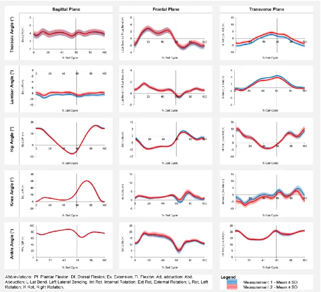
Figure 3 - Plots of joint angles waveforms during the gait cycle in the pain group. ... 31
Figure 4 - Plots of joint angles waveforms during the gait cycle in the control group. 50
Figure 5 - Plots of joint moments waveforms during the gait cycle in control group. ... 52
Figure 6 - Thoracic, lumbar and hip joint angles gait cycle waveforms (sagittal, frontal and transverse planes) in the pain and control group. ... 69
Figure 7 - Mean (SE) differences in the peaks of joint angles of the thoracic, lumbar and hip segments during gait in the pain and control group. ... 69
Figure 8 - Thoracic, lumbar and hip joint moments gait cycle waveforms (sagittal, frontal and transverse planes) in pain and control group. ... 70
Figure 9 - Mean (SE) differences in the peaks of joint moments of the thoracic, lumbar and hip segments during gait in the pain and control group. ... 70
Figure 10 - Residual rotations (º) of the thoracic (frontal) in x-axis and lumbar (transverse) in y-axis and residual rotations of the lumbar (transverse) in x-axis and hip (sagittal) in y-axis. ... 71
xvii
List of Tables
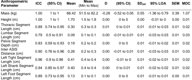
Table 1 - Reliability values for anthropometric measurements in the pain group. ... 28
Table 2 - Reliability values for time-distance parameters in the pain group. ... 28
Table 3 - Reliability values for kinematic parameters in the pain group. ... 29
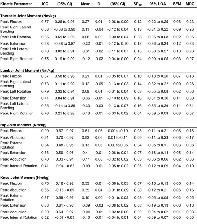
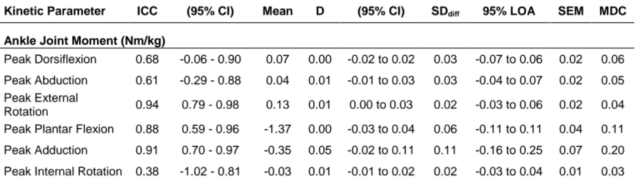
Table 4 - Reliability values for kinetic parameters in the pain group. ... 32
Table 5 - Reliability values for anthropometric and time-distance measurements in the control group. ... 47
Table 6 - Reliability values for kinematic parameters in the control group. ... 48
Table 7 - Reliability values for kinetic parameters in the control group. ... 51
Table 8 - Subjects characteristics’ and self-report measures in the pain and control group. ... 68
Table 9 - Gait parameters in the pain and control group. ... 68
Table 10 - Spearman correlation coefficients (rs), p values and correlation coefficients squared (Rs2) from thoracic, lumbar and hip residual rotations of the pain and control group. ... 72
xix
List of Abbreviations
2D - Two-dimensional
3D - Three-dimensional
3DGA - Three-dimensional gait analysis
ASIS - Anterior superior iliac spine
BMI - Body mass index
BPAQ - Baecke physical activity questionnaire
CI – Confidence interval CLBP - Chronic low back pain
D - Mean difference between measurements
GBD - Global burden of disease
GCVSPL – Generalized cross-validatory cubic spline smoothing routine
ICC - Intraclass correlation coefficient
LBP – Low back pain
LCS - Local coordinate system
LOA - Limits of agreement
MDC - Minimal detectable change
MSE – Mean standard error NRS - Numerical rating scale
OR - Odds ratio
PSIS - Posterior superior iliac spine
QBPDS - Quebec back pain disability scale
rs - Spearman correlation coefficients
Rs2 - Correlation coefficients squared
SDdiff - Standard deviation of the
difference
SE – Standard error
SEM - Standard error of measurement
SO - Segment optimization
STA - Soft tissue artifacts
TSK - Tampa scale of kinesiophobia
Chapter 1: General Introduction
1
General Introduction
3
Ch.1
General Introduction
Low back pain (LBP) is one of the most common health problems in society and causes considerable disability, work absenteeism, and use of health services (Cassidy, Côté, Carroll, & Kristman, 2005). A systematic analysis for the Global Burden of Disease (GBD) published in the Lancet (Vos et al., 2012) reported that LBP stands out as the leading musculoskeletal disorder because of a combination of high prevalence and greater disability weight associated with this health state. According to this report, LBP is one of the four most common disorders in all regions, and is the leading cause of years lived with disabilities (YLDs) in all developed countries. Low back and neck pain account for 70% of all YLDs from musculoskeletal disorders, and for every YLD due to neck pain there are 2.5 YLDs related to LBP. The burden as estimated in this report is substantially higher than previously assessed in the GBD 1990 and GBD 2000 rounds of estimations, which combined with the 33.3% increase in YLDs from 1990 to 2010 driven largely by population growth and ageing, have important implications for health systems. Estimates from GBD also identify low back and neck pain as the primary cause of YLDs in Portugal, with an increase of 13% from 1999 to 2013 (Burden global of Disease, 2013).
A meta-analysis on the clinical course of pain and disability in patients with acute LBP, confirm the earlier findings that typical course of acute LBP is initially favourable, i.e. is characterized by a marked reduction in the mean of pain and disability in the first six weeks (Costa et al., 2012). However, from this point forward, improvement slows down and thereafter only small reductions in mean pain and disability are apparent up to one year, when low to moderate levels of pain and disability are expected. A systematic review on the long-term course of LBP indicates that after an episode of low back pain, 42 to 75% of the patients still experience pain 12 months later (Hestbaek, Leboeuf-Yde, & Manniche, 2003). Moreover, 44% to 78% suffer from a relapse of back pain, 26 to 37% have relapses of work absence and 26% to 37% suffer from recurrent sick leave. The study also reports that the prevalence of LBP in cases with previous episodes is 56% (range 14–93%), compared with 22% (range 7–39%) for those without a prior history of LBP.
Based on time-related criteria, LBP can be classified as acute (up to six weeks), sub-acute (between six weeks and three months) and chronic (over three months) (Koes, van Tulder, & Thomas, 2006). Especially chronic low back pain (CLBP) has a large economic impact, mainly because of the above described sickness absence and
long-4
term disability and is considered to be one of the largest health related challenges in industrialized societies (Parthan, Evans, & Le, 2006). In Portugal, epidemiological findings have shown that musculoskeletal chronic pain is one of the most common complaints in general population (36.7%), with CLBP being among the main reasons for patients' sick health care (Azevedo, Costa-Pereira, Mendonça, Dias, & Castro-Lopes, 2012; Castro-Castro-Lopes, Saramago, Romão, & Paiva, 2010). A recent study conducted under the scope of EpiReumaPt found that among 10.661 subjects, 1487 self-reported CLBP, resulting in a prevalence of 10.4 % (95 % CI, 9.6 to 11.9 %) (Gouveia et al., 2016). This study also found that CLBP was associated with disability and with a high use of healthcare resources.
In the absence of a specific patho-anatomic diagnosis, approximately 90% of the individuals with LBP are labelled as “non-specific low back pain” or some equivalent term (Hancock, Maher, Laslett, Hay, & Koes, 2011) which, in essence, is a diagnosis based on exclusion of specific pathology (Koes et al., 2006). Consequently, a generic symptomatic treatment is applied and the results obtained are not satisfactory. A great variety of interventions, including multidisciplinary treatment, cognitive behavioural therapy and supervised exercise therapy, have been proposed for the treatment of non-specific CLBP (Airaksinen et al., 2006; Koes et al., 2010). However, the available evidence from placebo-controlled trials shows only small to moderate analgesic treatment effects, over and above placebo, for many interventions that are currently used in the management of non-specific acute or chronic LBP (Machado, Kamper, Herbert, Maher, & McAuley, 2009). The limitations of current approaches are further illustrated by the many systematic reviews that reveal that existing treatments for non-specific CLBP have, at best, only small effects (Deyo, 2004; Machado et al., 2009). One example is a systematic review that assessed the overall responses to treatments among non-specific LBP patients in 118 clinical trials (Artus, van der Windt, Jordan, & Hay, 2010). Results showed a similar pattern of initial improvement at 6 weeks followed by smaller improvement for both pain and functional disability at long-term follow-up. This was also shown by the pooled standardized mean difference for pain, which was 0.86 (95% CI, 0.65 to 1.07) at 6 weeks, 1.07 (95% CI, 0.87 to 1.27) at 13 weeks, 1.03 (95% CI, 0.82 to 1.25) at 27 weeks and 0.88 (95% CI, 0.60 to 1.1) at 52 weeks. The rational behind the modest effect of current interventions is not clear, however, one possible explanation is the heterogeneity in development of persistent pain trajectories between patients. Studies show that the course can differ per individual or group: some improve more rapidly, some more slowly, whereas others may fluctuate (Dunn, Jordan,
5
Ch.1
& Croft, 2006). The modest benefit may also be explained by patients’ response to treatment, since it can be assumed that individuals with different characteristics may respond differently to specific rehabilitation programmes.
Considering the high prevalence rates of CLBP and the limitations of current approaches, it is important to gain insight regarding the factors that may be associated with the development and course of this condition, as well as the ones influencing its outcome prognosis. This information is particularly important for further development of specific interventions based on previously identified modifiable prognostic factors. Literature has shown different groups of factors that can contribute to CLBP and disability, including psychosocial and biological ones (Wand & O’Connell, 2008). Psychological factors are an important part of the chronic LBP experience. They contribute to chronicity, explain a significant amount of the variance of different outcomes (e.g. disability, pain or return to work) among CLBP patients and have been identified as important barriers to pain resolution (Linton, 2000; Pincus, Burton, Vogel, & Field, 2002; Pincus, Vogel, Burton, Santos, & Field, 2006). Maladaptive coping strategies such as negative thinking, pathological fear and abnormal anxiety regarding pain, avoidant behaviour, catastrophizing and hypervigilance have been shown to be associated with high levels of pain, disability and muscle guarding (Linton, 2000; Main & Watson, 1996; Nachemson, 1999). Social factors such as the compensation system, workplace disputes and cultural issues affecting beliefs reinforce the psychological factors that can increase pain (Nachemson, 1999).These factors have been extensively studied and identified as key for the development and course of pain and disability in these patients. However, above described psychosocial factors seem to fall short as prognostic factors for recovery in CLBP. For instance, they seem to be of non-importance in a prognostic model on absolute and relative (30%) recovery on pain and disability in CLBP patients. In this study, at 5-month follow-up the prognostic factor most strongly associated with relative recovery in pain is Body Mass Index (BMI) of ≥ 25-29.9 kg/m2 (Odds Ratio (OR) 1.27 95% CI, 0.99 to 1.62) and higher work
participation at baseline (OR 1.27 95% CI, 0.93 to 1.73), while at 12-month follow-up is being married or living with one adult (OR 1.6 95% CI, 0.99 to 2.57) (Verkerk et al., 2015). Work participation (OR 1.34 95% CI, 0.93 to 1.93) (5-month follow-up), back pain intensity in the previous 3 months (OR 1.42 95% CI, 1.02 to 1.99) and BMI ≥ 30 kg/m2 (OR 1.74 95% CI, 1.10 to 2.76) (12-month follow-up), are the strongest
prognostic factors for absolute recovery (Verkerk et al., 2015). Factors of importance for recovery in disability at 5 and 12 months are younger age and higher scores on
6
disability and on the 36-Item Short-Form Health Survey at baseline. At 5-month follow-up, a shorter duration of complaints is a positive predictor, and having no comorbidity and less pain at baseline are additional predictors at 12-month follow-up (Verkerk & Luijsterburg, 2013).
Besides psychosocial factors, also biological ones are described to contribute to the development, persistence and recurrence of LBP. These factors are broader than potential nociceptive sources and include central modulation of pain and physical impairments (Hancock et al., 2011). Regarding the latter, people with LBP show notable limitations in both spinal and hip motion that compromise function, which may have impact on their quality of life (Shum, Crosbie, & Lee, 2005a). Studies that attempted to estimate these mobility impairments have focused on different functional activities and identified altered biomechanical patterns in LBP individuals during sit-to-stand and reverse (Shum et al., 2005a), putting on a sock (Shum, Crosbie, and Lee 2005b) and backward/forward bending (Shum, Crosbie, & Lee, 2010). In CLBP, motor control tasks seem to be altered, with patients demonstrating loss of variability during functional tasks and delayed reflexes (van Dieën, Cholewicki, & Radebold, 2003). This might also interfere with walking. Many CLBP individuals report problems with this complex activity, which is probably the reason why it has been the focus of studies, particularly with respect to its kinematics characteristics. Based on prior research, it appears that people with CLBP who are allowed to self-select walking speed, consistently walk slower (Lamoth et al., 2002; Lamoth, Stins, Pont, Kerckhoff, & Beek, 2008; Müller, Ertelt, & Blickhan, 2015), take shorter steps and have asymmetric step lengths, when compared with their healthy peers (Keefe & Hill, 1985; Vogt, Pfeifer, Portscher And, & Banzer, 2001). Regarding joint angles, literature shows conflicting results: some investigators have reported that people with CLBP display less axial rotation of the pelvis (Müller et al., 2015) or the lumbar segment (Gombatto et al., 2015), while others found no significant differences in absolute axial rotation of the trunk, thorax or pelvis between CLBP individuals and controls (Lamoth et al., 2002; Seay, Van Emmerik, & Hamill, 2011). Furthermore, one study reported that the degree of lumbar or thorax axial rotation depended on temporal parameters of the gait cycle (Crosbie, de Faria Negrão Filho, Nascimento, & Ferreira, 2013). Specifically, the authors verified that comparatively to healthy controls, recurrent LBP patients show less axial rotation of the thorax and lumbar during mid-stance and heel strike, respectively, but higher axial rotation of the lumbar during mid-stance. According to literature, the mentioned changes in trunk and pelvis mobility during gait in CLBP
7
Ch.1
individuals do not seem to be responsible for the development of LBP itself (Hodges & Tucker, 2011). They play a major role as an adaptive response to allow a short-term protection from further pain, injury, or both. This adaptation may have consequences that could lead to further problems in the long term (Hodges & Moseley, 2003; Hodges & Tucker, 2011).
To study the adaptive response to LBP, many researchers looked at different responses in variability and coordination of the trunk and pelvis during gait. Recent findings suggest that patients exhibit a reduced ability to adapt trunk–pelvis coordination in response to changes in gait velocity (Lamoth, Daffertshofer, Meijer, & Beek, 2006), display a more rigid and less flexible pelvis-thorax coordination (Lamoth et al., 2002), and have lower variability of trunk rotations, as a result of the coupling of deviations of residual rotations (in shape and amplitude) between pelvis and trunk (van den Hoorn, Bruijn, Meijer, Hodges, & van Dieën, 2012). Crosbie et al. (2013) also suggest that coordination between adjacent segments might be dependent of gait cycle phase, i.e. limited motion of the pelvis translates to reduced lumbar and lower thoracic angular displacement in LBP individuals at mid-stance sub-phase of the gait cycle. These findings are in disagreement with Vogt et al., (2001) who verified higher stride-to-stride variability and increased fluctuations in dynamic oscillations of angular displacement of thoracic and pelvic segments, in a sample of CLBP patients. These contradictory results may be explained by the fact that, in these studies, different methods were used to compute kinematics variability (Crosbie et al., 2013; Lamoth et al., 2006; van den Hoorn et al., 2012; Vogt et al., 2001). Moreover, the use of different walking surfaces (overground vs. treadmill), biomechanical models and the heterogeneous nature of LBP, may also have contributed to the conflicting results, even though it was suggested that these measures provide valuable information when assessing the quality of gait in these patients (Lamoth et al., 2002).
The kinematic analysis of functional activities is highly valuable. However, it remains descriptive and cannot fully explore the biomechanical mechanisms underlying changes in movement strategies and the nature of the loading patterns in the lumbar spine (Shum, Crosbie, & Lee, 2007). Previous studies that attempted to estimate kinetic variables in LBP individuals have mainly focused on functional activities that included flexion and extension of the trunk, namely lifting tasks (Kingma et al., 2001; Marras, Davis, Ferguson, Lucas, & Gupta, 2001), sit-to-stand and reverse (Shum, Crosbie, & Lee, 2009; Shum et al., 2007), as well as backward/forward bending (Shum, Crosbie, & Lee, 2010). Those studies found that, compared to healthy subjects, LBP
8
individuals had decreased sagittal joint moments acting on the lumbar spine at the end of the available range during forward/backward bending and sit-to-stand, had increased transverse plane joint moments during sit-to-stand. In addition, a decreased muscle power around the lumbar spine and hip was demonstrated in LBP individuals during sit-to-stand and stand-to-sit. This, as well as the above mentioned changes in kinematics and functional tasks, contribute to an explanatory theory that patients adopt a protective strategy in terms of reduced joint moments and powers acting on the spine and hips, in order to prevent further pain (Shum et al., 2007, 2010). In line with this hypothesis, previous studies also showed that LBP individuals recruit their muscles differently and have alterations of the flexion-relaxation response typically seen in asymptomatic counterparts (Alschuler, Neblett, Wiggert, Haig, & Geisser, 2009). Specifically, in LBP individuals flexion-relaxation was absent or significantly impaired (surface electromyographic activity persists at full trunk flexion) (McGorry & Lin, 2012), suggesting distinctive muscle activation patterns that may impose an altered load on the lumbar spine (Shum et al., 2007). According to our best knowledge, studies with CLBP individuals that focused on complex activities, such as gait, have limited their analysis to kinematic and electromyographic variables (Gombatto et al., 2015; Lamoth et al., 2006; van den Hoorn et al., 2012; Vogt et al., 2001). Combining information on the kinetics and kinematics of the trunk during gait is of importance, since it can help in better understanding the mechanisms behind the changes in movement patterns in CLBP.
Currently, three-dimensional (3D) analysis is considered a valid measurement tool to study change in motor adaptive patterns during gait and is classified as the ‘gold standard’ method (Meldrum, Shouldice, Conroy, Jones, & Forward, 2014). Validity refers to whether a given instrument or test measures what it aims to measure (Streiner, Norman, & Cairney, 2015). Another essential requirement of all outcome measures is its reproducibility. This concerns to the degree to which repeated measurements in stable study subjects provide similar results, and depends on the measurement error (how close the scores for repeated measurements are) and reliability (how well can subjects be distinguished from each other, despite measurement errors) (de Vet, Terwee, Knol, & Bouter, 2006). Information regarding the reproducibility of three-dimensional gait analysis (3DGA) still requires more investigation within healthy and clinical populations. Comparisons with asymptomatic participants have been made, profiles have been suggested and, in some cases, recommendations regarding intervention strategies were made, without taking into
9
Ch.1
account the measurement error and the minimal detectable change (MDC) of the measured gait variables.
Variability between ‘before’ and ‘after’ a given intervention may be due to treatment effects or measurement variation, or a combination of both. Knowledge on the error magnitude can enable clinical teams to minimise the risk of over-interpreting small differences as meaningful (Schwartz, Trost, & Wervey, 2004) and to have greater confidence that the treatment effect exceeds the measurement error. Additionally, the use of measurements with low reliability in clinical research may lead to underestimation or failure to detect significant effect sizes; with too much noise (error) drowning out real effects (McGinley, Baker, Wolfe, & Morris, 2009). To address change accurately in health-related outcomes, clinicians need measurement tools that show responsiveness and are able to detect minimal changes in performance over time (Streiner et al., 2015). This change must be large enough to be considered a “real” change and precise enough to detect small but important clinical changes over time considered to be important by patients and/or clinicians (Demoulin, Ostelo, Knottnerus, & Smeets, 2010; Terwee et al., 2007). The observed variability in gait data can be attributed to two sources: intrinsic variability or true variation in the patient’s gait pattern, and extrinsic variability due to methodological errors in marker placement, anthropometric measurements, or calibration of the motion capture system. Another source of variability in 3DGA is the “soft tissue artifacts” (STA), that arises from movement or deformation of the subcutaneous tissues associated with muscular contractions, skin movement and inertial effects (Cappozzo, Catani, Leardini, Benedetti, & Croce, 1996). The extent of STA for any movement depends upon the physical characteristics of individuals, marker locations and the nature of the movement task being performed (Peters, Galna, Sangeux, Morris, & Baker, 2010). While intrinsic variation cannot be reduced, the measurement variation that arises from extrinsic factors can be controlled (Schwartz et al., 2004). It is generally accepted that two major sources of error in 3DGA data are marker placement and STA, although other factors, such as inconsistent anthropometric measurements, variation in walking speed, data processing or measurement equipment errors, may also contribute to data variation (Monaghan, Delahunt, & Caulfield, 2007). Although 3DGA constitutes a complex procedure for daily clinic, the ability of clinicians to discern findings that are meaningful from those that are insignificant or artifactual is nonetheless essential.
A number of studies have evaluated the reliability of kinematic and kinetic parameters during gait in healthy and clinical populations, namely cerebral palsy, stroke,
10
adolescent idiopathic scoliosis, hip osteoarthritis, cervical spondylotic myelopathy and incomplete spinal cord injury. Approaches to the analysis of reliability in 3DGA have differed among studies. Some examined the reliability of kinematic and kinetic curves over the complete gait cycle (Delval et al., 2008; Meldrum et al., 2014; Schwartz et al., 2004; Steinwender et al., 2000), while others extracted key points from those curves, such as a peak value or a range (Fortin, Nadeau, & Labelle, 2008; Klejman, Andrysek, Dupuis, & Wright, 2010; McDermott, Bolger, Keating, McEvoy, & Meldrum, 2010). Key points have been considered more meaningful, as they are easier to compare and interpret than complete curves, and tend to include the most clinically relevant features of the curves (Redekop, Andrysek, & Wright, 2008). A systematic review based on the results of 15 studies using 3DGA, found highest reliability for kinematic parameters in the sagittal plane (intraclass correlation coefficient (ICC) 0.8), with the exception of pelvic tilt (ICC 0.6), and lowest reliability in the transverse plane (ICC <0.7) (McGinley et al., 2009). The authors reported standard error of measurement (SEM) values around 4º in the sagittal plane and 2º in the frontal plane, concluding that most kinematic parameters showed moderate to good reliability, but not small enough measurement errors that may be ignored in clinical interpretation. So, to study adaptive responses in CLBP during 3DGA, reliability and measurement error analysis have to be incorporated.
Although there is evidence that clinically acceptable errors are possible in 3DGA in both healthy individuals and patients (e.g. cerebral palsy or stroke), data on the reliability and measurement error of 3DGA in CLBP patients is lacking. According to a systematic review (Mieritz, Bronfort, Kawchuk, Breen, & Hartvigsen, 2012), studies that evaluated the reproducibility of 3D spinal motion analysis in CLBP patients focused on simple movements (e.g. flexion or extension of the trunk) and are difficult to interpret due to incomplete reporting of the studies’ populations, testing protocol, statistics and data presentation. The majority of the included studies did not report agreement parameters, which may question the performance of the evaluated instruments in clinical practice. Recent studies that aimed to examine the reliability and measurement error of 3D spinal motion parameters, verified that sagittal and frontal plane kinematics of the trunk (modelled as a whole segment) may be sufficiently reliable in measurements of groups of CLBP patients (Harsted, Mieritz, Bronfort, & Hartvigsen, 2016; Mieritz, Bronfort, Jakobsen, Aagaard, & Hartvigsen, 2014). Since reliability of measurement tools can be population (Streiner & Norman, 2008) and task specific, studies with the purpose of investigating test-retest reliability and MDC of 3DGA in a sample of CLBP patients are needed.
11
Ch.1
Additionally, to assess change in walking over time, clinical gait analysis typically seeks to compare between normal and abnormal gait (McGinley et al., 2009). Moreover, knowledge about reliability and MDC values from healthy population is extremely important since it can help clinicians and researchers interpret pathological data. As previously mentioned, several studies have investigated the reliability of 3DGA in healthy individuals and patients, revealing error values of less than 5º for all gait variables, excluding hip and knee rotation (McGinley et al., 2009). Likewise, moderate to good reliability for sagittal and frontal plane variables was reported, with the exception of pelvic tilt and knee varus/valgus in some studies. Two studies (Meldrum et al., 2014; Wilken, Rodriguez, Brawner, & Darter, 2012) provided absolute measures of measurement error and MDC values for kinematic and kinetic parameters in healthy individuals. Meldrum et al. (2014) reported low SEM (≤5˚) for the majority of the lower limb kinematic parameters and variable ICC values (0.14 to 0.92), while Wilken et al., (2012) reported good to excellent reliability of lower limb and trunk kinematics/kinetics across a range of controlled walking velocities, and low MDC values (approximately of 5º for joint angles). By adding trunk data, Wilken’s study made an important contribution to the knowledge on this topic. However, the authors considered the trunk as one rigid segment and excluded information regarding transverse plane kinetics, which may contribute to clinical reasoning and decision-making when dealing with movement disorders. Considering that sufficient evidence exists supporting that different regions of the trunk move differently, we can argue that one of the main limitations of the prior work is that the whole trunk was considered a single rigid segment. Thus, studies aimed at investigating reliability and MDC of kinematics and kinetics of multi-segment trunk in 3DGA in healthy and CLBP individuals are needed.
Thesis Aims and Methodology Synopsis
Although 3DGA has been used to measure kinematics in CLBP individuals, the reliability and measurement error of this evaluation procedure, in this specific population, has not been established. Additionally, trunk kinematics and kinetics can contribute to more detailed information on gait impairment. However, data on reliability and measurement error of multi-segment trunk on 3DGA is lacking on both healthy and CLBP individuals. Once we have the information on reliability and agreement, it will be possible to rigorously compare the gait of individuals with and without CLBP and to gain insight into the differences between their movement patterns.
12
Based on the lacking or conflicting knowledge described and discussed throughout this chapter, the main purpose of this thesis is to gain a better understanding about the differences in the biomechanics of the trunk and lower limbs during gait in CLBP and healthy individuals. Specifically, the established aims are:
To investigate test-retest reliability and MDC of 3DGA in a sample of CLBP patients.
To investigate test-retest reliability and MDC of 3DGA kinematic and kinetic data in a sample of healthy individuals, using a two rigid segment model for the trunk.
To compare lumbar and thoracic kinematics and kinetics between CLBP and healthy individuals during gait, taking into account the error values.
To assess the variability of movement between lumbar and thoracic segments, in association with joint moments and angles, in CLBP patients versus healthy individuals.
With our two final goals we expect to have a better understanding on the differences between the movement patterns’ of CLBP and healthy individuals, which will broaden future research and open up possibilities about how to change motor adaptive patterns and protective strategies in patients.
To achieve these aims, the thesis is divided into three distinct but related studies. In the first two, the focus lies on the reliability and measurement error studies. Two prospective (within assessor) test-retest studies, where participants (CLBP or healthy individuals) underwent two biomechanical gait assessments with a mean interval of one week, were conducted. Gait data were collected using a 13-camera opto-electronic system and three force platforms. Participants were instructed to walk during a few minutes at their preferred velocity and 10 gait cycles were selected for further processing. A Woltring generalized cross-validatory cubic spline smoothing routine was applied to kinematic and kinetic data. The marker set selection was based on previous reports (Leardini, Biagi, Merlo, Belvedere, & Benedetti, 2011; Seay, Selbie, & Hamill, 2008) and a 9 segments model (feet, shanks, thighs, pelvis, lumbar and thoracic segments) was built and optimized through segment optimization. Data analysis included ICCs, the mean difference between measurements (D), and the 95% Confidence interval (CI) for D, the standard deviation of the differences (SDdiff) and the
95% Bland and Altman limits of agreement (95% LOA) for anthropometric, time-distance and key kinematic/kinetic parameters. The third study followed a cross
13
Ch.1
sectional design, where gait of CLBP individuals was compared with matched controls. The data collection system, marker set, biomechanical model and laboratory procedures for data collection were the same as in the reliability studies. Filtering process and data optimization also followed the same procedures. Data analysis included the computation of time-distance parameters, peak values for hip and trunk joint angles/moments. Step-to-step variability of the thoracic, lumbar and hips was also calculated, and step-to-step deviations of these segments from their average pattern (residual rotations) were correlated to each other.
Thesis Outline
Chapter 2 presents a study in which reliability and minimal detectable change of 3DGA is tested in a sample of CLBP patients.
Chapter 3 reports the results of a study that evaluated the reliability and minimal detectable change of a two rigid segment model for the trunk during 3DGA, in sample of healthy individuals.
Chapter 4 describes the results of study that compared the variability of movement between lumbar and thoracic segments, in association with joint moments and angles, in CLBP patients versus healthy individuals. In this study the results of the lumbar and thoracic kinematics and kinetics are interpreted according to error values obtained in the reliability studies.
Chapter 5 addresses the main results of this thesis, discusses some methodological options, and makes some recommendations for further research.
References
Airaksinen, O., Brox, J. I., Cedraschi, C., Hildebrandt, J., Klaber-Moffett, J., Kovacs, F, et al. (2006). Chapter 4. European guidelines for the management of chronic nonspecific low back pain. European Spine Journal, 15 Suppl 2, S192–300. Alschuler, K. N., Neblett, R., Wiggert, E., Haig, A. J., & Geisser, M. E. (2009).
Flexion-relaxation and Clinical Features Associated With Chronic Low Back Pain. The
Clinical Journal of Pain.
Artus, M., van der Windt, D. A., Jordan, K. P., & Hay, E. M. (2010). Low back pain symptoms show a similar pattern of improvement following a wide range of primary care treatments: A systematic review of randomized clinical trials.
14
Azevedo, L. F., Costa-Pereira, A., Mendonça, L., Dias, C. C., & Castro-Lopes, J. M. (2012). Epidemiology of chronic pain: a population-based nationwide study on its prevalence, characteristics and associated disability in Portugal. The Journal of
Pain, 13(8), 773–83.
Cappozzo, A., Catani, F., Leardini, A., Benedetti, M. G., & Croce, U. Della. (1996). Position and orientation in space of bones during movement: experimental artefacts. Clinical Biomechanics (Bristol, Avon), 11(2), 90–100.
Cassidy, J. D., Côté, P., Carroll, L. J., & Kristman, V. (2005). Incidence and course of low back pain episodes in the general population. Spine, 30(24), 2817–2823. Castro-Lopes, J., Saramago, P., Romão, J., & Paiva, M. (2010). Pain proposal: A Dor
Crónica em Portugal, 1–12.
Crosbie, J., de Faria Negrão Filho, R., Nascimento, D. P., & Ferreira, P. (2013). Coordination of spinal motion in the transverse and frontal planes during walking in people with and without recurrent low back pain. Spine, 38(5), E286–92. da C Menezes Costa, L., Maher, C. G., Hancock, M. J., McAuley, J. H., Herbert, R. D.,
& Costa, L. O. P. (2012). The prognosis of acute and persistent low-back pain: a meta-analysis. CMAJ, 184(11), E613–24.
de Vet, H. C. W., Terwee, C. B., Knol, D. L., & Bouter, L. M. (2006). When to use agreement versus reliability measures. Journal of Clinical Epidemiology, 59(10), 1033–9.
Delval, A., Salleron, J., Bourriez, J.L., Bleuse, S., Moreau, C., Krystkowiak, P., Duhamel, A. (2008). Kinematic angular parameters in PD: reliability of joint angle curves and comparison with healthy subjects. Gait & Posture, 28(3), 495–501. Demoulin, C., Ostelo, R., Knottnerus, J. A., & Smeets, R. J. E. M. (2010). Quebec Back
Pain Disability Scale was responsive and showed reasonable interpretability after a multidisciplinary treatment. Journal of Clinical Epidemiology, 63(11), 1249–55. Deyo, R. A. (2004). Treatments for back pain: can we get past trivial effects? Annals of
Internal Medicine, 141(12), 957–8.
Dunn, K. M., Jordan, K., & Croft, P. R. (2006). Characterizing the Course of Low Back Pain: A Latent Class Analysis, American Journal of Epidemiology, 163(8), 754– 761.
Fortin, C., Nadeau, S., & Labelle, H. (2008). Inter-trial and test-retest reliability of kinematic and kinetic gait parameters among subjects with adolescent idiopathic scoliosis. European Spine Journal, 17(2), 204–16.
Global burden of Disease (2013). Portugal. Institute for Health Metrics and Evaluation website. Retrieved June 27, 2016, from http://www.healthdata.org/portugal.
Gombatto, S. P., Brock, T., DeLork, A., Jones, G., Madden, E., & Rinere, C. (2015). Lumbar spine kinematics during walking in people with and people without low back pain. Gait & Posture, 13–15.
Gouveia, N., Rodrigues, A., Eusébio, M., Ramiro, S., Machado, P., Canhão, H., & Branco, J. C. (2016). Prevalence and social burden of active chronic low back pain in the adult Portuguese population: results from a national survey.
15
Ch.1
Hancock, M. J., Maher, C. G., Laslett, M., Hay, E., & Koes, B. (2011). Discussion paper: what happened to the “bio” in the bio-psycho-social model of low back pain? European Spine Journal, 20(12), 2105–10. 3
Harsted, S., Mieritz, R. M., Bronfort, G., & Hartvigsen, J. (2016). Reliability and measurement error of frontal and horizontal 3D spinal motion parameters in 219 patients with chronic low back pain. Chiropractic & Manual Therapies, 24, 13.
Hestbaek, L., Leboeuf-Yde, C., & Manniche, C. (2003). Low back pain: what is the long-term course? A review of studies of general patient populations. European
Spine Journal, 12(2), 149–65.
Hodges, P. W., & Moseley, G. L. (2003). Pain and motor control of the lumbopelvic region: effect and possible mechanisms. Journal of Electromyography and
Kinesiology, 13(4), 361–70.
Hodges, P. W., & Tucker, K. (2011). Moving differently in pain: A new theory to explain the adaptation to pain. Pain, 152(3), S90–S98.
Keefe, F. J., & Hill, R. W. (1985). An objective approach to quantifying pain behavior and gait patterns in low back pain patients. Pain, 21(2), 153–61.
Kingma, I., Baten, C. T. M., Dolan, P., Toussaint, H. M., van Dieën, J. H., de Looze, M. P., & Adams, M. A. (2001). Lumbar loading during lifting: a comparative study of three measurement techniques. Journal of Electromyography and Kinesiology, 11(5), 337–345.
Klejman, S., Andrysek, J., Dupuis, A., & Wright, V. (2010). Test-retest reliability of discrete gait parameters in children with cerebral palsy. Archives of Physical
Medicine and Rehabilitation, 91(5), 781–7.
Koes, B. W., van Tulder, M., Lin, C.-W. C., Macedo, L. G., McAuley, J., & Maher, C. (2010). An updated overview of clinical guidelines for the management of non-specific low back pain in primary care. European Spine Journal, 19(12), 2075–94. Koes, B. W., van Tulder, M. W., & Thomas, S. (2006). Diagnosis and treatment of low
back pain. BMJ (Clinical Research Ed.), 332(7555), 1430–4.
Lamoth, C. J. C., Daffertshofer, A., Meijer, O. G., & Beek, P. J. (2006). How do persons with chronic low back pain speed up and slow down? Trunk-pelvis coordination and lumbar erector spinae activity during gait. Gait & Posture, 23(2), 230–9. Lamoth, C. J. C., Meijer, O. G., Wuisman, P. I. J. M., van Dieën, J. H., Levin, M. F., &
Beek, P. J. (2002). Pelvis-thorax coordination in the transverse plane during walking in persons with nonspecific low back pain. Spine, 27(4), E92–9.
Lamoth, C. J. C., Stins, J. F., Pont, M., Kerckhoff, F., & Beek, P. J. (2008). Effects of attention on the control of locomotion in individuals with chronic low back pain.
Journal of Neuroengineering and Rehabilitation, 5, 13.
Leardini, A., Biagi, F., Merlo, A., Belvedere, C., & Benedetti, M. G. (2011). Multi-segment trunk kinematics during locomotion and elementary exercises. Clinical
Biomechanics (Bristol, Avon), 26(6), 562–71.
Linton, S. J. (2000). A review of psychological risk factors in back and neck pain.
16
Machado, L. A. C., Kamper, S. J., Herbert, R. D., Maher, C. G., & McAuley, J. H. (2009). Analgesic effects of treatments for non-specific low back pain: a meta-analysis of placebo-controlled randomized trials. Rheumatology (Oxford, England), 48(5), 520–7.
Main, C., & Watson, P. (1996). Guarded movements: development of chronicity.
Journal of Musculoskeletal Pain, 4(4), 163–70.
Marras, W., Davis, K., Ferguson, S., Lucas, B., & Gupta, P. (2001). Spine loading characteristics of patients with low back pain compared with asymptomatic individuals. Spine, 26(23), 2566–74.
McDermott, A., Bolger, C., Keating, L., McEvoy, L., & Meldrum, D. (2010). Reliability of three-dimensional gait analysis in cervical spondylotic myelopathy. Gait &
Posture, 32(4), 552–8.
McGinley, J. L., Baker, R., Wolfe, R., & Morris, M. E. (2009). The reliability of three-dimensional kinematic gait measurements: a systematic review. Gait & Posture, 29(3), 360–9.
McGorry, R. W., & Lin, J.-H. (2012). Flexion relaxation and its relation to pain and function over the duration of a back pain episode. PloS One, 7(6), e39207.
Meldrum, D., Shouldice, C., Conroy, R., Jones, K., & Forward, M. (2014). Test-retest reliability of three dimensional gait analysis: including a novel approach to visualising agreement of gait cycle waveforms with Bland and Altman plots. Gait
& Posture, 39(1), 265–71.
Mieritz, R. M., Bronfort, G., Jakobsen, M. D., Aagaard, P., & Hartvigsen, J. (2014). Reliability and measurement error of sagittal spinal motion parameters in 220 patients with chronic low back pain using a three-dimensional measurement device. The Spine Journal, 14(9), 1835–43.
Mieritz, R. M., Bronfort, G., Kawchuk, G., Breen, A., & Hartvigsen, J. (2012). Reliability and measurement error of 3-dimensional regional lumbar motion measures: a systematic review. Journal of Manipulative and Physiological Therapeutics, 35(8), 645–56.
Monaghan, K., Delahunt, E., & Caulfield, B. (2007). Increasing the number of gait trial recordings maximises intra-rater reliability of the CODA motion analysis system.
Gait & Posture, 25(2), 303–15.
Müller, R., Ertelt, T., & Blickhan, R. (2015). Low back pain affects trunk as well as lower limb movements during walking and running. Journal of Biomechanics, 48(6), 1009–1014.
Nachemson, A. (1999). Back pain: delimiting the problem in the next millennium.
International Journal of Law and Psychiatry, 22(5-6), 473–90.
Parthan, A., Evans, C. J., & Le, K. (2006). Chronic low back pain: epidemiology, economic burden and patient-reported outcomes in the USA. Expert Review of
Pharmacoeconomics & Outcomes Research, 6(3), 359–69.
Peters, A., Galna, B., Sangeux, M., Morris, M., & Baker, R. (2010). Quantification of soft tissue artifact in lower limb human motion analysis: a systematic review. Gait
17
Ch.1
Pincus, T., Burton, A. K., Vogel, S., & Field, A. P. (2002). A systematic review of psychological factors as predictors of chronicity/disability in prospective cohorts of low back pain. Spine, 27(5), E109–20.
Pincus, T., Vogel, S., Burton, a K., Santos, R., & Field, A. P. (2006). Fear avoidance and prognosis in back pain: a systematic review and synthesis of current evidence. Arthritis and Rheumatism, 54(12), 3999–4010.
Redekop, S., Andrysek, J., & Wright, V. (2008). Single-session reliability of discrete gait parameters in ambulatory children with cerebral palsy based on GMFCS level.
Gait & Posture, 28(4), 627–33.
Schwartz, M. H., Trost, J. P., & Wervey, R. A. (2004). Measurement and management of errors in quantitative gait data. Gait & Posture, 20(2), 196–203.
Seay, J. F., Van Emmerik, R. E. A., & Hamill, J. (2011). Influence of low back pain status on pelvis-trunk coordination during walking and running. Spine, 36, E1070–9.
Seay, J., Selbie, W. S., & Hamill, J. (2008). In vivo lumbo-sacral forces and moments during constant speed running at different stride lengths. Journal of Sports
Sciences, 26(14), 1519–29.
Shum, G. L., Crosbie, J., & Lee, R. Y. (2009). Energy transfer across the lumbosacral and lower-extremity joints in patients with low back pain during sit-to-stand.
Archives of Physical Medicine and Rehabilitation, 90(1), 127–35.
Shum, G. L. K., Crosbie, J., & Lee, R. Y. W. (2005a). Effect of low back pain on the kinematics and joint coordination of the lumbar spine and hip during sit-to-stand and stand-to-sit. Spine, 30(17), 1998–2004.
Shum, G. L. K., Crosbie, J., & Lee, R. Y. W. (2005b). Symptomatic and asymptomatic movement coordination of the lumbar spine and hip during an everyday activity.
Spine, 30(23), E697–702.
Shum, G. L. K., Crosbie, J., & Lee, R. Y. W. (2007). Three-dimensional kinetics of the lumbar spine and hips in low back pain patients during sit-to-stand and stand-to-sit. Spine, 32(7), E211–9.
Shum, G. L. K., Crosbie, J., & Lee, R. Y. W. (2010). Back pain is associated with changes in loading pattern throughout forward and backward bending. Spine, 35(25), E1472–8.
Steinwender, G., Saraph, V., Scheiber, S., Zwick, E. B., Uitz, C., & Hackl, K. (2000). Intrasubject repeatability of gait analysis data in normal and spastic children.
Clinical Biomechanics (Bristol, Avon), 15(2), 134–9.
Streiner, D. L., & Norman, G. R. (2008). Health Measurement Scales: A Practical
Guide to their Development and Use (4th ed.). United Kingdom: Oxford University
Press.
Streiner, D. L., Norman, G. R., & Cairney, J. (2015). Health Measurement Scales: A
practical guide to their development and use (5 edition.). Great Clarendon Street,
18
Terwee, C. B., Bot, S. D. M., de Boer, M. R., van der Windt, D. a W. M., Knol, D. L., Dekker, J., … de Vet, H. C. W. (2007). Quality criteria were proposed for measurement properties of health status questionnaires. Journal of Clinical
Epidemiology, 60(1), 34–42.
van den Hoorn, W., Bruijn, S. M., Meijer, O. G., Hodges, P. W., & van Dieën, J. H. (2012). Mechanical coupling between transverse plane pelvis and thorax rotations during gait is higher in people with low back pain. Journal of
Biomechanics, 45(2), 342–7.
van Dieën, J. H., Cholewicki, J., & Radebold, A. (2003). Trunk muscle recruitment patterns in patients with low back pain enhance the stability of the lumbar spine.
Spine (Phila Pa 1976), 28(8), 834–841.
Verkerk, K., & Luijsterburg, P. (2013). Prognosis and Course of Disability in Patients With Chronic Nonspecific Low Back Pain: A 5- and 12-Month Follow-up Cohort Study. Physical Therapy, 93(12), 1603–1614.
Verkerk, K., Luijsterburg, P. a J., Heymans, M. W., Ronchetti, I., Pool-Goudzwaard, a L., Miedema, H. S., & Koes, B. W. (2015). Prognosis and course of pain in patients with chronic non-specific low back pain: A 1-year follow-up cohort study.
European Journal of Pain, 1–10.
Vogt, L., Pfeifer, K., Portscher And, M., & Banzer, W. (2001). Influences of nonspecific low back pain on three-dimensional lumbar spine kinematics in locomotion.
Spine, 26(17), 1910–9.
Vos, T., Flaxman, A. D., Naghavi, M., Lozano, R., Michaud, C., Ezzati, M., … Moradi-Lakeh, M. (2012). Years lived with disability (YLDs) for 1160 sequelae of 289 diseases and injuries 1990-2010: A systematic analysis for the Global Burden of Disease Study 2010. The Lancet, 380(9859), 2163–2196.
Wand, B. M., & O’Connell, N. E. (2008). Chronic non-specific low back pain - sub-groups or a single mechanism? BMC Musculoskeletal Disorders, 9, 11.
Wilken, J. M., Rodriguez, K. M., Brawner, M., & Darter, B. J. (2012). Reliability and Minimal Detectible Change values for gait kinematics and kinetics in healthy adults. Gait & Posture, 35(2), 301–7.
Chapter 2. Test-retest reliability and minimal detectable change of three-dimensional gait analysis in chronic low back pain patients
2
Test-retest reliability and minimal
detectable change of
three-dimensional gait analysis in
chronic low back pain patients
Rita Fernandes, Paulo Armada da Silva,
Annelies Pool-Goudzwaard,
Vera Moniz Pereira and António P. Veloso
Based on:
Gait Posture. 2015 Oct;42(4): 491-7.
21
Ch.2
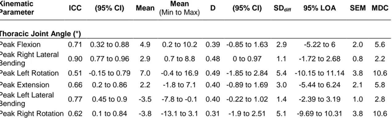
Abstract
Background and Aim: Three-dimensional gait analysis can provide detailed data on gait impairment in CLBP patients. However, data about reliability and measurement error of 3DGA in this population is lacking. The aim of this study is to investigate test-retest reliability and minimal detectable change of 3DGA in a sample of CLBP patients.
Methods: A test-retest study was conducted with a sample of 14 CLBP patients that underwent two biomechanical gait assessments with an interval of 7.6±1.8 days. Anthropometric and time-distance parameters, as well as peak values for lower limb and trunk joint angles and moments, were computed. ICC3,k and their 95% confidence
intervals were calculated. SEM, MDC and limits of agreement (LOA) were also estimated.
Results: The obtained ICC values demonstrate high test-retest reliability for most joint angles, with low SEM (<2.5º) values. Although joint moments showed lower reliability than joint angles, the majority of the ICCs were above 0.7 and the SEM and MDC values were low (≤0.06 Nm/kg and ≤0.18 Nm/kg). Bland-Altman plots with 95% LOA revealed a good agreement and time-distance parameters were all highly repeatable (ICCs > 0.86).
Conclusions: The results of this study show high test-retest reliability for lower limb and trunk joint angles, and time-distance parameters during gait in CLBP individuals, together with a low measurement error. These results also support the use of this method in clinical assessments of CLBP patients’ gait patterns.
Keywords
23
Ch.2
2.1 Introduction
Chronic Low back pain is a common health condition in western industrialised countries with an estimated prevalence of 20.1±9.8% (Hoy et al., 2012). Patients often report difficulties during daily activities, such as gait. Studies have reported that gait coordination is changed in CLBP patients: they walk slower, take shorter steps and have asymmetric step lengths when compared with their healthy peers (Keefe & Hill, 1985; Vogt, Pfeifer, Portscher And, & Banzer, 2001). Chronic low back pain patients also have difficulty in moving from pelvis-trunk in-phase to anti-phase (pelvis and trunk moving in the same or in opposite directions, respectively) as walking speed increases (Lamoth et al., 2002) and consequently show lower variability of trunk rotations, possibly adopting a protective movement strategy to diminish pain (Huang et al., 2011).
In clinical settings, gait evaluation in CLBP patients is frequently carried out by observation and functional tests (Andersson, Lin, & Smeets, 2010), or is included in specific disability questionnaires (Malliou, Gioftsidou, Beneka, & Godolias, 2006), which only provide limited information. In contrast, although time consuming, 3DGA can provide detailed quantitative data concerning gait impairment (Baker, 2013). As an advantage in CLBP patients, 3D instruments can obtain real-time information on 3D lumbar spine kinematics and kinetics without any known risk to the patients (Mieritz, Bronfort, Jakobsen, Aagaard, & Hartvigsen, 2014). Thus, 3DGA can assist in reaching clinical functional diagnoses and can be useful to evaluate the outcome of therapeutic interventions (Mieritz et al., 2014). However, as with any analysis tool, reliability and measurement error emerge as critical factors in its applicability to clinical decision-making (McDermott, Bolger, Keating, McEvoy, & Meldrum, 2010). Since low reliability in clinical research may lead to underestimation or failure to detect significant effect sizes (McGinley, Baker, Wolfe, & Morris, 2009), we have to strive for good reliability. In addition, knowledge of the error’s magnitude can minimise the risk of over-interpreting small differences as meaningful (de Vet et al., 2006) and contribute to the certainty that a measured intervention effect exceeds the measurement error.
Data on reliability and measurement error of 3DGA in CLBP patients is lacking, although evidence exist that clinically acceptable errors are possible in 3DGA in healthy individuals and in patients with cerebral palsy or stroke (McGinley et al., 2009). The few studies that evaluated reliability and measurement error of 3D spinal motion analysis in CLBP patients (Mieritz et al., 2014; Mieritz, Bronfort, Kawchuk, Breen, & Hartvigsen, 2012) focused on simple activities and are difficult to interpret due to