Nota Técnica
*e-mail: [email protected]
DEVELOPMENT AND VALIDATION OF A HIGH PERFORMANCE LIQUID CHROMATOGRAPHIC METHOD FOR DETERMINATION OF CYCLOSPORINE-A FROM BIODEGRADABLE INTRAOCULAR IMPLANTS
Juliana Barbosa Saliba, Armando da Silva Cunha Junior e Elionai Cassiana de Lima Gomes
Faculdade de Farmácia, Universidade Federal de Minas Gerais, Av. Pres. Antônio Carlos, 6627, 31270-901 Belo Horizonte - MG, Brasil Herman Sander Mansur
Departamento de Engenharia Metalúrgica e de Materiais, Universidade Federal de Minas Gerais, Av. Pres. Antônio Carlos, 6627, 31270-901 Belo Horizonte - MG, Brasil
Gisele Rodrigues da Silva*
Faculdade de Farmácia, Universidade Federal de São João Del Rei, Rua Sebastião Gonçalves Coelho 400, 35501-296 Divinópolis - MG, Brasil
Recebido em 21/1/10; aceito em 18/6/10; publicado na web em 9/11/10
DEVELOPMENT AND VALIDATION OF A HIGH PERFORMANCE LIQUID CHROMATOGRAPHIC METHOD FOR DETERMINATION OF CYCLOSPORINE-A FROM BIODEGRADABLE INTRAOCULAR IMPLANTS. An HPLC method was developed and validated aiming to quantify the cyclosporine-A incorporated into intraocular implants, released from them; and in direct contact with the degradation products of PLGA. The separation was carried out in isocratic mode using acetonitrile/water (70:30) as mobile phase, a C18 column at 80
oC and UV detection at 210 nm. The method provided selectivity based on resolution
among peaks. It was linear over the range of 2.5-40.0 µg/mL. The quantitation and detection limits were 0.8 and 1.2 µg/mL, respectively. The recovery was 101.8% and intra-day and inter-day precision was close to 2%.
Keywords: cyclosporine-A; biodegradable intraocular implant; validation.
INTRODUCTION
Intraocular implants are controlled (sustained)-drug delivery sys-tems designed for the treatment of several chronic ocular diseases of the posterior segment of the eye. They can be used for the treatment of uveitis and cytomegalovirus retinitis, in which repeated local ad-ministration of drugs is not effective. In diseases such as proliferative vitreoretinopathy and diabetic retinopathy, that frequently require a surgical procedure, intraocular devices implanted adjunctively during surgery may improve recovery. Moreover, the intraocular implants are necessary to treat chronic diseases with no satisfactory therapies, such as geographic atrophy in age-related macular degeneration and macular edema.1
Generally, the intraocular implants are introduced into the vitreous, which is located posterior to the lens and anterior to the retina. Despite the invasive characteristics of the implantation te-chnique, the implants present several advantages that overlap the inconveniences of the implantation procedure. These advantages include: the overcoming of the blood-retina barrier, allowing drug delivery at therapeutic levels directly into the targeted site; prolonged drug delivery; and reduction of side effects frequently observed with intravitreal injections and systemic administration.2-4
The non-biodegradable implants based on ethylene vinyl acetate and/or polyvinylalcohol have been reported by several studies as a successful long-term drug delivery system of ganciclovir,5,6 luocino-lone acetonide,7 cyclosporine-A,8 and betamethasone.9 On the other hand, the major disadvantage of the non-biodegradable systems is the need of the system removal by a second surgery to avoid any risk of ibrous encapsulation formation. In this way, biodegradable systems have gained interest to eliminate the removal step.10The poly (D,L-lactide-co-glycolide) (PLGA) (Figure 1a) is a classic example
amongst the synthetic biodegradable polymers that has been well applied as ocular drug delivery systems,11-14 due to its satisfactory biocompatibility and absence of signiicant in vivo toxicity.4,15
Intraocular implants controlled (sustained)-drug delivery sys-tems based on biodegradable polymers are frequently loaded to corticosteroids, immunosuppressive drugs and/or anti-metabolic
substances. Cyclosporine-A has been widely used in the treatment of various forms of chronic diseases affecting the posterior segment of the eye, because of its potent immunosuppressive activity, with anti-inlammatory properties (Figure 1b).16
determination of cyclosporine-A described in literature are based on high performance liquid chromatography coupled with mass spectro-metry (HPLC-MS).19-21 These methods are applied to quantify either isolated cyclosporine-A or in contact with different kinds of drugs in the same biological sample. The mass spectrometric detection is a re-ference technique for the determination of cyclosporine-A in complex biological samples, because it provides speciic measurement of this drug over a wide analytical range.19,22,23 Nowadays, HPLC-MS has matured to a true alternative to radioimmunoassay in cyclosporine-A monitoring practices.22 On the other hand, HPLC-MS and RIA do not represent viable analytical methods to quantify the drug in routine analysis of quality control of cyclosporine-A in pharmaceutical forms. It is desirable to develop a simple, rapid and reliable HPLC with ul-traviolet detection for assaying the immunosuppressive drug released from pharmaceutical formulations and present in non-complex samples.
Some methods reported in literature for assaying cyclosporine-A are based on the high performance liquid chromatography with ultraviolet detection (HPLC-UV). Bonifacio et al.24 developed and validated an HPLC-UV method for simultaneous determination of the immunosuppressive drug as well as its major impurities in capsules and its generic versions. Baldeli et al.25 reported the de-velopment and validation of an HPLC-UV method for concomitant measurement of everolimus and cyclosporine-A concentration in whole blood. Jaiswal et al.26 determined the cyclosporine-A loaded biodegradable polymeric nanoparticles by an HPLC method at a wavelength detection of 210 nm. Moreover, the United States Phar-macopeia27 described an isocratic HPLC-UV method for assaying cyclosporine-A in capsules and injections using a mixture of ace-tonitrile, water, methanol and phosphoric acid (550:400:50:5.5) as mobile phase, at wavelength of 210 nm and a column that contains dimethylsilane chemically bonded to porous silica particles. All the HPCL-UV methods indicated could be successfully applied to assay cyclosporine-A in different pharmaceutical preparations, manufactured likely by different processes and excipients; but the chromatographic conditions of the described methods were not suitable to quantify unequivocally cyclosporine-A released from biodegradable intraocular implants derived from PLGA.
Recently, we have developed and characterized an intraocular implant based on PLGA and cyclosporine-A intended to be used in the treatment of posterior ocular diseases.16 In this study, our main goal was the development and validation, through speciicity, limits of detection and quantitation, linearity, precision and accuracy, of a simple and reliable HPLC-UV method to quantify the cyclosporine-A incorporated into biodegradable intraocular implants synthesized by PLGA. Additionally, the analytical method was applied to assay the cyclosporine-A released from these polymeric devices and in direct contact with the degradation products of PLGA.
EXPERIMENTAL
Materials and reagents
Cyclosporine-A reference standard was purchased from Sigma Pharma (Brazil) (MW 1202, 99% of purity). Poly(D,L-lactide-co-glycolide) in ratio of 75:25 [PLGA (75:25)] was purchased from Boehringer Ingelheim (Germany). All the solvents and reagents used in buffer solutions, in the preparation of the implants, and mobile phase were HPLC or analytical grade. Water was distilled, deionized and iltered through a 0.22 µm ilter (Millipore, USA).
Preparation of the biodegradable intraocular implants
Firstly, a 25% (w/w) concentration of cyclosporine-A and PLGA
(75:25) was dissolved in an appropriate quantity of acetonitrile, as organic solvent. The solution was placed in a freezer under -80 oC. Afterwards, the frozen solution was lyophilized during 48 h. The in-traocular implants were prepared by molding the lyophilized mixture into rods using a Telon® sheet heated on a hot plate at a temperature from 100 to 120 °C.16 The average weight of implants was 5.9 ± 0.1 mg (n = 10). The implants presented cylindrical shape with approxi-mately 4.00 mm of length and 1.00 mm of diameter.
Instrumentation and analytical conditions
A Waters® system equipped with an autosampler model 717plus (Waters, USA) and a pump model 515 (Waters, USA) was used. An ultraviolet detector model 2487 (Waters, USA) was used in conjunction with Eppendorf CH-500 column oven. Millenium model 2.15.01 (Waters, USA) was used for data acquisition. A Chromolith Performance® 100 RP-18e column, 250 mm × 4.6 i.d. and 5 µm particle size (Merck, Germany), was used and maintained at 80 ºC. The wavelength of 210 nm and the automatic injector it-ted at 10 µL were set. The mobile phase consisit-ted of acetonitrile and ultrailtrated water (70:30, v/v) was used at the low rate of 1.0 mL/min.
Preparation of solutions
Cyclosporine-A standard solution: appropriate amounts of cyclo-sporine-A reference compound was dissolved in 20% of acetonitrile in volumetric lask. Phosphate buffer solution (PBS, pH = 7.4) was added to complete the lask volume. All the standard solutions were iltered through a 0.45 µm i lter (Sartorius, Germany). The concen-µm i lter (Sartorius, Germany). The concen-m ilter (Sartorius, Germany). The concen-tration of cyclosporine-A in each solution prepared is deined in the description of the parameter of validation.
Phosphate buffer solution was prepared by dissolving 0.64 g of sodium chloride, 0.075 g of potassium chloride, 0.048 g of calcium chloride, 0.03 g of magnesium chloride, 0.39 g sodium acetate and 0.17 g of sodium citrate in 1000 mL of water. The pH was adjusted to 7.4 ± 0.1 with sodium hydroxide 1% (w/V).28
Method validation
The method was validated according to the International Con-ference on Harmonization guidelines for validation of analytical procedures.29
Speciicity
The intraocular implants were dissolved in 10 mL of acetonitrile and transferred to a volumetric lask (50 mL). PBS was added to complete the lask volume. Speciicity was evaluated comparing the chromatographic peaks of cyclosporine-A and PLGA (75:25).
Solutions containing the degradation products derived from the PLGA (75:25) were collected after 40 days of incubation of it in PBS at 37 ºC. Speciicity was evaluated comparing the chromatographic peaks of cyclosporine-A reference standard, at 40.0 µg/mL, and of degradation products from PLGA (75:25).
To achieve the speciicity of the method, no peak, with the same retention time of cyclosporine-A, is allowed.
Linearity
regres-sion analysis by the least square regresregres-sion method. The correlation coeficient was calculated.
Limit of detection (LOD) and limit of quantitation (LOQ)
The LOD and LOQ were determined based on the signal to noise method. Cyclosporine-A reference standard solutions at 0.5, 0.8, 1.2 and 1.5 µg/mL were prepared and evaluated. Each solution was inject 3 times in the chromatographic system (n = 3 for each concentration). Precision
The intraocular implant was dissolved in 10 mL of acetonitrile and transferred to a volumetric lask (50 mL). PBS was added to complete the lask volume (n = 8). Cyclosporine-A reference standard solutions at 40.0 µg/mL were prepared. The intra-day precision (repeatabili-ty) was evaluated by analyzing the obtained solutions. Similarly, the inter-day precision (intermediate precision) was evaluated in 2 consecutive days. The precision was expressed as relative standard deviation (RSD) amongst responses.
Accuracy
Solutions containing the degradation products derived from the PLGA (75:25) were collected after 40 days of incubation of it in PBS at 37 ºC. Accuracy was studied by recovery of known amount of cyclosporine-A reference standard in contact with the solution previously prepared. Cyclosporine-A reference standard, at 40 µg/ mL, was dissolved in solution containing the degradation products of PLGA. A blank solution containing cyclosporine-A reference standard, at 40 µg/mL, was also prepared. Solutions were prepared in triplicate with 5 injections of each solution (n = 15). The percent recovery of added cyclosporine-A was calculated comparing peak areas of the resultant solutions with reference standard cyclosporine-A solutions at the same concentration.
Determination of cyclosporine-A content in the biodegradable intraocular implants
The biodegradable intraocular implants were dissolved in 10 mL of acetonitrile and transferred to a volumetric lask (50 mL) (n = 10). PBS was added to complete the lask volume. The amount of cyclosporine-A was determined by high performance liquid chroma-tographic method described above. The obtained amount of the drug was expressed as the percentage content of the pre-indicated value (25%). The relative standard deviation was calculated.
Determination of released cyclosporine-A from biodegradable intraocular implants
The United States Pharmacopeia states in the general chapter <1092> the dissolution procedure: “sink conditions are deined as the volume of medium at least three times that required in order to form a saturated solution of drug substance. When sink conditions are present, it is more likely that dissolution results will relect the properties of the dosage form”.27
The in vitro release of cyclosporine-A from biodegradable intrao-cular implants was carried out under sink conditions during 23 weeks. As the aqueous solubility of cyclosporine-A is 42 ± 1.0 µg/mL at 37 oC,30 sink conditions are achieved with at least 3 mL. The intraocular implants were placed in different tubes containing 2 mL of PBS (n = 3). Those tubes were placed inside an incubator (Tecnal model TE 424) set at 37 oC and 30 rpm. At predetermined intervals, the entire medium was sampled and 2 mL of fresh PBS was added to each tube. The release proile was evaluated as the cumulative percentage of cyclosporine-A released in the medium. The amount of cyclosporine-A in the PBS
solution was detected and quantiied by validated high performance liquid chromatographic method described above.
RESULTS AND DISCUSSION
Optimization of the chromatographic conditions
In this study, an HPLC-UV method was developed and validated for determination of cyclosporine-A content incorporated into intrao-cular implants derived from PLGA. Moreover, the analytical method was also developed and validated for assaying the immunosuppressive drug released from them and in direct contact with the degradation products of PLGA.
The optimal absorption wavelength for detection of cyclosporine-A was chosen based on the higher detector response for this drug. Therefore, the absorption wavelength for detection was 210 nm.
As described in literature,24 high temperature is imperative for as-saying cyclosporine-A in order to assure fast equilibration of individual conformers and therefore to achieve satisfactory peak shape. In this work, different temperatures were tested. However, the temperature of 80 oC contributed to the attainment of a narrow peak shape and a high resolution among cyclosporine-A and by-products of PLGA peaks.
Different proportions of acetonitrile and water, as mobile phase, were tested. The selected proportion was 70:30 in accordance to Jaiswal et al..26 It was observed that increasing the amount of acetonitrile in mobile phase reduced the retention time of cyclosporine-A, a highly hydrophobic drug; and maintained the resolution among peaks of drug and degradation products of PLGA. In addition, the use of this mobile phase in the chromatographic separation of cyclosporine-A and by-products of the polymer reduced the drug peak broadening effect. Method validation
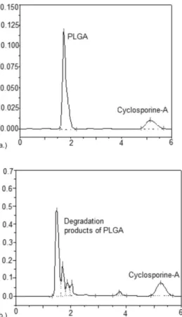
It was observed that PLGA (75:25) and cyclosporine-A eluted at approximately 1.0 min and 5.3 min, respectively (Figure 2a). Under the described HPLC conditions, the degradation products of PLGA and cyclosporine-A eluted at approximately 2.0 and 5.2 min, respectively (Figure 2b). The chromatographic peaks were completely resolved and any substance presented the same retention time of the immu-nosuppressive drug, allowing the unequivocal determination of the drug. Therefore, the method showed speciicity for cyclosporine in the presence of the PLGA and the degradation products of this polymer.
A calibration curve for concentration of the drug versus area of peaks was plotted and the obtained data were subjected to regression analysis by the least square method. The calibration curve was linear over the range of 2.5 to 40.0 µg/mL. The linearity could be deined by following equation y = 37224x – 72255, where y and x were area and concentration (µg/mL), respectively. The correlation coeficient (r) of the obtained curve was 0.9999, indicating highly signiicant correlation between concentration and peak area. The signiicance of the intercept obtained in the calibration curve was tested and this parameter was not statistically signiicant (p > 0.05), consequently, it can be considered that the curve passes through the origin.
The LOD was 0.8 µg/mL and this concentration displayed a signal-to-noise ratio of 3:1. The LOQ was 1.2 µg/mL and this
con-centration displayed a signal-to-noise ratio of 10:1 and RSD of 1.89 and 1.92%, respectively (n = 3 for each concentration).
In the intra-day and inter-day precision analyses (n = 8 for each day), the mean content of cyclosporine incorporated into the polyme-ric implants was 95.78% (RSD = 1.80%) and 96.65% (RSD = 1.65%), respectively. RSD values attested the precision of the HPLC method.
Determination of cyclosporine-A in the biodegradable intraocular implants and released from them
The validated HPLC method described was applied to assay cyclosporine-A content in the intraocular devices. The amount of cyclosporine-A incorporated into the polymeric matrix was of 1.404 mg (n = 10), corresponding to the concentration of 95.2% of the drug. Therefore, the units were inside of the range of 90-110% of the pre-indicated amount of cyclosporine-A. The relative standard deviation for replicates was 1.89%.
Figure 3 shows in vitro release proile of cyclosporine-A from biodegradable intraocular implants and in contact with the degradation products of PLGA. During the irst 13-week period, no signiicant
amounts of cyclosporine-A was released from the devices. In this irst stage, it was considered that the aqueous permeability in the polymeric matrix was not enough to provide the release of the cyclosporine. In a second stage that occurs between the 14th and the 23th week of the test, about 8.4% of cyclosporine-A was released from intraocular implants under sink conditions.16 In this second stage, the dissolution and diffusion of the drug were attributed to the increased aqueous permeability due to hydrolytic degradation of the surface of PLGA It was not observed the cyclosporine-A burst delivery, representing an advantage of the system. CONCLUSION
It was developed an HPLC-UV method for assaying cyclosporine-A incorporated into biodegradable intraocular implants based on PLGA, and released from them. The method also provided unequivo-cal determination of cyclosporine-A in contact with the degradation products of PLGA (75:25). The developed method showed to be speciic, linear, precise and accurate.
ACKNOWLEDGEMENTS
The authors would like to acknowledge the inancial support from the following institutions: CAPES/MEC, CNPq/MCT and FAPEMIG (Brazil).
REFERENCES
1. Yasukawa, T.; Ogura, Y.; Sakurai, E.; Tabata, Y.; Kimura, H.; Adv. Drug Delivery Rev.2005, 57, 2033.
2. Amo, E. M.; Urtti, A.; Drug Discov Today2008, 13, 135.
3. Hughes, P. M.; Olejnik, O.; Chang-Lin, J.; Wilson, C. G.; Adv. Drug Delivery Rev.2005, 57, 2010.
4. Yasukawa, M. D.; Kimura, H.; Tabata, Y.; Ogura, Y.; Adv. Drug Delivery Rev.2001, 52, 25.
5. Sanborn, G. E.; Anand, R.; Torti, R. E.; Nightingale, S. D.; Cal, S. X.; Yates, B.; Ashton, P.; Smith, T.; Arch Ophthalmol. 1992, 110, 188. 6. Kappel, P. J.; Charonis, A. C.; Holland, G. N.; Narayanan, R.; Kulkarni,
A. D.; Yu, F.; Boyer, D. S.; Engstrom, R. E.; Kuppermann,B. D.;
Oph-thalmology2006, 113, 673.
7. Jaffe, G. J.; Yang, C-S.; Wang, X-C.; Cousins, S. W.; Gallemore, R. P.; Ashton, P.; Ophthalmology1998, 105, 46.
8. Jaffe, G. J.; Martin, D.; Callanan, D.; Pearson, P. A.; Levy, B.; Com-stock, T.; Ophthalmology2006, 113, 1020.
9. Okabe, J.; Kimura, H.; Kunou, N.; Okabe, K.; Kato, A.; Ogura, Y.;
Invest. Ophthalmol. Vis. Sci.2003, 44, 740.
10. Felt-Baeyens, O.; Eperon, S.; Mora, P.; Limal, D.; Sagodira, S.; Breton, P.; Simonazzi, B.; Gurny, R.; Int. J. Pharm. 2006, 322, 6.
11. Kunou, N.; Ogura, Y.; Hashizoe, M.; Honda, Y.; Hyon, S.; Ikada, Y.; J.
Controlled Release1995, 37, 143.
12. Kuppermann, B. D.; Blumenkranz, M. S.; Haller, J. A.; Williams, G. A.; Weinberg, D. V.; Chou, C.; Whitcup, S. M;. Arch Ophthalmol. 2007,
125, 309.
13. Fialho, S. L.; Rego, M. G. B.; Siqueira, R. C.; Jorge, R.; Haddad, A.; Rodrigues, A. L.; Maia-Filho, A.; Silva-Cunha, A. J.; Curr. Eye Res. 2006, 31, 525.
14. Zhou, T.; Lewis, H.; Foster, R. E.; Schwendeman, S. P.; J. Controlled Release1998, 55, 281.
15. Avitable, T.; Marano, F.; Castiglione, F.; Bucolo, C.; Cro, M.; Ambrosio, L.; Biomaterials 2001, 22, 195.
16. Saliba, J. B.; Faraco, A. A. G.; Yoshida, M. I.; Vasconcelos, W. L.; Silva-Cunha, A.; Mansur, H. S.; Mater. Res. 2008, 11, 207.
17. Morse, G. D.; Holdsworth, M. T.; Walshe, J. J.; Ther. Drug Monit.1989,
11, 238. Figure 2. (a) Chromatogram of the PLGA (75:25) and cyclosporine-A. (b)
Chromatogram of the degradation products of PLGA (75:25) and cyclospo-rine-A. Chromatographic conditions: C18 column 250 × 4.6 mm at 80 ºC; ACN and water (70:30, v/v); 1.0 mL/min of low rate; wavelength of 210 nm
Figure 3. Cumulative release of cyclosporine-A (CyA) from biodegradable
intraocular implant in PBS (pH = 7.4). The values are shown as mean ± SD
18. Huupponen, R.; Hirvisalo, E-L.; Neuvonen, P.; Ther. Drug Monit.1997,
19, 446.
19. Salm, P.; Taylor, P. J.; Lynch, S. V.; Warnholtz, C. R.; Pillans, P. I.; Clin. Biochem. 2005, 38, 667.
20. Ansermot, N.; Fathi, M.; Veuthey, J. L.; Desmeules, J.; Rudaz, S.; Hochstrasser, D.; Clin. Biochem. 2008, 41, 728.
21. Bogusz, M. J.; Enazi, E. A.; Hassan, H.; Abdel-Jawaad, J.; Ruwaily, J. A.; Al-Tufail, M.;J. Chromatogr., B: Anal. Technol. Biomed. Life Sci.
2007, 850, 471.
22. Seger, C.; Tentschert, K.; Stöggl, W.; Griesmacher, A.; Ramsay, S. L.;
Nat. Protoc.2009, 4, 526.
23. Kaiser, P.; Akerboom, T.; Wood, W. G.; Reinauerm, H.; Ger. Med. Sci.
2006, 4, 41.
24. Bonifacio, F. N.; Giocanti, M.; Reynier, J. P.; Lacarelle, B.; Nicolay, A.;
J. Pharm. Biomed. Anal.2009, 49, 540.
25. Baldelli, S.; Zenoni, S.; Merlini, S.; Perico, N.; Cattaneo, D.; Clin. Chim. Acta2006, 364, 354.
26. Jaiswal, J.; Kumar, S. G.; Kreuter, J.; J. Controlled Release2004, 96, 169.
27. The United States Pharmacopeia, 31th ed., United States Pharmacopeial
Convention: Rockville, 2008.
28. Reynolda, J. E. F.; Martindalethe extra pharmacopoeia, 29th ed.,
Phar-maceutical Press: London, 1989.
29. International Conference on Harmonisation of Technical Requirements for Registration of Pharmaceuticals for Human Use; Validation of
Ana-lytical Procedures: Text and Methodology, 2005.