P
ROTEIN
F
OLDING
,
M
ETAL
I
ONS
AND
C
ONFORMATIONAL
S
TATES
THE CASE OF A DI
-
CLUSTER FERREDOXIN
Dissertation presented to obtain the PhD degree in Biochemistry at the Instituto de Tecnologia Química e Biológica,
Universidade Nova de Lisboa
Sónia Cristina Alves Dickson Leal Solano
Supervisor
Cláudio Emanuel Moreira Gomes
Opponents
Pernilla-Wittung Stafshede and Eduardo P. Melo
Instituto de Tecnologia Química e Biológica, Universidade Nova de Lisboa
The case of a di-cluster Ferredoxin
by Sónia S. Leal
Second Edition, December 2008
ITQB - Protein Biochemistry Folding and Stability Laboratory
This dissertation describes the work performed under the supervision of Cláudio M. Gomes, in the Protein Biochemistry Folding and Stability Laboratory, at the Instituto de Tecnologia Química e Biológica, from October 2004 to May 2008.
The studies presented here aim at understanding the role of metal ions in protein stability and on the folding-unfolding pathways of a zinc-containing ferredoxin holding a [3Fe-4S] and a [4Fe-4S] cluster.
I would like to express my sincere appreciation to whom professionally but in many cases, also personally helped and supported me during my Ph.D contributing to make my aspiration a pleasant stage of my life.
Cláudio M. Gomes, my supervisor for his knowledge, encouragement, and patience that made this work possible. I also want to thank him for his enthusiasm for science and continuous new challenges that always kept me motivated. He has been an inspiring example of commitment and pragmatism that I would much ambition for myself.
My wonderful and dear colleagues at the Protein Biochemistry Folding and Stability group, for their friendship, constant support and help in the laboratory and for providing a most pleasant working environment: Barbara Henriques, Raquel Correia, Hugo Botelho, Vesna Prosinecki and João Rodrigues.
To Rita Rocha from PBFS group at ITQB for the studies on the stability of the di-cluster ferredoxin isoforms from Sulfolobus metallicus
To Vitor H. Teixeira, António Baptista and Cláudio Soares, from ITQB for the molecular dynamics simulations on the di-cluster ferredoxin isoforms from
Sulfolobus metallicus
To Smilja Todorovic from ITQB, Ingo Zebger and Peter Hildebrandt from Technische Universität, Berlin, and Daniel Murgida from the Universidad de Buenos Aires for the FT-IR and RR studies.
To Carlos Salgueiro from the Departamento de Quimíca, Faculdade de Ciências e Tecnologia, Universidade Nova de Lisboa, for the NMR study and useful advices.
To Carlos Frazão, David Aragão, Ricardo Coelho and Maria Arménia Carrondo from ITQB for the crystallographic structure of the di-cluster ferredoxin from
Acidianus ambivalens
To Manuela Regalla from ITQB for sequencing the di-cluster ferredoxin isoforms from Sulfolobus metallicus
To Arnulf Kletzin for receiving me in his laboratory at Darmstadt University and for his collaboration on molecular biology experiments.
To Miguel Teixeira for kindly providing the soluble extract of Acidianus
Fundação para a Ciência e Tecnologia is acknowledged for financial support, by awarding a PhD Grant SFRH/BD/18653/2004
Leal S.S., Teixeira M., Gomes C.M. (2004)
Studies on the degradation pathway of iron-sulfur centres during unfolding of a hyperstable ferredoxin: cluster dissociation, iron release and protein stability
J. Biol. Inorg. Chem. 9: 987-996. Leal S.S. and Gomes C.M. (2005)
Linear three-iron centres are unlikely clusters degradation intermediates during unfolding of iron-sulfur
Biol. Chem.386: 1295-1300.
Rocha R.M., Leal S.S., Teixeira V.H., Regalla M., Huber H., Batista A.M., Soares C.M., Gomes C.M. (2006)
Natural domain design: enhanced thermal stability of zinc lacking ferredoxin isoforms shows that a hydrophobic core efficiently replaces the structural metal site
Biochemistry45: 10376-10384. Leal S.S. and Gomes C.M. (2007)
Towards the understanding of the folding process and cofactor assembly in iron-sulfur proteins: structural characterisation of a ferredoxin molten globule state
Proteins 68: 606-616.
Todorovic S., Leal S.S., Salgueiro C.A., Zebger I., Hildebrandt P., Murgida D.H., Gomes C.M. (2007)
A spectroscopic study of the temperature induced modifications on ferredoxin folding and iron-sulfur moieties
Biochemistry46: 10733-10738
Frazão, C., Aragão, D., Coelho, R., Leal S.S., Gomes, C.M., Teixeira, M., Carrondo, M.A. (2008)
Crystallographic analysis of the intact metal centres [3Fe-4S]1+/0 and [4Fe-4S]2+/1+ in a Zn2+ - containing ferredoxin”
FEBS Lett. 582: 763-767
Leal S.S. and Gomes C.M. (2008)
On the relative contribution of ionic interactions over iron-sulfur clusters to ferredoxin stability
BBA – Proteins and Proteomicsin Press doi:10.1016/j.bbapap.2008.05.001
Other publications not included in this thesis
Boscolo B., Leal S.S., Ghibaud E.M., Gomes C.M. (2007)
Lactoperoxidase folding and catalysis relies on the stabilization of the α-helix rich core domain
Metal ions are present in over thirty percent of known proteins. Apart from a well established function in catalysis and electron transfer, metals and metal centres are also important structural elements which may as well play a key role in modulating protein folding and stability. In this respect, cofactors can act not only as local structural stabilizing elements in the native state, contributing to the maintenance of a given specific structural fold, but may also function as potential nucleation points during the protein folding process.
This dissertation addresses the role of iron-sulfur clusters and a zinc centre in the folding and overall stability of an Archaeal family of di-cluster ferredoxins commonly found among hyperthermophiles. These ferredoxins are small proteins of about 100 amino acids that present a [3Fe-4S] and a [4Fe-4S] centre, as well as an N-terminal extension of ~30 residues that holds a zinc centre and wraps the Fe-S containing core. Interestingly, some highly identical ferredoxin isoforms are simultaneously expressed in some organisms that either contain or lack the zinc site. In this respect, the conformational properties of the zinc-containing (FdA) and zinc-lacking (FdB) isoforms present in Sulfolobus metallicus were used as models, to investigate the effect of the absence of this site on ferredoxin folding and stability. The studies have indicated that FdB assumes a fold identical to that of the Zn2+ containing isoform. The thermal stability of the isoforms was investigated in a broad pH range (2<pH<10), and surprisingly the Zn2+ lacking isoform was always found to be more stable than its Zn2+ containing counterpart: a ΔTm ≈ 9°C is determined at pH 7 a difference which becomes even more
significant at extreme pH values, reaching a ΔTm≈ 24°C at pH 2 and 10. The
contribution of the Zn2+ site to ferredoxin stability was further resolved using selective metal chelators: during thermal unfolding the zinc scavenger TPEN significantly lowers the Tm in FdA (≈ 10°C) whereas it has no effect in FdB. This
formation of a novel local stabilizing hydrophobic core and illustrate a strategy of natural fold design.
One of the aspects focused in this work was the behavior of the Fe-S clusters during ferredoxin unfolding. This fallowed previous studies to this dissertation that suggested that upon protein unfolding the iron-sulfur clusters degraded via linear three-iron sulfur centre species with a transient absorption spectra resembling the one observed in purple aconitase. In order to appraise this hypothesis we proceeded with a detailed kinetic and spectroscopic investigation on the alkaline chemical denaturation of the protein, in an attempt to elucidate the degradation pathway of the iron-sulfur centres in respect to protein unfolding events. For this purpose we investigated cluster dissociation, iron release and protein unfolding by complementary biophysical techniques. We found that shortly after initial protein unfolding, iron release proceeds monophasically at a rate comparable to that of cluster degradation, and that during the process no typical EPR features of linear three-iron sulfur centres are observed. Further, it was observed that EDTA prevents formation of the intermediate and that sulfide significantly enhances its intensity and lifetime, even after protein unfolding. Altogether, our data suggests that iron sulfides, which are formed from the release of iron and sulfide resulting from cluster degradation during protein unfolding in alkaline conditions, are in fact responsible for the observed intermediate spectral species, thus disproving the hypothesis suggesting the presence of a linear three-iron centre intermediate.
sulfides at pH 10, produced from the degradation of the iron centres, and not from rearrangements leading to linear three-iron centres. Thus, at this point, any relevant functional role of linear three-iron centres as cluster degradation intermediates in iron-sulfur proteins remains elusive.
In order to evaluate the relative contribution of iron–sulfur clusters in respect to ionic interactions to the overall protein stability, we have investigated the interplay between structural features and metal cofactors integrity as a function of pH in the ferredoxin model. Changes in protonation affect both the stability and the conformational dynamics of the protein fold. In the pH 5.5–8 interval, the protein has a high melting temperature (Tm ~ 120°C), which decreases towards pH
extremes. Acidification triggers events in two steps: down to the isoelectric point (pH 3.5) the Fe-S clusters remain unchanged, the secondary structure content increases and the single Trp becomes more solvent shielded, denoting a more compact fold. Further acidification down to pH 2 sets off exposure of the hydrophobic core and Fe-S cluster disintegration, yielding a molten globule state. The relative stabilising contribution of the clusters becomes evident when stabilising ionic interactions are switched off as a result of poising the protein at pH 3.5, at an overall null charge: under these conditions, the Fe-S clusters disassemble at Tm= 72 °C, whereas the protein unfolds at Tm= 52°C. Overall, this
ferredoxin denotes a considerable structural plasticity around its native conformation, a property which appears to depend more on the integrity of its metal clusters rather than on the status of its stabilising electrostatic interactions. The latter however play a relevant role in determining the protein thermal stability.
simultaneous to alterations on the secondary structure. Upon cooling, an apo-ferredoxin state is obtained, with characteristics of a molten globule: compactness identical to the native form; similar secondary structure evidenced by far-UV CD; no near-UV CD detected tertiary contacts; and an exposure of the hydrophobic surface evidenced by 1-anilino naphthalene-8-sulfonic acid (ANS) binding. In contrast to the native form, this apo ferredoxin state undergoes reversible thermal and chemical unfolding. Its conformational stability was investigated by guanidinium chloride denaturation and this state is ~1.5 kcal mol-1 destabilised in respect to the holo ferredoxin. The single tryptophan located nearby the Fe-S pocket probed the conformational dynamics of the molten globule state: fluorescence quenching, red edge emission shift analysis and resonance energy transfer to bound ANS evidenced a restricted mobility and confinement within a hydrophobic environment. This rather structured, stable and flexible apo molten globule state lead us to speculate on the possible physiological relevance of this conformational state in the folding of Fe-S cluster proteins and speculate on the possible relevance of this apo state as a on-pathway apo conformation for metal center insertion.
Subsequently, we have assigned in detail the specific alterations occurring on the secondary structure during the formation of the molten globule state, and we have individually screened the transformations occurring on the [3Fe-4S] and [4Fe-4S] clusters along the process. Our main goal with this work was to infer on the possible relevance of this molten globule state in the folding pathway and discriminate the contribution of each cluster for the overall stability of the ferredoxin. In agreement, thermal perturbation of the ferredoxin was investigated employing a toolbox of spectroscopic methods. FTIR and visible CD were used for assessing changes of the secondary structure and coarse alterations of the
[3Fe-4S] and [4Fe-4S] cluster moieties, respectively. Fine details of the
disassembly of the metal centers were revealed by paramagnetic NMR and resonance Raman spectroscopy. Overall, thermally-induced unfolding of AaFd is initiated with the loss of α-helical content at relatively low temperatures (Tmapp ~ 44 °C) followed by the disruption of both iron-sulfur clusters
(Tmapp ~ 58 °C) and a change, rather than a significant net loss, of secondary
structure (Tmapp ~ 60 °C). In fact, this latter process triggers a secondary structure
reorganization that leads to an increase in the β-structure content. The combined spectroscopic approach here reported illustrates how changes in the metalloprotein organization are intertwined with disassembly of the iron-sulfur centers, denoting the conformational interplay of the protein backbone with cofactors. In addition it seems to evidence that both clusters disintegrate in a single event and therefore most likely contribute equally to overall stability. Moreover, the pathway for the formation of this molten globule state clearly evidenced the formation of non-native β-structure along the process which strongly suggests that this conformation is most likely an off-pathway specie.
Os iões metálicos são parte integrante de mais de trinta por cento de todas as proteínas identificadas. Para além do seu proeminente envolvimento em funções de catálise e transferência de electrões, os metais e os centros metálicos são também importantes elementos estruturais, podendo simultaneamente desempenhar um papel chave no enrolamento e estabilidade das proteínas. De facto, os cofactores podem não só actuar como importantes elementos locais estabilizadores do estado nativo contribuindo para a manutenção específica da estrutura, mas também funcionar como determinantes pontos de nucleação durante o processo de enrolamento das proteínas.
Esta dissertação visa essencialmente o estudo do papel dos centros de ferro e enxofre mas tambem do centro de zinco no enrolamento e estabilidade de ferredoxinas de sete ferros. Estas proteínas pertencem á família de ferredoxinas, vulgarmente encontradas em organismos hipertermofilicos da ordem Archaea e caracterizadas por conterem um centro [3Fe-4S] e um centro [4Fe-4S], bem como um centro de zinco numa extensão de ~30 a.a no N-terminal. Curiosamente, isoformas sequencialmente muito idênticas destas ferredoxinas mas que não contêm o centro de zinco são simultaneamente expressas. No sentido de investigar o efeito da ausência deste metal na estrutura e estabilidade da ferredoxina, as propriedades conformacionais destas isoformas expressas em
Sulfolobus metallicus com o centro de zinco (FdA) e que não apresentam o
centro (FdB) foram utilizadas como modelo. Os estudos indicam que a FdB assume um enrolamento estrutural muito semelhante á isoforma que contem o centro de zinco FdA. A estabilidade térmica das isoformas foi investigada a diferentes pHs no intervalo 2 <pH <10 e surpreendentemente a FdB foi para todas as condições testadas sempre mais estável que a sua isoforma, a FdA que contem o centro de zinco: a pH 7 foi observado um ΔTm ≈ 9°C, uma diferença
que se revelou ainda mais prenunciada a pHs extremos atingindo um ΔTm≈ 24°C
Tm na FdA (≈ 10°C), não provocando qualquer alteração na FdB. Isto sugere que
o centro de zinco contribui para a estabilidade da FdA mas também que a estratégia estrutural alternativa da FdB sem o centro de zinco é mais estável. Analise á sequência e modelo estrutural da FdB sugere que o aumento de estabilidade desta isoforma pode resultar de contactos to tipo van der Waals entre os resíduos que ocupam a mesma região espacial onde os ligandos do zinco se encontram na FdA. Estes contactos favorecem a formação de um centro hidrofóbico estabilizador no local e ilustra uma estratégia natural de enrolamento.
provavelmente estão na origem da formação das espécies espectrais intermediarias observadas. Neste sentido os dados apresentados claramente estão em desacordo com a hipótese que sugeria a formação de um centro linear intermediário de três ferros. No seguimento destas evidências com a ferredoxina alargamos este estudo a outras proteínas no sentido de esclarecer duvidas relativamente á possível formação de um centro linear de três ferros durante a desnaturação de proteínas de Fe-S. Nesse sentido, analisamos a cinética de desnaturação química de proteínas com centros de Fe-S contendo e não contendo sulfureto inorgânico bem como proteínas com apenas centros de ferro, na presença e ausência de sulfuretos exógenos. De facto, os resultados claramente evidenciam que é possível reproduzir em todas as proteínas estudadas as espécies espectrais intermediarias associadas á formação de um centro linear de três ferros. A libertação de ferro e a cinética de desnaturação proteica claramente indicam que a formação destas espécies espectrais resultam da formação de sulfuretos de ferro que são produzidos em solução alcalina e não devido ao rearranjo dos centros de Fe-S com formação de um centro linear de três ferros.
No sentido de avaliar o envolvimento das ligações iónicas relativamente á contribuição dos centros de Fe-S para a estabilidade geral da ferredoxina, investigamos a interacção existente entre as características estruturais da proteína e a integridade dos centros de Fe-S em função do pH. Os resultados sugerem que alterações no estado de protonação da proteína afectam a estabilidade e a dinâmica conformacional da estrutura da ferredoxina. No intervalo de pH entre 5.5 e 8 a ferredoxina evidenciou uma média elevada na temperatura de transição (Tm ~120 °C) que contudo diminui significativamente
proteína é incubada ao pH correspondente ao seu ponto isoeléctrico (pH 3.5) e as interacções iónicas nativas são “desligadas”: nestas condições os centros de Fe-S dissociam-se a uma temperatura de transição Tm= 72 °C, enquanto que
outros elementos estruturais apresentam uma temperatura de transição significativamente inferior com um Tm= 52°C. No geral a ferredoxina evidencia
uma elevada plasticidade estrutural em relação á estrutura nativa, uma propriedade que aparenta estar mais dependente da integridade dos centros de Fe-S do que no estado das suas interacções iónicas. No entanto as interacções iónicas nativas evidenciaram um papel determinante na estabilidade térmica da proteína.
A inserção biológica dos centros de Fe-S envolve a interacção de chaperons
moleculares com uma dada forma proteica parcialmente enrolada. Neste sentido o estudo de formas não nativas em proteínas de Fe-S pode contribuir para uma melhor compreensão do processo de enrolamento destas proteínas bem como do processo de inserção dos centros. Assim procedemos á investigação da formação e caracterização de um estado apo molten globule na ferredoxina de sete ferros. Estudos biofísicos revelaram que a pH 2.5 a proteína mantém a integridade dos centros metálicos e a sua estrutura nativa. No entanto ocorrem na estrutura da proteína uma serie de modificações com o aumento da temperatura: nomeadamente os centros de Fe-S bem como o de zinco dissociam-se alem de ocorrer uma perda acentuada dos contactos terciários e alterações ao nível da estrutura secundária. Após o arrefecimento obtém-se uma forma apo da ferredoxina com todas as características de um estado molten
globule: compacticidade idêntica ao estado nativo, elevado conteúdo de estrutura
diferentes propriedades de fluorescência deste resíduo evidenciam que o Trp apresenta uma mobilidade muito restrita num ambiente altamente hidrofóbico. Este estado molten globule bastante estruturado e estável levou-nos a especular na possível relevância fisiológica deste estado configuracional no enrolamento e inserção dos centros de Fe-S nestas proteínas. Neste sentido, avaliamos posteriormente em detalhe as alterações que ocorrem ao nível da estrutura secundária da ferrdoxina durante a formação do estado molten globule e monitorizamos individualmente as transformações que ocorrem nos respectivos centros [3Fe-4S] e [4Fe-4S] durante o processo. O principal objectivo com este trabalho foi simultaneamente inferir na possível relevância deste estado molten globule durante o enrolamento da proteína e descriminar individualmente a contribuição relativa de cada centro de Fe-S para a estabilidade global da proteína. Neste sentido, a estrutura secundária da ferredoxina foi detalhadamente monitorizada por FT-IR durante a desnaturação térmica da proteína. A dissociação do centro [3Fe-4S] e do [4Fe-4S] foi individualmente analisada através de espectroscopia de RMN e RR. Os resultados evidenciaram que a desnaturação térmica da ferredoxina tem início com a perda de α-hélices a relativamente baixa temperatura (Tmapp ~ 44 °C) e que é seguida da
desintegração simultânea de ambos os centros (Tmapp ~ 53-60 °C). Esta
dissociação despoleta alterações estruturais mais significativas na matriz da ferredoxina incluindo a perda de contactos terciários (Tmapp ~ 58 °C) e uma
alteração no conteúdo da estrutura secundária (Tmapp ~ 60 °C). De facto este
ferredoxina e portanto é pouco provável que possa representar a forma apo da ferredoxina onde os centros são inseridos.
1
PROTEIN FOLDING, STABILITY AND METAL IONS: AN INTRODUCTION1.1 OVERVIEW……… 3 1.2 THE PROTEIN FOLDING EVENT………….………...
|Setting off protein folding: mechanisms to fold………
|Two state versus multi-state protein folding………....
|Role of intermediates………..…
|Off-pathway phenomena: Misfolding and aggregation effects….…....
|Assistants in folding………...
4
7 9 11
13 14
1.3 PROTEIN CONFORMATIONAL STATES,DYNAMICS AND STABILITY………
|The native state………...……….………
|The molten globule state……….
|Bulk of unfolded states: outcome on refolding pathway……….…
|Hyperthermostability in proteins………..………..
20
21 23 26 27
1.4 PROTEINS ALLIANCE WITH METAL IONS……….…..………
|Residues involved in cross-linking with metal ions………..……...
|Metal ions in proteins folding………..………...
30
32 34
1.5 REFERENCES……… 36
2
IRON-SULFUR PROTEINS AND THE ARCHAEAL DI-CLUSTER FERREDOXINS2.1 IRON-SULFUR CLUSTERS AND PROTEINS………
|Function and structural diversity………...……….
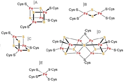
|Assembly of Fe-S clusters: an evolutionary perspective…….…..……
|Iron-sulfur clusters and hosting fold………...………...
49
50 53
55
2.2 THE ARCHAEAL [3FE-4S][4FE-4S] FERREDOXINS ………...
|Crystal structure of a di-cluster ferredoxin holding a zinc centre…….
|Stability and unfolding of di-cluster ferredoxins: state of the art……...
60
62
64
3
CENTRES DURING UNFOLDING OF DI-CLUSTER FERREDOXIN: CLUSTER DISSOCIATION, IRON RELEASE AND PROTEIN STABILITY3.1 SUMMARY……………… 73
3.2 INTRODUCTION…………….…………..… 74 3.3 MATERIALS AND METHODS…………….… 75 3.4 RESULTS…………….……….
|Ferredoxin alkaline chemical unfolding……….
|Kinetics of cluster dissociation, iron release and protein unfolding…..
|EPR analyses of iron-sulfur cluster degradation……..…...………
|Cluster degradation and polypeptide unfolding…………...………
|The effect of EDTA on metal centres and protein stability…………....
|Chemical nature of the 610 nm transient species………...………
|Formation of iron sulfides (FexSy) during ferredoxin unfolding…...….. 77
77
78
80
80 82 84 86
3.5 DISCUSSION…………….…………..……. 88 3.6 REFERENCES……… 88
3.7 ACKNOWLEDGMENTS………... 90
4
LINEAR THREE-IRON CENTRES ARE UNLIKELY CLUSTER DEGRADATION INTERMEDIATES DURING UNFOLDING OF IRON-SULFUR PROTEINS4.1 SUMMARY……………… 93
4.2 INTRODUCTION……………… 93
4.3 MATERIALS AND METHODS………….….. 95
4.4 RESULTS
|Kinetics of Iron centre degradation……….………..
|Effect of exogenous sulfide……….……….………..
|Spectral decomposition of the intermediate species………..…
|Protein unfolding and iron release………..……..
96
96
97 98
99
4.5 DISCUSSION……….. 101
4.6 REFERENCES……… 102
5
ENHANCED THERMAL STABILITY OF A ZINC-LACKING FERREDOXIN ISOFORM SHOWS THAT A HYDROPHOBIC CORE EFFICIENTLY REPLACES THE STRUCTURAL METAL SITE5.1 SUMMARY... 107 5.2 INTRODUCTION……… 108 5.3 MATERIALS AND METHODS……… 110 5.4 RESULTS……….
|Electrostatic effects on the thermal stability of Fe-S clusters…….……..
|Conformational changes upon altering net charge through equilibrium pH titrations………..…..
|Influence of net charge on Fe-S clusters disruption and hydrophobic core exposure………
|Contributions of protein folding and Fe-S clusters to thermal stability at zero netcharge………...…
114
114
115
117
119
5.5 DISCUSSION………. 120 5.6 REFERENCES………... 123 5.7 ACKNOWLEDGMENTS …………..………... 125
6
ON THE RELATIVE CONTRIBUTION OF IONIC INTERACTIONS OVER IRON-SULFUR CLUSTERS TO FERREDOXIN STABILITY6.1 SUMMARY... 129 6.2 INTRODUCTION……… 129
6.3 MATERIALS AND METHODS……… 130 6.4 RESULTS………
|Electrostatic effects on the thermal stability of Fe-S clusters…………..
|Conformational changes upon altering net charge through equilibrium pH titrations………..……..
|Influence of net charge on Fe-S clusters disruption and hydrophobic core exposure……….……
|Contributions of protein folding and Fe-S clusters to thermal stability at zero netcharge………..
132
133
135
137
138
7
STRUCTURAL CHARACTERISATION AND IMPLICATIONS ON PROTEIN FOLDING AND FE-S CENTRE ASSEMBLY7.1 SUMMARY... 145 7.2 INTRODUCTION………. 146 7.3 MATERIALS AND METHODS……….……… 148 7.4 RESULTS………..
|Ferredoxin retains native like structure at pH 2.5……….……...
|Ferredoxin thermal transition occurs in two steps……….……..
|Apo-ferredoxin is a molten globule……….……...
|Conformational dynamics of the molten globule state………….………...
|Conformationalstability of the molten globule……….………
151
151
151
154 156 159
7.5 DISCUSSION………. 160 7.6 REFERENCES………... 164
8
A SPECTROSCOPIC STUDY OF THE TEMPERATURE INDUCED MODIFICATIONS ON FERREDOXIN FOLDING AND IRON-SULFUR MOIETIES8.1 SUMMARY... 169 8.2 INTRODUCTION……… 170 8.3 MATERIALS AND METHODS……….……… 171 8.4 RESULTS………..
|Monitoring secondary structure alterations: FT-IR and far-CD…………
|Appraisal on the forming molten globule………...………..
|Course alterations on the di-clusters integrity during molten globule formation: visible-CD and RR ……….
|Probing individually Fe-S clusters bridges to the ferredoxin:
a 1H-NMR evaluation……….………...………
173
174
175
176
178
8.5 DISCUSSION………. 180 8.6 REFERENCES………... 182 8.7 ACKNOWLEDGMENTS………..……… 183
9
FE-S CLUSTERS AND FERREDOXIN FOLDINGGENERAL DISCUSSION AND CONCLUSIONS... 187
1
P
ROTEINF
OLDING,
S
TABILITY ANDM
ETALI
ONS:
AN
I
NTRODUCTIONCONTENTS
1.1
|
OVERVIEW……….…………..31.2
|
THE PROTEIN FOLDING EVENT………...…….4|Setting off protein folding: mechanisms to fold………7
|Two state versus multi-state protein folding……...………….……….9
|Role of intermediates………...……….………..11
|Off-pathway phenomena: misfolding and aggregation effects....13
|Assistants in folding……….………...……14
1.3
|
PROTEIN CONFORMATIONAL STATES,DYNAMICS AND STABILITY………...…20
|The native state………....21
|Themolten globule state………...………....23
|Bulk of unfolded states: outcome on refolding pathways……...26
|Hyperthermostability in proteins……….…....27
1.4
|
PROTEINS ALLIANCE WITH METAL IONS…………...….……..30|Residues involved in cross-linking with metal ions………….…….32
|Metal ions in proteins folding………..………….34
1.1
|
O
VERVIEWProtein folding is a puzzling process. Proteins appear to fold into a unique native conformation, in spite of an astronomical number of alternative configurations.In regard, the introduction of a theoretical framework based on the global properties of the energy landscape helps to explain the simplest models of protein folding and provides an estimable approach for the understanding of more complex protein molecules. This approach to the folding process has in fact already contributed to a substantial improvement in protein structure prediction and design [1-4], as well as in the construction of metal sites [5], in addition to a emergent ability in engineering efficient foldable artificial polymers for practical applications [6-9]. Even so, a full comprehension of much of the priorities of folding forces and a quantitative understanding of the factors that define single low-energy fold remains yet rather limited. In fact, it appears that not all amino acids and therefore guiding folding forces, are equally important in specifying which fold is adopted; as proteins with high sequence identity can result in very distinct folds [10].
One of the main goals of protein folding studies relies not only in the ability to predict the protein structure from its amino acid sequence but also, and in particular, to quite understand the pathways and mechanisms of the folding process. In fact, a detailed knowledge on protein folding can greatly contribute to a better comprehension of several human pathologies associated with misfolded and aggregated proteins [11], as well as improve our understanding on the role of co-factors in protein folding, stability as well as metals assembly. In this respect, the elucidation of the kinetic and thermodynamic properties of a protein
in vitro can be a step forward on the way to characterize its complete folding
pathway. Subsequent steps can comprise the characterization of transiently formed intermediates and of the transition states between the various states of the protein and examining the factors that may influence in vivo folding, like crowding effects of the cellular environment and the presence of folding assistants [12].
regarding the instituted approaches to the phenomena. The contextualization of protein folding events within the biological and evolutionary scenario is also discussed.
1.2
|
THEP
ROTEINF
OLDINGE
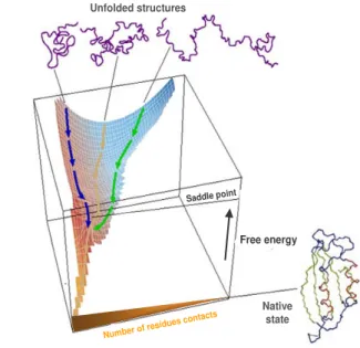
VENTinteractions present in the functional native state are not in conflict and do not give rise to the concurrent presence of competing interactions that may result in distinct ground states, like it was observed for a random polymer. Instead, folding interactions must be mutually supportive and cooperatively lead to a single low-energy structure. By avoiding the frustrating conflicts between different energetic biases, proteins have most likely evolved a funnelling energy landscape that optimizes native structure-seeking interactions, while selecting against interactions leading to non-productive traps.
Unfolded structures
Transition State
Number of residue s contacts
Free energy
Native state
Saddle point
Unfolded structures
Transition State
Number of residue s contacts
Free energy Native state Unfolded structures Transition State
Number of residue s contacts
Free energy Native state Unfolded structures Transition State
Number of residue s contacts Unfolded structures
Transition State
Number of residue s contacts Unfolded structures
Transition State
Number of residue s contacts Unfolded structures
Transition State
Number of residue s contacts Unfolded structures
Transition State
Number of residue s contacts Unfolded structures Transition State Unfolded structures Transition State Unfolded structures Transition State
Number of residue s contacts
Free energy
Native state
Saddle point
Figure 1.1 | Schematic energy landscape for protein folding.
The surface is derived from a computer simulation of the folding of a highly simplified model of a small protein. The surface ‘funnels’ the multitude of denatured conformations to the unique native structure, where highly simplified trajectories are indicated. The critical region on a simple surface such as this one is the saddle point corresponding to the transition state, the barrier that all molecules must cross if they are to fold to the native state. Adapted from [28]
Once the energetic frustrations associated with conflicting interactions have been minimized, the topology of the protein becomes the key determinant of the folding mechanism, encoding the interplay between stabilizing interactions and chain entropy. The funneled organization of the energy landscape therefore dominates the kinetics of folding, and is responsible for the robust ability of proteins to fold. In fact, it seems rather evident that this process is extremely efficient for those special sequences that have been selected during evolution to fold to globular structures, where only a very small number of all possible conformations need to be sampled during the search process. However, the funnel theory does not necessarily implicate a single pathway; rather, a multiplicity of downhill folding routes is likely to co-exist in parallel, via distinct mechanisms [31]. In agreement, the heterogeneity in the folding kinetics which is in some cases observed when the process is monitored using distinct probes, or a non-exponential kinetics, has been interpreted as an evidence for a downhill folding mechanism [32]. In addition, it is established that in some cases, the same protein may fold via different pathways for the reason that the folding conditions changed, and can affect differently the stabilities of the diverse substructures along the folding pathway [33-35]. Also, the binding of cofactors can be accountable to induce a change in the folding pathway of the protein [36]. Moreover, the occurrence of mutations may be overcome by a simple shift in the folding route [37, 38], as long as native interactions prevail over the non-native ones [39]. Evolution is likely to have selected a funneling landscape with multiple folding pathways as a folding model, as a way to ensure successful in vivo protein folding under the varying conditions observed in the cellular context.
Although detailed mechanisms of folding for complex proteins is still to be achieved, in general the funnel landscape is likely to be enforced in all proteins. Such common patterns can be inferred from the perfect funnel models — as most naturally occurring proteins has to have sufficiently reduced energetic frustration, or otherwise would had been deleted by evolution. The funnel landscape idea implies the notion, as a general guideline, that topology determines the mechanism of folding, and that the topography of the global landscape has been determined by evolutionary pressure [24]
Protein folding is addressed by multiple approaches, both experimental and theoretical or computational. Among these are the experimental monitoring of protein unfolding and refolding using biophysical techniques, frequently involving the use of site-directed mutagenesis to probe the roles of individual residues during the folding process [44]. Theoretical approaches are also important in the understanding of the protein folding event, particularly those based on computer simulations of the events occurring during folding, or more often unfolding, as this process is easier to simulate and can be related to the complementary folding reaction [21, 45]. The ultimate objective of such studies is to define the complete energy landscape for the folding reaction, and to understand in detail how this is defined by the primary sequence.
|
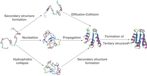
Setting off protein folding: mechanisms to foldGiven the diversity of protein structures and the evolutionary pressure on function, a unique mechanism for folding, although rather appealing, seems nevertheless very unlikely. In fact, a variety of distinct folding mechanisms has been described even for small single-domain proteins, where secondary structures can form before or after collapse and side-chains can order before or after the main chain topology. In this regard, several models have emerged in order to aid in categorizing the different mechanisms of proteins folding (figure 1.2).
The diffusion-collision model, proposes that precedence is given to the formation
as a scaffold for the build up of the rest of the structure [48], whereas other views suggests that proteins may fold via the stepwise assembly of structural subunits called foldons [49]. In the hydrophobic collapse models, a non-specific hydrophobic collapse is expected to occur as the initial step [50]. Clearly, a unified mechanism able to describe the protein folding reaction cannot be envisaged. Even so, an investigation on possible common features in folding reaction has suggested the possibility that some distinct pathways can be in fact the manifestation on an underlying common feature [51].
Secondary structure formation
Hydrophobic collapse
Nucleation
Secondary structure formation Diffusion-Collision
Propagation
Tertiary structure Formation of Secondary structure
formation
Hydrophobic collapse
Nucleation
Secondary structure formation Diffusion-Collision
Propagation
Tertiary structure Formation of
Figure 1.2 | Pathways for protein folding.
In the framework model, precedence is given to the formation of secondary structural units. In the hydrophobic collapse model, precedence is given to an initial chain collapse. In the nucleation-condensation model, an extended nucleus is formed early during folding. Molten globule-like intermediates accumulate during the folding of many proteins. For some proteins, particularly those following nucleation mechanisms, a molten globule intermediate does not usually accumulate. Although different folding pathways are usually discussed in the context of different proteins, can a single protein utilize fundamentally different folding pathways in different folding conditions? For example at very low temperatures, at which hydrophobic interactions are weakened, a protein could conceivably switch from a hydrophobic collapse to a framework mechanism. Adapted from [52]
particularly folding mechanism is directly associated with the amino-acid sequence or with any specific structural features in the protein.
|
Two state versus multi-state protein foldingThe most elementary models of protein folding are the apparent two-state systems. In this minimalist framework, protein unfolding and refolding are considered monophasic processes, where only the unfolded state (U) and the folded native state (F) are populated (figure 1.3 a|). Simultaneously, a single energetic barrier separates the folded from the unfolded state (figure 1.3 b|).
P o pul a ti on folded Reaction coordinate U F a| Free energy Reaction coordinate U F TS b| P o pul a ti on folded Reaction coordinate U F a| P o pul a ti on folded Reaction coordinate U F a| Free energy Reaction coordinate U F TS b| Free energy Reaction coordinate U F TS b|
Figure 1.3 | Outline of a hypothetical two-state protein folding reaction
a| Side view of a cooperative folding/unfolding process where only the unfolded (U)
and the folded (F) state are populated species b| Thermodynamic profile where a
single energy barrier separates the unfolded (U) from the folded (F) state with the
transition state (TS) as point along the reaction coordinate with the highest free
energy.
hand, fast kinetics can reveal intermediates in apparent two-state unfolding reactions, by showing unfolding intermediates in the msec time range even though both the major folding and unfolding reactions follow a single exponential time course in the second’s time range. In agreement, NMR methods were able to identify intermediates in otherwise thought to be two-state folding reactions [57]. These evidences appear to suggest that the two-state folding model, at least in some cases maybe under estimated. In fact, a nonlinear energy relationship in protein unfolding from an apparent two-state folding protein has been associated with the existence of unstable intermediates [58-60]. Moreover, these high-energy intermediates can be stabilized relatively to the native state by a change in the folding conditions [61, 62] or by mutation [63], and therefore an apparent two-state folding protein can be turned into a multi-two-state folder. However, is still very difficult in these conditions to evaluate whether the intermediate or transition state that is being stabilized is on the same pathway as it was before the folding perturbation, or if in fact it is just the exploitation of an alternative folding route [35].
Conversely, several proteins have been shown to fold typically to their native states via a populated intermediate [64-67]. Interestingly, in RNase H from
Escherichia coli, it was also possible to alter the folding kinetics, converting a
the native three-dimensional structure. Accordingly, the two-state folding process probably represents a merely simplified version of hierarchical folding. In overview, it appears that the folding pathway of a given protein is strongly dependent on the folding conditions and not exclusively on the characteristics of the protein itself: given that it is possible to convert an apparent two-state folder to a multi-sate folding reaction, and vice-versa. In agreement, it comes out that intermediates can act as valuable signposts for identifying possible changes in the apparent multiplicity of folding pathways.Moreover, it seems very likely that an apparent two-state folding pathway does not imply for certain that no intermediate exists, but simply that no intermediate could be detected. Therefore, it is most likely that the folding mechanism is in fact a unified one that depends on the native backbone topology and proceeds through a range of multi-states, with no single sequential route [52, 71].
|
Role of intermediatesThe role of intermediate states on folding pathways has been at the least controversial. It hasbeen suggested that the presence of intermediate stateson folding pathways may slow the folding process, as these constitute a energetic trap on the pathway that otherwise needs to be reversed in order to get the proper fold [72, 73]. As well, and in complete disagreement, it was proposed that the partially folded states can in fact enhance the rate of protein folding, by guiding the folding polypeptide chains to low-energy transition states that otherwise may not become accessible directly from the unfolded state [74]. Moreover, it has been noted that intermediates must play a productive role to a correct folding [75-77], but also pointed out that intermediates can be off-pathway species leading to misfolded species [78, 79].
determined by the same cooperative interactions that determine the target native structure. Several experimental data was shown to support this view of protein folding in which all of the molecules in a refolding population fold essentially through the same intermediate structures [80, 81]. On the other hand, intermediates are often interpreted not has discrete pre-determined conformations in the folding pathway, but in terms of an ensemble of conformations, where the transition from one ensemble of structures to the next one on the folding pathway can happen on independent parallel routes [82, 83]. This model of multiple pathways further suggests that specific populated intermediates do not need necessarily to exist and that partially folded conformations can in fact represent the slower multi-state fractions [84-86]. This heterogeneity has been evidenced within several proteins as distinct coexisting subpopulations [87-90] and also within families of proteins with similar folds [91]. In order to address these conflicting evidences on folding intermediates, a convergent hypothesis was recently proposed that merges the opposite explanations [92]. This unifying approach suggests that these discrepant interpretations can be resolved by
modifying the predetermined pathway model to include probabilistic misfolding
errors that can block the forward progress of normally occurring intermediates.
Chance misfolding errors can corrupt different intermediates and insert optional
error-repair barriers at different points in a pathway. When the error probability is
zero at all steps of the pathway, folding appears to be a two-state process. When
it is unity at one particular step, three-state folding occurs. Any other values or
combinations will produce mixed behavior in which different population fractions
display different naturally occurring but partially corrupted intermediates, or none
at all, and fold at different rates. This heterogeneous behavior, when detected by
the usual spectroscopic observations of kinetic phases, will appear to represent
multiple alternative pathways. This hypothesis seems able to explain a varied folding behavior of proteins quite generally grounded in the observation that
folding errors are ubiquitous. Well known misfolding errors include prolyl and
prolyl peptide bond mis-isomerization, transient aggregation, formation of
non-native hydrophobic clusters, disulfide shuffling, cofactor misligation, and perhaps
nonnative domain docking modes. These errors are optional, not intrinsic to the
folding conditions. This goes in agreement with the evidence that the folding conditions can preferentially influence the sub-populations that become most populated during the folding pathway [33], thus implying that the folding pathway and observed intermediates for a given protein can be different under different conditions. Altogether, these findings seem to substantiate that the task of monitoring and characterizing intermediates can be a rather slippery job. As a result, it is not yet possible to ascertain if folding intermediates are a mere result of protein folding that characterizes a given folding pathway or if in fact they play a determinant role in guiding and defining the pathway.
|
Off-pathway phenomena: misfolding and aggregationA misfolded protein is a protein that failed to fold properly. The misfolding process results when the protein acquires a number of persistent non-native interactions that affect its overall architecture and/or its properties in a biologically significant manner, like the loss of function. In addition, misfolded conformations often expose hydrophobic amino acid residues and segments of unstructured polypeptide backbone to the solvent thus promoting inter-chain hydrogen bonding and hydrophobic interactions that can lead to aggregation.
be observed in soluble proteins with no recognized connection to any known disease, frequently just by lowering the pH or increasing the temperature above that required for unfolding [95, 96]. In agreement, it is also possible in many cases to reproduce under laboratory conditions the structural transitions of the disease-associated molecules, by exposing the folded proteins to mildly denaturing conditions [97]. Altogether, these evidences suggest that the ability to form amyloid fibrils can in fact be an intrinsic property of polypeptide chains. In agreement with this suggestion is also the fact that the intermolecular bonds that stabilize amyloid fibrils are known to involve the peptide backbone, which is common to all proteins. Therefore, amyloid conformation can be a possible common state for all proteins regarding that favourable conditions are provided [98]. Thus, except when the protein is exposed to specific conditions, the peptide backbone is not accessible to form the inter-chain hydrogen bonds associated with amyloid fibrils.
The reason why only a few proteins actually form amyloid aggregates in vivo is likely to be influenced by the protein amino-acid sequence that defines the degree of exposed surfaces prone to aggregation in the native state [99], or become exposed by specific conditions in the cell. It is interesting to speculate that avoidance of aggregation, particularly to highly insoluble amyloid fibrils, might be an equally important driving force in the evolutionary design of natural proteins. Biology must has found a way to avoid the formation of this unwanted material under normal physiological conditions where proteins that persist to form amyloid aggregates must likely represent the exception that need further evolutionary improvements. In fact, many factors must be involved in this protective mechanism, but the selection of sequences during evolution that can fold efficiently to a globular form in which the polypeptide chain and the hydrophobic residues are hidden in the interior is likely to be particularly important [100, 101].
|
Assistants in foldingvivo folding often requires assistance to either promote proper folding, improve the yield in many folding reactions or to play a role in a post-translational quality control system and maintain the proper conformation of proteins under changing environmental conditions. Such helping factors can include a vast network of
i)molecular chaperones [103], as well as the effect of ii)chemical chaperoning
[104] in addition to the ambiguous influence of the iii) molecular crowding
environment in the cell over proteins folding and assembly [105].
i)Molecular chaperones are a group of structurally diverse and mechanistically
of chaperones is associated with the ability to hold nascent and newly synthesized chains in a flexible state competent for subsequent folding.
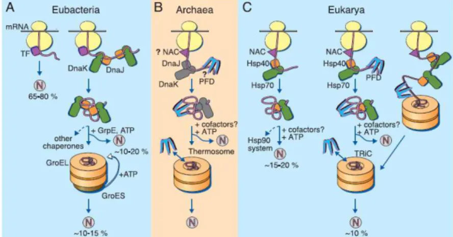
Figure 1.4 | Models for the chaperone-assisted folding of newly synthesized polypeptides in the cytosol.
(A) Eubacteria. TF, trigger factor; N, native protein. Nascent chains probably interact
generally with TF, and most small proteins (~65 to 80% of total) fold rapidly upon synthesis without further assistance. Longer chains (~10 to 20% of total) interact subsequently with DnaK and DnaJ and fold upon one or several cycles of ATP-dependent binding and release. About 10 to 15% of chains transit the chaperonin system GroEL and GroES for folding. GroEL does not bind to nascent chains and is thus likely to receive an appreciable
fraction of its substrates after their interaction with DnaK. (B) Archaea. PFD, prefoldin;
NAC, nascent chain associated complex. Only some archaeal species contain DnaK/DnaJ. The existence of a ribosome-bound NAC homolog, as well as the interaction of PFD with
nascent chains, has not yet been confirmed experimentally. (C) Eukarya - the example of
the mammalian cytosol. Like TF, NAC probably interacts generally with nascent chains. The majority of small chains may fold upon ribosome release without further assistance. About 15 to 20% of chains reach their native states in a reaction assisted by Hsp70 and Hsp40, and a fraction of these must be transferred to Hsp90 for folding. About 10% of chains are co- or posttranslationally passed on to the chaperonin TRiC in a reaction mediated by PFD. Adapted from [103]
and the TCP-1 ring complex (TriC) respectively. The assistance from the chaperonins is achieved via an active ATP-dependent mechanism that undergoes large scale ATP-driven conformational changes crucial for their protein folding function in contrast to the ribosome binding chaperones, usually addressed as holders and folding catalysts that act via a passive, ATP-independent mechanism. Approximately ten percent of all newly synthesized polypeptides transit to chaperonins in the cell [107, 108] where only a very few were identified with an absolute chaperonin dependence for correct folding [109]. The majority of these proteins were identified with complex topologies that often fold slowly and are strongly aggregation prone, owing to the exposure of extensive hydrophobic surfaces in their non-native states. These evidences seem to substantiate the role of chaperonins in preventing aggregation, but in addition it was also evidenced that chaperonins can also speed up the folding reaction substantially [110]. This acceleration or increased efficiency in protein folding has been widely explained by the affect of confinement of proteins in the cage of the chaperonins. This effect is proposed to smooth the energy landscape of folding for larger proteins, either by preventing the formation of certain kinetically trapped intermediates or by facilitating their progression toward the compact native state. In addition, an alternative model has also been proposed, where the chaperonins are proposed to speed up folding by a mechanism of “iterative annealing” [111]. In this mechanism the chaperonin is suggested to facility folding by cycles of unfolding kinetically trapped states, fallowed by repartitioning of the unfolded protein between productive and non-productive folding pathways.
To ensure an efficient use of the cytosolic folding machinery in thousands of different proteins in the cell, protein synthesis must be coordinated with the activities of the various chaperone systems in stabilizing nascent chains and promoting proper folding. In this respect, the mechanistic principles underlying this functional cooperation are not yet well understood and careful must be taken when performing in vitro studies to approach in vivo activity [112].
ii) Chemical chaperones, are a group of small molecular weight compounds that
over proteins, or by ligands, inhibitors and cofactors that afford a more specific action over a particular target protein. Osmolytes includes polyols, sugars, methylamines, free aminoacids or its derivatives. These molecules can promote protein folding and increase thermodynamic stability from the resulting hydration effect on proteins. This effect relies on the concept that the interaction of the polypeptide with osmolytes is mostly unfavourable, which indirectly promotes preferential protein hydration by water molecules. As a result the free energy of both the native and the unfolded state increase. Nonetheless the raised energy is much more significant in the unfolded state on the account of the increased protein surface area exposed and therefore favours folding-promoting contacts and the native state configuration [113]. Osmolytes can therefore provide some protection to proteins against denaturation, and in fact some of the osmolytes are significantly present or untaken by certain organisms in order to adapt to harsh environments or stressful conditions [114, 115]. Moreover it was also describe the ability of these molecules to even compensate molecular chaperoning deficiency [116] or regulate the folding activities of molecular chaperones under stressful conditions [117]. On the other hand, chemical chaperoning by ligands, inhibitors and cofactors that bind reversibly to a specific protein are also suggested to be able to restore protein function and folding. This heterogeneous group of molecules bind weakly to a specific target protein (due to the specificity of action are often classified as pharmacological chaperones) that either can induce protein refolding, stabilisation, or contribute to structuring a particular region or domain within the protein. This effect is attained when the binding of the chemical agent to the protein has an energetic contribution favouring the native state. In this situation the absence of the binding factor would imply the loss of stabilizing interactions which increases the number of possible conformations accessible to that conformation and therefore result in protein destabilization and misfolding.
iii) Macromolecular crowding is the term attributed to the influence that excluded
1.3
|
P
ROTEINSC
ONFORMATIONALS
TATES
D
YNAMICS ANDS
TABILITYProteins are known to undergo conformational transitions, and to be able to withstand some structural variability, which frequently depends on the protein environment. This amazing property of proteins is proven vital to regulate biological activity and targeting of proteins to different cellular locations in vivo. In fact, biological systems have probably become more robust with the ability to control and regulate the various states accessible to a given polypeptide chain, at given times and under given conditions.
Accordingly with their structural features, proteins can generally be classified within four major conformational states [125]: i. the native state, in which the side chains are tightly interdigitated, leading to a densely packed overall three-dimensional structure; ii) the molten globule state, corresponding to a structured, compact intermediate state with relatively high content of native-like secondary structure, native-like topology and some tertiary contacts, but lacking the tight packing and rigid tertiary structure found in the native state; iii) pre-molten globule
states, that relies on ensembles of relatively unstructured intermediates that have
regions of at least transient native-like secondary structure and are condensed relative to the unfolded state; iv) the unfolded state, which typically encompasses a very large ensemble of disordered conformations.
Although, the most stable and biologically active form of the protein under relevant conditions is generally considered the native state; It follows that protein function in some cases can arise from any of the four distinct conformational states or with transitions between them [126, 127]. In agreement, a growing number of intrinsically disorder proteins with biological function has been identified, experimentally characterised and classified in a database [128]. Presently, these proteins are often considered common in nature [125, 129, 130] and typically involved in regulation, signalling and control pathways [131, 132] where conformational changes associated with function may also be brought about by alterations in environmental or cellular conditions.
disorder conformational structure. In fact, “the high proportion of gene sequences in the genomes of all organisms argues for important, as yet unknown functions, since there could be no other reason for their persistence throughout evolution” [133]. Within this scenario, it appears that proteomics studies will have to be re-evaluating since whether the lack of specific three-dimensional structure occurs wholly or in part, such proteins do not fit the standard paradigm that an organised structure is a prerequisite to function [134]. Instead, the function of a protein seems to depend on its ability to adopt a specific conformation, structured or not. On the other hand, proteins can shift through these various conformational states as a result of changes in pH [135, 136], temperature or pressure [137], solvent alterations [138, 139] as well as ligand binding [140] and protein-protein interactions [141].Despite the fact that environmental conditions strongly influence the structural features of proteins, almost all studies on protein folding do not mimic the cellular environment. In fact, as previously mentioned, the inside of cells is extremely crowed achieving concentrations of 300-400gL-1 [142], whereas most studies on proteins are performed in dilute solutions < 1 gL-1. Given that macromolecular crowding has been suggested to influence the structure and compactness of proteins [143, 144] , it may be quite possible that some considered disordered proteins, are just artefacts resulting from the way those proteins are studied. Eventually, some putatively disordered proteins will be structured inside the cell, while others will not.
|
The native state|
The molten globule stateMolten globules are conformations with physical properties between those of the native and the unfolded states, and it is frequently assumed that most proteins can form such species if the appropriate experimental conditions can be found. In fact, changes in the protein environment can reduce, abolish or alter part of the native conformational interactions of the protein. These conformational alterations that concurrently can define the conversion to a molten globule, can be usually obtained under mildly denaturing conditions, such as pH extremes [151, 152], low concentrations of denaturants (chemicals, salts, alcohols, sugars) [153], thermal perturbation [154], or by the removal of prosthetic groups (like metal ions) [155, 156].
The definition of molten globule state covers an ensemble of sub-conformations ranging from those that are quite compact and afford substantial native-like contacts, to those that are less-structured and retain relatively few native contacts. Therefore it does not look surprising that experimental evidence regarding thermodynamic analyses and detailed structural characterization on molten globules forms can greatly diverge. In fact, for some molten globules it was possible to detect a significant calorimetric transition relatively to the unfolded state, while for others no difference in the heat capacity and enthalpy change could be observed, as well only for the most structured cases it was possible to detect a cooperative transition for the unfolding of a molten globule. These thermodynamic differences are inherent to the diverse residual structure present in molten globules forms, particularly on the degree of collapse of the hydrophobic core. In agreement, theoretical calculations evidenced that the differences in the heat capacity between two conformational states (like the molten globule to the unfolded sate) is proportional to the change in solvent-accessible apolar area with the transition [157]. This suggests that only molten globule forms that retain significant hydrophobic core will be able to show a calorimetric transition.
structure present in molten globules is significantly less stable than the corresponding regions in the native state). Besides, molten globule states ought to present a near-native level of compactness typically expanded no more than twenty percent. In addition, these partially folded conformations must reveal an increased solvent-exposed hydrophobic surface area relative to the native state, where hydrophobic residues which are buried in the native state have become exposed to solvent. This last characteristic of molten globules most likely affords an explanation for the fact that the transition of molten globule forms to the unfolded state usually represents a minor enthalpic transition when compared with the unfolding of the native state to the molten globule state.
unfolding conditions, within the same protein [168]. This points out for the plasticity of molten globule forms even within the same protein and suggests that ultimately the folding/unfolding route can lead to an off or on-pathway molten globule conformation. These species can therefore represent a folding intermediate for some proteins under particular conditions, but are not necessarily a mandatory intermediate for all proteins to fold. Nevertheless it is important to always keep in mind that most of the studies in protein folding do not mimic the cellular environment. The biological stage includes a multitude of chaperones that can lead some proteins to different folding routes; besides the heavy molecular crowding that is present in the cell is known to also be able to influence proteins conformational states by stabilizing the native state, or in contrast, induce the molten globule formation [169].
Unfolded
Folded Molten globule
F U F
U
Unfolded Unfolded
Folded Folded Molten globule
Molten globule
F
U FF
U U F
U FF
U U
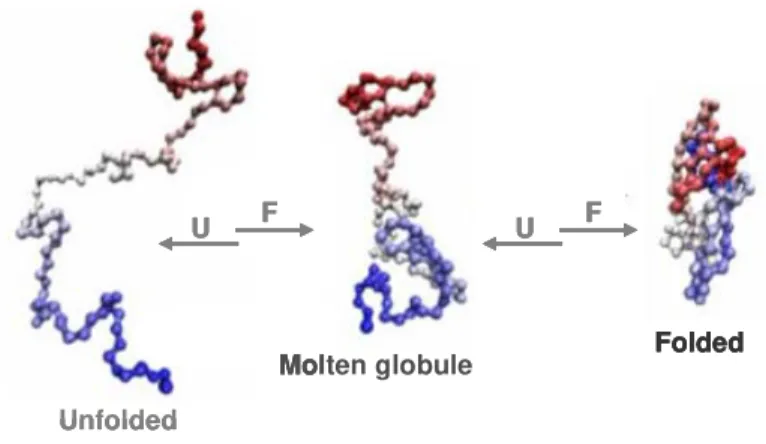
Figure 1.5 | Scheme on protein folding via a molten globule state as a general strategy for protein assembly.
U – unfolding; F - folding
diseases [28, 177]. The experimental and computational study of molten globules continues to be a stimulating area of research within the field of protein science. Elucidating the structural features of these partially folded conformers can provide important insights into the determinants of protein topology, the mechanism of protein folding, and biological function.


![Figure 2.2 | Evolutionary scheme of the 2x[4Fe–4S] ferredoxin fold.](https://thumb-eu.123doks.com/thumbv2/123dok_br/15769789.641157/81.918.169.637.216.536/figure-evolutionary-scheme-x-fe-s-ferredoxin-fold.webp)


