Molecular cloning and functional analysis of the fatty acid-binding protein
(
Sp-FABP
) gene in the mud crab (
Scylla paramamosain
)
Xianglan Zeng, Haihui Ye, Ya’nan Yang, Guizhong Wang and Huiyang Huang
College of Ocean and Earth Sciences, Xiamen University, Xiamen, China
Abstract
Intracellular fatty acid-binding proteins (FABPs) are multifunctional cytosolic lipid-binding proteins found in verte-brates and inverteverte-brates. In this work, we used RACE to obtain a full-length cDNA of Sp-FABP from the mud crab Scylla paramamosain. The open reading frame of the full length cDNA (886 bp) encoded a 136 amino acid polypeptide that showed high homology with related genes from other species. Real-time quantitative PCR identified variable levels ofSp-FABP transcripts in epidermis, eyestalk, gill, heart, hemocytes, hepatopancreas, muscle, ovary, stomach and thoracic ganglia. In ovaries,Sp-FABP expression increased gradually from stage I to stage IV of devel-opment and decreased in stage V.Sp-FABP transcripts in the hepatopancreas and hemocytes were up-regulated af-ter a bacaf-terial challenge withVibrio alginnolyficus. These results suggest that Sp-FABP may be involved in the growth, reproduction and immunity of the mud crab.
Keywords: fatty acid-binding protein, immunity, ovary development, real-time quantitative PCR,Scylla paramamosain.
Received: July 5, 2012; Accepted: October 15, 2012.
Introduction
Fatty acid-binding proteins (FABPs) are small (14-15 kDa), ubiquitous, multigenic cytosolic proteins that bind non-covalently to hydrophobic ligands, mainly fatty acids (FAs) (Esteves and Ehrlich, 2006). Apart from functioning as energy sources, FAs can act as signaling molecules (Sumidaet al., 1993; Graberet al., 1994; Nunez, 1997) and regulate Na+, K+, Ca2+and Cl-ion channels (Ordwayet al., 1991; Kang and Leaf, 1996; Xiaoet al., 1997; Liu et al., 2001). FAs also have a role in gene transcription, especially genes that encode proteins involved in lipid metabolism
(DeWille and Farmer 1993; Martin et al., 1997; Clarke
2000; Louetet al., 2001). FABPs are therefore indirectly involved in biological responses mediated by FAs.
Since the isolation of the first invertebrate FABP from the desert locust,Schistocerca gregaria,by Haunerl and Chisholm (1990), a growing number of FABPs have been identified in invertebrates. In vertebrates and inverte-brates, FABPs have a wide range of crucial biological roles, including the regulation of cellular lipid homeostasis, cell growth and differentiation, cellular signaling, gene tran-scription and cytoprotection (Zimmerman and Veerkamp, 2002). Studies in knockout mice have confirmed the impor-tance of FABPs in the uptake and transport of long-chain fatty acids and their interaction with other transport sys-tems and enzymes (Coburnet al., 2000). Moreover, studies
with the Chinese mitten crab Eriocheir sinensis have
shown thatEs-FABPexpression levels vary with the stage
of ovarian development (Gonget al., 2010).
FABPs may have a role in the immune reactions of invertebrates and vertebrates. Sm14, the first
platy-helminth FABP isolated from the parasite Schistosoma
mansoni(Moser et al., 1991), is a highly immunogenic peptide that offers important protection against experi-mental infections in cattle and other animals (Tendleret
al., 1996). Homologous proteins such as Sj-FABPc from
Schistosoma japonicum(Beckeret al., 1994), Fh15 from Fasciola hepatica (Rodríguez-Pérez et al., 1992) and
FgFABP from Fasciola gigantic (Estunningsih et al.,
1997) also provide protection from challenge with infec-tious agents. In crustaceans, FABPs are known to be
cor-related with immunity. InLitopenaeus vannamei(Zhaoet
al., 2007), Penaeus stylirostris (Dhar et al., 2003),
Procambarus clarkii (Zeng and Lu, 2009) and Fenneropenaeus chinensis(Wanget al., 2008; Renet al., 2009) the expression levels of FABPs were up-regulated after a challenge with infectious agents.
Mud crabs (Scyllaspp.) are a group of four commer-cially important Portunid species that are found in intertidal and subtidal, sheltered, soft-sediment habitats, particularly
mangroves, throughout the Indo-Pacific region (Le Vayet
al., 2008). In this report, we provide the first description of the cDNA structure, phylogenetic relationships and tissue distribution of an intracellular FABP from the mud crab Scylla paramamosain. The levels ofSp-FABPexpression
Send correspondence to Haihui Ye. College of Ocean and Earth Sciences, Xiamen University, 361005 Xiamen, China. E-mail: haihuiye@xmu.edu.cn.
in different stages of ovarian development and after micro-bial infection were also examined.
Material and Methods
Tissue preparation
Healthy adult female crabs were purchased from a lo-cal market in Xiamen, Fujian Province, China. Samples from ten tissues (epidermis, eyestalk, gills, heart, hemo-cytes, hepatopancreas, muscle, ovary, stomach and thoracic ganglia) were collected. The ovarian samples were col-lected based on the classification of Shangguan and Liu (1991) for ovarian developmental stages I (undeveloped), II (early-developing), III (developing), IV (nearly ripe) and V (ripe). All tissues were immediately frozen in liquid ni-trogen and stored at -80 °C until nucleic acid extraction.
For the immune challenge,S. paramamosain crabs
from Dongshan farm in Zhangzhou, Fujian Province,
Chi-na, were injected with Vibrio alginnolyficus (1 x 10⁷
CFU/mL; 20mL) at the base of the right fourth pleopod
(Chenget al., 2004). Control crabs were injected with an equal volume of sterile saline solution. A total of 24 crabs per group were used, with three crabs for each time interval. At 0 (basal), 3, 6, 12, 24, 48, 72 and 96 h post-injection the hepatopancreas and hemocytes were collected from indi-viduals injected with saline (control) orV. alginnolyficus and preserved with RNAsafer stabilizer reagent (TaKaRa, Japan).
Nucleic acid extraction
RNA was extracted using Trizol reagent (Invitrogen, USA) according to the manufacturer’s protocol. The RNA concentration and quality were assessed spectrophotomet-rically based on the absorbance of 260 nm (NanoDrop Technologies, Inc., USA) and by agarose gel
electrophore-sis, respectively. Total RNA was reverse transcribed using
a PrimeScript RT-PCR kit with oligo (dT)18 primers
(TaKaRa, Japan).
Full-length cDNA cloning
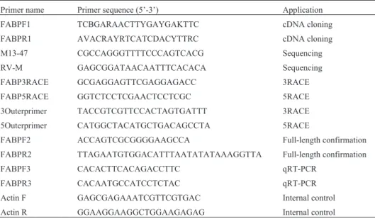
To clone the cDNA, FABPsequences were
down-loaded from NCBI and aligned using ClustalX. A pair of degenerate primers, FABPF1 and FABPR1 (Table 1), was designed based on the conserved regions. The PCR was done in an ABI 2720 Thermal Cycler in a total volume of
25mL containing 2.5 mL of 10x PCR buffer (containing
Mg2+), 2.0mL of dNTP mix (2.5 mM each), 1mL of each primer (10mM), 2mL of cDNA (500 ng/mL), 0.125mL of
Taq polymerase (5 U/mL; TaKaRa), and 16.375 mL of
RNase-free water. The PCR conditions were as follows: 94 °C for 5 min, 32 cycles of 94 °C for 30 s, 46 °C for 30 s and 72 °C for 30 s, with a final extension at 72 °C for 10 min. The PCR products were assessed visually after electrophoresison 1.2% agarose gels and those of appropri-ate size were purified, ligappropri-ated into a pMD19-T Vector
(TaKaRa) and then transformed in Escherichia coli by
overnight culture. Positive clones with inserts of the pre-dicted size were sequenced using the primers M13-47 and RV-M (Table 1) at Sangon Biotech Co., Ltd (Shanghai, China).
The 3’ and 5’ end fragments were completed by 3’ and 5’ rapid amplification of cDNA ends (RACE) with a 3’, 5’ full RACE kit (TaKaRa). Specific primers based on the initial sequence (FABP3RACE and FABP5RACE), to-gether with a 3’ outer primer and a 5’ outer primer (Ta-ble 1), were used in the PCRs. The full length ofSp-FABP was assembled by piecing together the 3’ and 5’ ends and the initial sequence. The sequence of the full-length cDNA was verified by using a pair of specific primers (FABPF2
Table 1- Primers used in this study.
Primer name Primer sequence (5’-3’) Application
FABPF1 TCBGARAACTTYGAYGAKTTC cDNA cloning
FABPR1 AVACRAYRTCATCDACYTTRC cDNA cloning
M13-47 CGCCAGGGTTTTCCCAGTCACG Sequencing
RV-M GAGCGGATAACAATTTCACACA Sequencing
FABP3RACE GCGAGGAGTTCGAGGAGACC 3RACE
FABP5RACE GGTCTCCTCGAACTCCTCGC 5RACE
3Outerprimer TACCGTCGTTCCACTAGTGATTT 3RACE
5Outerprimer CATGGCTACATGCTGACAGCCTA 5RACE
FABPF2 ACCAGTCGCGGGGAAGCCA Full-length confirmation
FABPR2 TTAGAATGTGGACATTTAATATATAAAGGTTA Full-length confirmation
FABPF3 CACACTTCACAGACCTTC qRT-PCR
FABPR3 CACAATGCCATCCTCTAC qRT-PCR
Actin F GAGCGAGAAATCGTTCGTGAC Internal control
and FABPR2; Table 1) designed based on the preliminary sequencing results.
Homology and phylogenetic analysis
TheSp-FABPnucleotide and deduced amino acid se-quences were compared to those reported for other organ-isms using the BLAST algorithm at the National Center for Biotechnology Information. The amino acid sequences of
FABP fromS. paramamosainand representative taxa were
retrieved from NCBI GenBank and analyzed using ClustalX software. The open reading frame (ORF) of the
cloned Sp-FABP cDNA was determined with the ORF
Finder, and SignalP 4.0 software was used to identify the putative signal peptide. Hydrophobic regions were pre-dicted with Protscal. A neighbor-joining (NJ) phylogenetic tree was constructed using MEGA software v. 5.0 based on 1000 bootstraps.
Real-time quantitative PCR analysis
Total RNA levels of various mud crab tissues, of ova-ries at different stages of development and of the hepa-topancreas and hemocytes after bacterial challenge were examined by real-time quantitative PCR (qRT-PCR). The
final volume of each qRT-PCR was 20mL and contained
10mL of 2 x SYBR Premix ExTaq(TaKaRa), 1mL of
di-luted cDNA template, 0.5 mL of each primer (10 mM
FABPF3 and FABPR3; Table 1) and 8mL of PCR-grade
water. Ab-actin fragment was amplified using the primer pair Actin F and Actin R (Table 1) and served as an internal control (Huanget al., 2012). The cDNA template PCR con-ditions were as follows: 95 °C for 30 s, 50 cycles of 95 °C for 10 s, 60 °C for 30 s, 72 °C for 20 s and a final extension at 72 °C for 10 min. All samples were run in triplicate and theSp-FABPexpression levels were calculated by the 2-DDCt comparative CT method. The results were expressed as the mean±SD (standard deviation) of triplicate determinations and shown as the n-fold difference relative tob-actin. Sta-tistical comparisons were done using Studentst-test and a value of p < 0.05 indicated significance.
Results
Cloning and identification ofSp-FABPcDNA
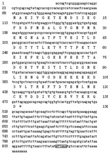
A full-length (885 bp)FABPcDNA (Sp-FABP) was
isolated from the ovaries of female mud crabs (GenBank:
JQ824129). The sequence of theSp-FABPgene contained
an ORF of 411 bp (including the stop codon), with 5’ and 3’ untranslated regions of 67 bp and 407 bp, respectively (Figure 1). A single polyadenylation signal (ATTAAA) was observed 856 bp upstream of the 12 bp poly (A) tail. The ORF coded for a polypeptide of 136 amino acids, with a calculated molecular mass of 15, 381.67 Da and an
isoelectric point of 5.55. Analysis of theSp-FABPcDNA
sequence using ClustalX revealed significant similarity to
the sequences of otherFABPs included in the NCBI
data-base. No signal peptide was identified by the SignalP 4.0 Server.
Homology and phylogenetic analysis ofSp-FABP
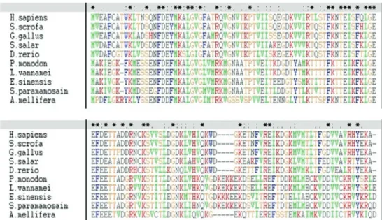
ClustalX alignment of the deduced amino acid se-quence with other related sese-quences revealed a high degree
of similarity: 85% identity with the shrimp Penaeus
monodon, 83% identity with the Chinese mitten crab E. sinensis and 60% identity with the ant Acromyrmex echinatior(Figure 2).
An NJ phylogenetic tree was constructed based on re-ported FABP sequences using MEGA5.0 software (Figu-re 3). The (Figu-reliability of the branching was tested by bootstrap resampling (with 1000 pseudo-replicates). Two distinct sister groups were observed, with a tree topology that agreed with traditional taxonomic relationships. The first group contained invertebrate FABPs (fromE. sinensis, S. paramamosain, Penaeus monodon, Litopenaeus vannameiandApis mellifera) while the second group con-tained vertebrate FABPs (Danio rerio,Salmo salar,Gallus gallus,Homo sapiensandSus scrofa).
A homology model of Sp-FABP predicted using the SWISS-MODEL database revealed conservation of the ter-tiary structure, with the 10 anti-parallelb-strands forming a barrel and a clamshell-like structure.
Sp-FABPexpression in different tissues and in ovaries at various reproductive stages
qRT-PCR was used to investigate the distribution of Sp-FABPmRNA in different tissues and to assess the ex-pression of this gene in different female reproductive stages.Sp-FABPshowed variable levels of expression in a wide variety of tissues, including epidermis, eyestalk, gill, heart, hepatopancreas, hemocytes, muscle, ovary, stomach
and thoracic ganglia (Figure 4).Sp-FABPtranscripts were constitutively expressed in mud crab ovary, although the level of expression varied with the stage of ovarian
matura-tion. The expression ofSp-FABPincreased from
reproduc-tive stage I to stage IV, when it reached a peak, and then decreased significantly at stage V (Figure 5).
Sp-FABPexpression in hepatopancreas and
hemocytes after a bacterial challenge
To gain insight into the involvement of FABP in the
crab immune response, the expression profiles ofSp-FABP
were assessed by qRT-PCR after a bacterial challenge. The hepatopancreas showed an increase in the level of Sp-FABPtranscripts at all time intervals after the bacterial challenge, especially at 3 h; after 3 h, the expression of Sp-FABP gradually decreased, but the levels were still higher than in the control group (Figure 6). In hemocytes,
Figure 2- ClustalX alignment of vertebrate and invertebrate FABP amino acid sequences. Alignment shows the following sequences (GenBank acces-sion numbers in parentheses):Apis mellifera(NP_001011630.1), Danio rerio(NP_999972.1), Eriocheir sinensis(ADM64456.1), Gallus gallus
(NP_990639.1), Homo sapiens (AAB87141.1), Litopenaeus vannamei (ADK66280.1), Penaeus monodon (ABE77154.1), Salmo salar
(NP_001135371.1),Scylla paramamosain(JQ824129) andSus scrofa(NP_001020400.1).
Figure 3- Neighbor-joining phylogenetic tree of representative vertebrate and invertebrate FABP amino acid sequences. Bootstrap values support-ing the branch points are expressed as the percentage of 1000 replicates. The following organisms with FABPs were included in the analysis:Apis mellifera,Danio rerio,Eriocheir sinensis,Gallus gallus,Homo sapiens,
Litopenaeus vannamei, Penaeus monodon, Salmo salar, Scylla paramamosainandSus scrofa. See Figure 2 legend for GenBank acces-sion numbers.
there was a slight increase in the level ofSp-FABP tran-scripts at 3, 6 and 12 h post-challenge and a marked in-crease at 24, 48, 72 and 96 h post-challenge, with a peak at 72 h (Figure 7).
Discussion
FABPs belong to a large family of ubiquitous, low-molecular-mass, small cytosolic lipid-binding proteins re-sponsible for the non-covalent binding of hydrophobic lig-ands, primarily fatty acids (Zimmerman and Veerkamp, 2002). The biological roles of these proteins include a wide range of processes such as the transport, cellular uptake and cytoplasmic use of FAs, and FA-mediated regulation of gene expression (Esteves and Ehrlich, 2006). FABPs have been extensively studied in vertebrates whereas consider-ably less is known about these proteins in invertebrates.
In the current study, the full-lengthSp-FABPcDNA
encoded a putative FABP of 136 amino acids with a theo-retical molecular mass similar to that of other FABPs
(127-136 amino acids) (Chenet al., 2006). The ClustalX
alignment of Sp-FABP and nine other reported vertebrate and invertebrate FABP sequences revealed high identity (63-85%) among invertebrate sequences. Three-dimensio-nal homology modeling revealed that several key tertiary structures of Sp-FABP were similar to those of vertebrate FABPs, such as the ten anti-parallelb-strands, their resul-tant barrel with a clamshell-like structure, and the barrel cap consisting of a pair ofa-helices that enclose the cavity of the FABP lipid-binding site (Chmurzynska, 2006). These results confirmed the identity of the cloned Sp-FABP. Phylogenetic analysis revealed two distinct sis-ter groups within the NJ tree: group 1 contained inverte-brate FABPs whereas group 2 contained verteinverte-brate FABPs. Evolutionarily, Sp-FABP is highly homologous to other FABPs and all FABPs are remarkably conserved.
Variable levels ofSp-FABPtranscripts were detected in epidermis, eyestalk, gill, heart, hemocytes, hepatopan-creas, muscle, ovary, stomach and thoracic ganglia. The presence ofFABPin all of the tissues examined suggested
thatFABPwas ubiquitous and indispensable to crustacean
growth. The expression levels were remarkably high in gill and stomach, which may be important for the following reasons. In crustaceans, the gill is a multifunctional organ, responsible for respiratory gas exchange, hemolymph acid-base and osmo-ionic regulation, and the excretion of nitrogenous metabolites (Freire et al., 2008), while the stomach is an important organ of the digestive system.
Hence,Sp-FABPmay be involved in the maintenance and
adjustment of essential physiological and pathological pro-cesses in the gill and stomach.
Ovarian expression of Sp-FABP depended on the
stage of ovarian maturation, with the level of expression in-creasing from stage I to stage IV (peak expression occurred in this stage) followed by a significant decrease in stage V,
in agreement with a previous study (Gonget al., 2010).
During development, the ovary accumulates substantial
Figure 5-Sp-FABPexpression in different stages of ovarian develop-ment, as determined by qRT-PCR. The columns are the mean±SD (n = 3).
Figure 6- HepatopancreaticSp-FABPexpression after a bacterial chal-lenge withV. alginnolyficus, as determined by qRT-PCR. The relative abundance of theSp-FABPtranscripts is expressed as the mean±SD (n = 3). *p < 0.05 and *p < 0.01 compared to the corresponding controls.
amounts of nutritional substrates, especially FAs
(Milla-mena, 1989; Shangguan and Liu, 1991; Mourenteet al.,
1994; Chenget al., 2001). FAs accelerate the expression of
FABP (Kaikauset al., 1993a,b) because the tissue FABP
content is associated with the rate of FA uptake and utiliza-tion (Ockneret al., 1980; Glatz and Vusse, 1990; van Breda et al., 1992). The variation inSp-FABPexpression levels withdevelopmental stage suggests that FABP may be asso-ciated with ovarian maturation in the mud crab.
Although FABPs have been implicated in invertebrate immunity, there has not yet been any clear demonstration of a link between these two phenomena. In crustaceans, the hepatopancreas not only initiates the humoral immune re-sponse but also contains highly specialized cells and phagocytes that function in the cellular immune response (Panet al., 2005). The invertebrate innate immune system provides the major defense mechanisms against pathogenic agents and the immune responses occur mainly in
hemolymph (Somboonwiwat et al., 2006). The
hepato-pancreas and hemocytes are presumably the primary sites for the production of immune response factors. To investigate the relationship between FABP and the immune response, we initially injected bacteria into crabs and examined the
ex-pression ofSp-FABPafter the challenge.Sp-FABP
expres-sion was up-regulated in hepatopancreas and hemocytes
af-ter challenge with V. alginnolyficus, indicating that
Sp-FABPis inducible and is involved in the immune
re-sponse. Transcripts ofSp-FABPreached a peak at 3 h and
then gradually decreased in hepatapancreas, while in
hemocytes Sp-FABPexpression was significantly
up-re-gulated at 24, 48, 72 and 96 h post-challenge, with a peak at 72 h. Together, these findings indicate thatV. alginnolyficus
stimulated FABP expression as part of the host defense
against infection, with the response being acute in hepatapancreas and gradual but persistent in hemocytes.
Crustacean diseases have received increasing atten-tion because pathogens such as bacteria and viruses can ad-versely affect the commercial breeding of these species. FABP may also be involved in hemocyte production since
hemocyte numbers are higher in crayfish Pacifastacus
leniusculusand shrimpP. monodon(Söderhäll, 2006). The
precise role and mechanism ofFABPin the immune
re-sponse to a bacterial challenge remains to be determined. Such information would improve our understanding of pro-cesses linked to cellular damage and repair in crabs, and help in the development of more effective methods for miti-gating crab diseases.
In conclusion, the results of this study provide
evi-dence for a role ofSp-FABPin lipid metabolism during
ovarian development and in the defense response after a bacterial challenge.
Acknowledgments
This research was supported by the National Natural Science Foundation of China (grants 40406030 and 41076081).
References
Becker MM, Kalinna BH, Waine GJ and McManus DP (1994) Gene cloning, overproduction and purification of a function-ally active cytoplasmic fatty acid-binding protein (Sj-FABPc) from the human blood fluke Schistosoma japonicum. Gene 148:321-325.
Chen Z, Xu L and Shan A (2006) Fatty acid-binding proteins (FABPs) and their genes of FABPs. J Northeast Agric Univ 37:689-692.
Cheng W, Liu CH, Yeh ST and Chen JC (2004) The immune stimulatory effect of sodium alginate on the white shrimp
Litopenaeus vannamei and its resistance against Vibrio alginolyticus. Fish Shellfish Immunol 17:41-51.
Cheng YX, Li SJ, Wang GZ, Chen XL, Lin QW and Xu J (2001) Changes in lipid class and fatty acid compositions in the ovaries and hepatopancreas of the mud crabScylla serrata
during the vitellogensis of the second ovarian maturation. Acta Oceanolog Sin 22:66-77.
Chmurzynska A (2006) The multigene family of fatty acid-bin-ding proteins (FABPs): Function, structure and polymor-phism. J Appl Genet 47:39-48.
Clarke SD (2000) Polyunsaturated fatty acid regulation of gene transcription: A mechanism to improve energy balance and insulin resistance. Br J Nutr 83:59-66.
Coburn CT, Knapp Jr FF, Febbraio M, Beets AL, Silverstein RL and Abumrad NA (2000) Defective uptake and utilization of long chain fatty acids in muscle and adipose tissues of CD36 knockout mice. J Biol Chem 275:32523-32529.
DeWille JW and Farmer SJ (1993) Linoleic acid controls neonatal tissue-specific stearoyl-CoA desaturase mRNA levels. Biochim Biophys Acta - Lipids Lipid Metab 1170:291-295. Dhar AK, Dettori A, Roux MM, Klimpel KR and Read B (2003)
Identification of differentially expressed genes in shrimp (Penaeus stylirostris) infected with White Spot Syndrome Virus by cDNA microarrays. Arch Virol 148:2381-2396. Esteves A and Ehrlich R (2006) Invertebrate intracellular fatty
acid binding proteins. Comp Biochem Physiol C Toxicol Pharmacol 142:262-274.
Estunningsih SE, Smooker PM, Wiedosari E, Widjajanti S, Vaiano S, Partoutomo S and Spithill TW (1997) Evaluation of antigens ofFasciola giganticaas vaccines against tropi-cal fasciolosis in cattle. Int J Parasitol 27:1419-1428. Freire CA, Onken H and McNamara JC (2008) A
structure-function analysis of ion transport in crustacean gills and ex-cretory organs. Comp Biochem Physiol A Mol Integr Physiol 151:272-304.
Glatz JFC and Vusse GJ (1990) Cellular fatty acid-binding pro-teins: Current concepts and future directions. Mol Cell Biochem 98:237-251.
Gong YN, Li WW, Sun JL, Ren F, He L, Jiang H and Wang Q (2010) Molecular cloning and tissue expression of the fatty acid-binding protein (Es-FABP) gene in female Chinese mitten crab (Eriocheir sinensis). BMC Mol Biol 11:e71. Graber R, Sumida C and Nunez EA (1994) Fatty acids and cell
signal transduction. J Lipid Mediat Cell Signal 9:91-116. Haunerl NH and Chisholm JM (1990) Fatty acid binding protein
Huang JR, Huang HY, Ye HY and Li SJ (2012) Cloning and anal-ysis ofb-actin gene fromScylla paramamosain. J Xiamen Univ (Nat Sci) 51:274-279.
Kaikaus RM, Chan WK, de Montellano PRO and Bass NM (1993a) Mechanisms of regulation of liver fatty acid-binding protein. Mol Cell Biochem 123:93-100.
Kaikaus RM, Sui Z, Lysenko N, Wu NY, Ortiz de Montellano PR, Ockner RK and Bass NM (1993b) Regulation of pathways of extramitochondrial fatty acid oxidation and liver fatty acid-binding protein by long-chain monocarboxylic fatty ac-ids in hepatocytes. Effect of inhibition of carnitine pal-mitoyltransferase I. J Biol Chem 268:26866-26871. Kang JX and Leaf A (1996) Evidence that free polyunsaturated
fatty acids modify Na+channels by directly binding to the channel proteins. Proc Natl Acad Sci USA 93:3542-3546. Le Vay L, Lebata MJH, Walton M, Primavera J, Quinitio E,
Lavilla-Pitogo C, Parado-Estepa F, Rodriguez E, Ut VN and Nghia TT (2008) Approaches to stock enhancement in man-grove-associated crab fisheries. Rev Fish Sci 16:72-80. Liu L, Barrett CF and Rittenhouse AR (2001) Arachidonic acid
both inhibits and enhances whole cell calcium currents in rat sympathetic neurons. Am J Physiol-Cell Physiol 280:C1293-C1305.
Louet JF, Chatelain F, Decaux JF, Park EA, Kohl C, Pineau T, Gi-rard J and Pegorier JP (2001) Long-chain fatty acids regulate liver carnitine palmitoyltransferase I gene (L-CPT I) expres-sion through a peroxisome-proliferator-activated receptora
(PPARa)-independent pathway. Biochem J 354:189-197. Martin G, Schoonjans K, Lefebvre AM, Staels B and Auwerx J
(1997) Coordinate regulation of the expression of the fatty acid transport protein and acyl-CoA synthetase genes by PPARaand PPARgactivators. J Biol Chem 272:28210-28217.
Millamena OM (1989) Effect of fatty acid composition of broodstock diet on tissue fatty acid patterns and egg fertil-ization and hatching in pond-reared Penaeus monodon. Asian Fish Sci 2:127-134.
Moser D, Tendler M, Griffiths G and Klinkert MQ (1991) A 14-kDaSchistosoma mansonipolypeptide is homologous to a gene family of fatty acid binding proteins. J Biol Chem 266:8447-8454.
Mourente G, Medina A, Gonzalez S and Rodriguez A (1994) Changes in lipid class and fatty acid contents in the ovary and midgut gland of the female fiddler crab Uca tangeri
(Decapoda, Ocypodiadae) during maturation. Mar Biol 121:187-197.
Nunez EA (1997) Fatty acids involved in signal cross-talk be-tween cell membrane and nucleus. Prostaglandins Leukot Essent Fatty Acids 57:429-434.
Ockner RK, Lysenko N, Manning JA, Monroe SE and Burnett DA (1980) Sex steroid modulation of fatty acid utilization and fatty acid binding protein concentration in rat liver. J Clin In-vest 65:1013-1023.
Ordway RW, Singer JJ and Walsh JV (1991) Direct regulation of ion channels by fatty acids. Trends Neurosci 14:96-100. Pan D, He N, Yang Z, Liu H and Xu X (2005) Differential gene
expression profile in hepatopancreas of WSSV-resistant shrimp (Penaeus japonicus) by suppression subtractive hy-bridization. Dev Comp Immunol 29:103-112.
Ren Q, Du ZQ, Zhao XF and Wang JX (2009) An acyl-CoA-binding protein (FcACBP) and a fatty acid acyl-CoA-binding protein
(FcFABP) respond to microbial infection in Chinese white shrimp,Fenneropenaeus chinensis. Fish Shellfish Immunol 27:739-747.
Rodríguez-Pérez J, García-Blanco MA and Hillyer GV (1992)
Fasciola hepatica: Molecular cloning, nucleotide sequence, and expression of a gene encoding a polypeptide homolo-gous to aSchistosoma mansonifatty acid-binding protein. Exp Parasitol 74:400-407.
Shangguan B and Mand Liu ZC (1991) Histological studies on ovarian development inScylla serrata. J Fish China 15:96-103.
Söderhäll I, Tangprasittipap A, Liu HP, Sritunyalucksana K, Pra-sertsan P, Jiravanichpaisal P and Söderhäll K (2006) Char-acterization of a hemocyte intracellular fatty acid-binding protein from crayfish (Pacifastacus leniusculus) and shrimp (Penaeus monodon). FEBS J 273:2902-2912.
Somboonwiwat K, Supungul P, Rimphanitchayakit V, Aoki T, Hirono I and Tassanakajon A (2006) Differentially ex-pressed genes in hemocytes of Vibrio harveyi-challenged shrimpPenaeus monodon. J Biochem Mol Biol 39:26-36. Sumida C, Graber R and Nunez E (1993) Role of fatty acids in
sig-nal transduction: Modulators and messengers. Prostaglan-dins Leukot Essent Fatty Acids 48:117-122.
Tendler M, Brito CA, Vilar MM, Serra-Freire N, Diogo CM, Almeida MS, Delbem AC, Da Silva JF, Savino W and Garratt RC (1996) ASchistosoma mansonifatty acid-bin-ding protein, Sm14, is the potential basis of a dual-purpose anti-helminth vaccine. Proc Natl Acad Sci USA 93:269-273. van Breda E, Keizer HA, Vork MM, Surtel DAM, de Jong YF, van der Vusse GJ and Glatz JFC (1992) Modulation of fatty-acid-binding protein content of rat heart and skeletal muscle by endurance training and testosterone treatment. Pflugers Archiv Eur J Physiol 421:274-279.
Wang B, Li F, Luan W, Xie Y, Zhang C, Luo Z, Gui L, Yan H and Xiang J (2008) Comparison of gene expression profiles of
Fenneropenaeus chinensis challenged with WSSV and
Vibrio. Mar Biotechnol 10:664-675.
Xiao YF, Gomez AM, Morgan JP, Lederer WJ and Leaf A (1997) Suppression of voltage-gated L-type Ca2+currents by poly-unsaturated fatty acids in adult and neonatal rat ventricular myocytes. Proc Natl Acad Sci USA 94:4182-4187. Zeng Y and Lu CP (2009) Identification of differentially
ex-pressed genes in haemocytes of the crayfish (Procambarus clarkii) infected with white spot syndrome virus by suppres-sion subtractive hybridization and cDNA microarrays. Fish Shellfish Immunol 26:646-650.
Zhao ZY, Yin ZX, Weng SP, Guan HJ, Li SD, Xing K, Chan SM and He JG (2007) Profiling of differentially expressed genes in hepatopancreas of white spot syndrome virus-resistant shrimp (Litopenaeus vannamei) by suppression subtractive hybridization. Fish Shellfish Immunol 22:520-534. Zimmerman AW and Veerkamp JH (2002) New insights into the
structure and function of fatty acid-binding proteins. Cell Mol Life Sci 59:1096-1116.
Internet Resources
ORF Finder, http://www.ncbi.nlm.nih.gov/gorf (June 5, 2012). SignalP 4.0 software, http://www.cbs.dtu.dk/services/SignalP
Protscal, http://www.expasy.ch/tools/protscale.html (June 5, 2012).
MEGA software ver. 5.0, http://www.megasoftware.net (June 5, 2012).
SWISS-MODEL database, http://swissmodel.expasy.org (June 5, 2012).
Associate Editor: Carlos R. Machado


