Physica A 352 (2005) 535–546
Gastrulation as a self-organized symmetry
breaking process
Alcides Castro-e-Silva
a,, Ame´rico T. Bernardes
baDepartamento de Fı´sica, Instituto de Cieˆncias Exatas, Universidade Federal de Minas Gerais,
Caixa Postal 702, 30.123-970, Belo Horizonte/MG, Brazil
bDepartamento de Fı´sica, Universidade Federal de Ouro Preto, Campus do Morro do Cruzeiro,
35.400-000, Ouro Preto/MG, Brazil
Received 14 June 2004 Available online 14 March 2005
Abstract
Among the stages a fertilized egg undergoes until reaching its final shape, gastrulation represents the first step in breaking its initial symmetry. This process is of enormous importance in development of the embryos sagittal symmetry plane or dorso-ventral axis. Gastrulation also results in the appearance of three regions, the germ layers, from which all of the organs and systems of the organism originate. In this paper, following the hypothesis which affirms that pattern formation at some stages of organism development are due to morphogens gradient, we introduce a model which mimics the early stages of gastrulation of many multicellular organisms. In this model, the cause of symmetry breaking is given by the intrinsic dynamics of the system.
r2004 Published by Elsevier B.V.
PACS:87.18.La; 05.45.a; 02.70.Uu
1. Introduction
From the latin morphe, shape, form, and genesis, to create, to generate; morphogenesis means the generation of new shapes and structures during the www.elsevier.com/locate/physa
0378-4371/$ - see front matterr2004 Published by Elsevier B.V. doi:10.1016/j.physa.2004.10.039
development of organisms. Morphogenesis as a whole is an unsolved problem. It is remarkable that, after some weeks of incubation, an egg turns into an entire bird. In a short period of time, one single cell suffers transformations and reorganizations which lead to a final arrangement where many different cells, tissues and organs work together in a cooperative and orchestrated way. Morphogenesis, as described below, generally is constituted as a sequence of four basic stages, one of them, gastrulation, which is the earliest epithelial folding process. In this paper, we focus our attention on the basic mechanisms which cause the initial stages of rearrangement of embryo’s cells. One of the first attempts to explain morphogenesis was thehomunculus, which was supposed to be a completely formed organism, in reduced scale, stored in the spermatozoid. When the egg was fertilized, the
homunculus was transferred to the egg, and with it, all information necessary to
‘‘build’’ a new organism. From this point of view, there was no increase in complexity.
Current knowledge extends far beyond thehomunculuspicture and many aspects of development are quite well understood. Unlike unicellular organisms, which reproduce by fission or conjugation, the majority of multicellular ones do not spring fully formed (of course there are exceptions like Hydra, an example of an animal which reproduce by budding). Generally, these organisms reach their final shape under a relatively slow process of change, with a progressive increase of complexity. In most cases, the development of a multicellular being starts with a single cell, the fertilized egg calledzygote. It is important to emphasize that there are many different pathways for the development of an organism. As pointed out by Keller et al.[1]: ‘‘this diversity of morphogenic mechanism is driven by the evolution of diverse reproductive strategies and life histories, which may entail the evolution of different egg sizes, amounts and proportions of yolk, and developmental rates.’’ However, even after taking into account this diversity of mechanisms and processes, a general view of morphogenesis can be defined following four basic stages (most pattern of morphogenesis can be set using a combination and/or variation of these four cases. More than this, the stages overlapping is also common.)[2,3].
(1) Right after the formation of thezygote, cleavagetakes place. At this stage, the egg undergoes an extremely rapid series of mitotic divisions, leading to a cluster of cells called themorula. At the end ofcleavage, the cells (now calledblastomeres) tend to form a hollow sphere known as theblastula. Its interior is called theblastocoel.
(2) Once the rate of mitotic divisions has slowed down, or even stopped, the blas-tomeres start to change their relative positions. This process of cell rearrangement is calledgastrulation. These cellular migrations lead to an invagination of the blastula, forming the gastrula, a bilaterally symmetric three-layered structure. These germ
layersare responsible for the origin of all organs, tissues and systems of the embryo.
(3) The next step is organogenesis. After the formation of the three layers, the cells interact with each other producing the organs. Many organs originate from cells of different layers and it is common that the outside of an organ is derived from one layer while the inside comes from another one. During organogenesis, the cells can perform long migrations from their original place to their final location.
(4) The final stage is growth and maturation, where most of the organs and systems, already formed, acquire their functional capabilities. At this point, another very important cell differentiation takes place, when a portion of egg cytoplasm are set apart to originate thegermcells, the precursors of thegametes. All the other cells are called somaticcells. The germ cells typically migrate to thegonads, where they differentiate into gametes in a process called gametogenesis. The production of the gametes is only completed when the organism reaches its sexual maturity, usually after its birth.
This paper is focused on the final steps of stage 1 and the beginning of stage 2 described above, that is:how the blastula turns itself into a gastrula?. The importance of gastrulation can be viewed in distinct ways. From the developmental biology point of view, gastrulation creates a spatial arrangement, originated by blastula folding, which makes possible the formation of the three layers as well as the contact between cells of different layers. At this stage, many biochemical processes lead to complex cells movements [1,4–6]. During gastrulation, embryo suffers a drastic reduction in symmetry. For many species, blastula can be viewed as a hollow sphere (some cases as a cylinder), which has an infinite number of symmetry axes; so, gastrulation reduces the number of symmetry axes to one. The question we are concerned about is: is it possible that this spatial symmetry breaking happens without any external agent and without even a privileged internal axis? In this paper, following the hypothesis which affirms that pattern formation at some stages of organisms development are due to morphogens gradient, we present a model where such type of breaking of symmetry can be achieved without any kind of external agent; all the information necessary to reduce the symmetry lies on the dynamics of the system, and only local rules are required. From this point of view, gastrulation occurs as a robust and self-organized process.
In the last decade, many different models have been proposed, dealing with the different stages of morphogenesis. The very first attempt to give a mathematical view to the biological patterns observed in Nature was done by D’Arcy Thompson, in his
classic On Growth and Form, which connected mathematics, geometry and
have a simplified version of those Potts models, where each site represents a cell type. As in other models inspired in magnetism, cell adhesion is simulated by ‘‘ferromagnetic interaction’’ between cells of the same type. Movements of the cells are governed by cell adhesion and diffusion of chemical substances, which is simulated in our model by a time/position varying ‘‘magnetic field’’. The balance of this paper is structured as follows: in the next section we introduce our model. After that, we present our results and discussions.
2. The model
In our model, the embryo as well as the medium where it grows are represented in a cubic latticeN3:Each site of this lattice represents either a cell, part of the embryo, or a portion of that medium. Each site bears an integer valued variableSi;the state of theith site.Si¼0 represents an empty exterior site, whileSi¼1 represents a cell (which will become a blastomere), andSi¼2 site indicates the blastocoel.
The growth process starts with the central site occupied by a cell (Si¼1), representing the zygote; all the other sites are empty (Si¼0). The zygote undergoes successive mitotic divisions, which is accomplished by replicating the cells, thus generating cells of the same type. At step one the central site splits in two, the original occupied site is kept and the new cell is placed at one of the 26 empty nearest or next-nearest neighbors, chosen at random. In the second step, the two cells split into four, with the new cells being placed at one of the available neighbors. The system grows under this dynamics until step k. When there is no empty neighbor available, the new cell division promotes a spatial rearrangement as follows: it pushes one of the 26 neighbors from its site and takes its place. The displaced cell does the same with its other neighbors, and the process goes on until it finds an empty site, in a three-dimensional random walk. After ksteps we have a set of 2k cells.
The embryo is now ready to undergo gastrulation via changes in the cellular arrangement. Like in other models, these movements are modelled via a Monte Carlo dynamics, minimizing a Potts-like energy given by
H¼ J1X
i;j
dS
i;SjJ2
X
i;j
MidSi;1; (1)
whereJ1andJ2are parameters andMirepresents the concentration of morphogens at site i. We choose this Hamiltonian in order to describe the two ways in which embryonic cells obtain spatial information: (a) through direct intercellular interaction and (b) from a gradient of diffusible substances (morphogens), released by them.
The first term in this Hamiltonian is a local energy component representing the interactions between a cell and its neighbors. The sum is performed over the 26 nearest and next-nearest neighbors [19]. This interaction is supported by the differential adhesion hypothesis, which explains cell movements as a minimization of contact energy at cell interfaces. This hypothesis was successfully used to model cell sorting, where the cells migrate over distances much greater than one cell diameter, to restore disrupted patterns or create new ones[20,21].
The second term, and, in particular, the parameter J2;plays a crucial role in the model, being the main factor that makes possible self-organization and spontaneous symmetry breaking, since it can force the movement of cells to a region where one has a high concentration of morphogens. In our model, the fieldMican be seen as a morphogenic ‘‘field’’ representing the concentrations of morphogenic substances dissolved in the medium. The morphogenic field is responsible for the transmission of the spatial information over the entire embryo, in a manner very different from
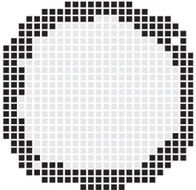
Fig. 1. Cross-section of a three-dimensional simulation of blastomeres migration under the minimization ofH, without morphogenic field (J2¼0).J1¼2:0 andT¼0:2:Since there is no preferential region of
cell–cell interaction [22]. In embryos, the release of morphogens, and consequent creation of a morphogen gradient are due to many factors, related to the relative position of the cells [22]. So, this diffusion process (or change in morphogen concentration) is related to the diffusion or production of chemical substances when, for example and particularly significant for our model, a tension is produced in a membrane, changing its curvature. In our model, and representing the initial symmetry of the system, all sites have the same morphogen concentration in the beginning of the simulation. Movements of the skin cells may change the morphogen concentration, which will in turn influence the evolution of the cell configuration. Such approach is strongly supported by Cummings’ model of gastrulation[23–26]. In his work, the Gaussian curvature of a cell layer is a function of a relative concentration of morphogens in the medium.
The Monte Carlo dynamics is performed using the following rules: (1) Initially, all theMi are set to zero.
(2) ASi¼1 (blastomere) site and one of its neighborsSj are randomly chosen. (3) If this neighbor isSj¼0 or 1 the choice is discarded, and we return to step (2). (4) IfSj¼2 we try to move the cellSi¼1 to positionj. The difference in energy
DE between the new and the old configuration is calculated and the movement is
performed with probability 1 if DEp0; and eDE=T if DE40: Temperature here
represents the flexibility of the system. Like in other models, which show a qualitative picture of the dynamics, it can be adjusted in order to give us the possible results that can be obtained with this model.
(5) If the movement is accepted, the blastomere Si occupies the site j. This movement leads to a rearrangement of the embryo’s cells: site j pushes one of its neighbors, randomly chosen between its neighbors and avoiding the position i, producing a sequence of movements that reaches the embryo’s surface. This happens when a siteS¼1 moves to a siteS¼0:
(6) After moving inwards, the blastomere (initially ati) pulls one of its neighbors of the same type 1, randomly chosen, which in turn pulls one neighbor and so on, as a stripe of S¼1 cells, avoiding rupture of the membrane. If during this rearrangement the skin breaks (the contact between S¼0 and 2 sites), the movement is not accepted.
site j and a predefined distance (typically the radius of the embryo before gastrulation begins).
(8) Return to step (2).
Only sites with Si¼1 are capable of movement, since they represent the blastomeres. Moreover, the blastomeres can only move to a site with Si¼2; the blastocoel. This is done in order to allow the blastula to fold, and to prevent it from falling apart, since at this time, there is no further production of cells.
3. Results and discussion
The results shown here were obtained with the parameter valuesJ1¼2:0;T ¼0:2; N¼31; k¼13 (213 cells), using 1000 Monte Carlo steps. Larger systems, with N¼101;k¼15;were also simulated, with the same qualitative results.
Fig. 1shows the result of a simulation withJ2¼0;i.e., without the morphogenic field M. Here, the dynamics simply reduces the contact energy among the cells, a particular case of cell sorting[20]. In this picture, it can be seen that the blastomeres spread all over the surface, in a qualitatively homogeneous fashion. Thus, a Hamiltonian defined only by a contact energy is not capable of yielding a spherical symmetry breaking, since all sites with blastomeres experience similar fluctuations, and have the same probability to move.
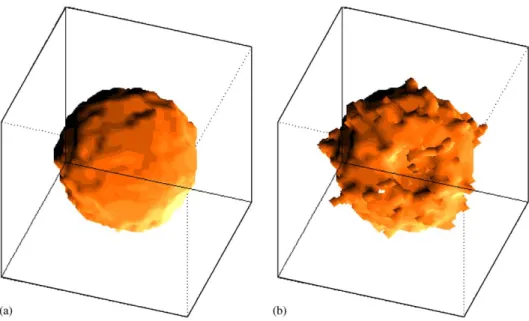
The behavior of the system is drastically changed when M plays a role in the dynamics.Figs. 2a and bshow an external 3d view of the system, before and after
Fig. 2. Artistic view of the external region of the system before (a) and (b) after minimization ofH. The parameters here are the same as inFig. 1except the presence of morphogenic fieldJ2¼1:0:It can be seen
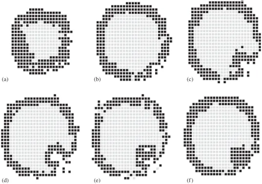
relaxation, now with the presence morphogens (J2 ¼1:0).Fig. 2a is asphericalcell aggregate, which, after the system reaches equilibrium, changes to the form shown in Fig. 2b. One can clearly see that an invagination has appeared on its surface. A better way to see this invagination is slicing the system, as shown inFig. 3.Fig. 3a shows the border of the system; the concentration of blastomeres is high. Proceeding inwards, one sees inFig. 3b the border of the invagination (right-bottom part of the figure). While the membrane is homogeneous in almost all regions of this figure, signs of different behavior are observed in the right-bottom region. Figs. 3c–e represent slices in the middle of the system; the invagination is evident. Finally,Fig. 3f shows the other border of the gastrula.
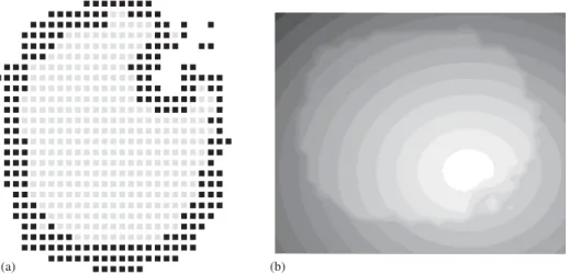
Figs. 4a and bshow, respectively, one slice passing through the same position in the medium S and in the morphogenic field M. In this way, we can see how the morphogens are distributed in the medium. The contours increase from black (low concentration) to white (high concentration). Note that the difference inMbetween neighboring sites is of the order of unity. The white region ofFig. 4bcoincides with the invagination. The figures help to understand the most important characteristics of the model: although there is no a priori a preferential region for gastrula formation, it is rapidly created due to the fluctuations in the concentration of morphogens. Once the process begins, it inhibits any other invagination on the surface of the system. Moreover, the initial process creates a flux of morphogens to
(a) (b) (c)
(d) (e) (f )
the invagination region. The higher the concentration of morphogens, the faster is the invagination. In other words, when one cell moves, the concentration of morphogens around the region where the movement took place increases, also increasing the probability of a new movement in this region. At the same time, there is a decrease in such concentrations in regions far from the origin of the movement and then the probability of any cell belonging to those regions to move is reduced. This mechanism prevents the formation of more than one invagination region.
It is important to mention that the dynamics here reported is in accordance with the basic assumptions of previous works [23]. The creation of this invagination region occurs in a self-organized way, since it is not a result of any external agent, or even an internal privileged one. This means that it arises from the interaction of all the elements of the system, in non-linear dynamics. The main component responsible for self-organization is the morphogenic field M, which represents a memory of cellular movements.
From another point of view, one cell movement implies in an energy change, or, in a Hamiltonian’s change, that, for its turn, reflects in the subsequent cell movements, creating a feedback loop between the cells and the morphogenic substances released by them. This loop is the key feature that makes possible the system self-organization. It resembles some other non-equilibrium processes, with self-catalytic loops. In those systems, the dynamics is governed by their own products, accelerating or slowing down themselves. Many works propose that self-organized processes driven by self-catalytic loops are responsible for the emergence of order and complexity in natural systems[27].
The model exhibits other remarkable features. The first one is its robustness, that is, the immunity to small changes in the parameters. This is a very desirable property for biological systems, once it introduces a kind of homeostasis, the capacity to
(a) (b)
assimilate some perturbations introduced by environment or another source, without causing catastrophic damage. The second characteristic is that the system evolves to an equilibrium state. In the same way that the system ‘‘chooses’’ the region where the invagination occurs, it also defines this equilibrium state. After reaching this state, no further movements are accepted.
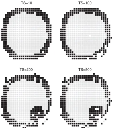
Such phenomenon can be seen inFigs. 5and6.Fig. 5shows a cross section of the system at different times. Fig. 6 shows the evolution of the coordinates of the system’s center of mass. Initially, the system experiences small changes, representing attempts to gastrulation. Once the region of the invagination is ‘‘chosen’’, a rapid rearrangement is produced, and changes occur in the CM coordinates in the interval of 100–200 MCS. Finally, the process stops. Only few movements are accepted. This occurs because the skin was stretched to its limit. More movements would break it. Some care must be taken with this kind of assumption. The most relevant aspect is showing that a mechanical force is responsible for stopping the entire process and thus leading the system to reach an equilibrium state. This process resembles those which occur in living beings. In those cases, what is observed is a competition between curvature and an elastic tension, where cell adhesion plays a fundamental role [1]. In real systems, the skin is not stretched to its maximum, like in our
TS=200 TS=10
TS=500 TS=100
Fig. 5. Time evolution of cellular movements. The parameters areJ1¼2:0;J2¼1:0 andT¼0:2:Note
simulations. Several models [16,17,21], where deformable cells are represented by many lattice sites, can represent this situation and could be an improvement of our model for future works.
Another important remark is the influence of temperature parameter T. As mentioned above, in this model Tis not a real temperature, but gives some sort of softness to the system. HigherTmakes the system easier to deform; lowerTacts in the opposite way. However, the influence of Tis very weak if compared with the other parameters. This can be explained by a spatial argument: since only cellsS¼1 may move and since these cells are linked forming a skin of cells, it makes their mobility much more dependent on the geometry and the morphogenic field than on the temperature. In other words, mobility is not an advantage when there is no space to move.
The model introduced in this work does not have either the ability or the purpose of explaining the development process beyond the beginning of the gastrulation. It is an approximation even in this respect. It answers the question of how one region can behave differently from the rest, when all, initially, are under the same constraints. Of course, other models answer the same problem, by using mechanical properties or
0 100 200 300 400 Monte Carlo Steps
36.9 37.0 37.1
Zcm 36.1 36.1 36.2
Ycm 35.5 35.6 35.7
Xcm
Fig. 6. Evolution of the system’s center of mass, given by its spatial three-dimensional coordinatesXcm;
the action of chemical substances. Our basic proposal was to emphasize the role of morphogens in this process. More than this, we would like also to emphasize that the final product, the final configuration of the system, is obtained by the cooperative action of many simple agents, our ‘‘cells’’, which obey very simple rules imposed by our Hamiltonian. From this point of view, each cell might be considered as an automaton programmed to execute simple set of movements. However, this dynamics becomes highly organized and coordinated when seen as a whole.
Acknowledgements
The authors acknowledge R. Dickman for discussions and a careful reading of the manuscript. ATB acknowledges the kind hospitality of the Departamento de Fı´sica-UFMG. The simulations were performed in the Digital work-stations at Physics Departments of UFMG and UFOP. This work was partially supported by the Brazilian Agencies CNPq, FINEP and FAPEMIG.
References
[1] R. Keller, L.A. Davidson, D.R. Shook, Differentiation 71 (2003) 171–205. [2] D. Voet e J.G. Voet, Biochemistry, Wiley, New York, 1990.
[3] S.F. Gilbert, Developmental Biology, Sinauer Associates Inc., Massachusetts, 1994.
[4] R. Keller, L. Davidson, A. Edlung, T. Elul, M. Ezin, D.R. Shook, P. Skoglund, Philos. Trans. R. Soc. London B 355 (2000) 897–922.
[5] R. Keller, Science 298 (2002) 1950.
[6] I.C. Scott, D.Y.R. Stainier, Nature 425 (2003) 461.
[7] D. Thompson, On Growth and Form, Cambridge University Press, Cambridge, 1961. [8] A.M. Turing, Philos. Trans. R. Soc. London B 237 (1952) 37–72.
[9] S. Sawai, Y. Maeda, Y. Sawada, Phys. Rev. Lett. 85 (2000) 2212–2215.
[10] W. Stein e F.J. Varela, Thinking About Biology, Addison-Wesley, New York, 1993. [11] A.V. Spirov, Int. J. Bifurcat. Chaos 5 (1998) 991.
[12] D. Drasdo, G. Forgacs, Dev. Dyn. 219 (2000) 182–191.
[13] Y. Jiang, H. Levine, J. Glazier, Biophys. J. 75 (991) (1998) 2615–2625. [14] M.S. Alber, Y. Jiang, M.A. Kiskowski, Physica D 191 (2004) 343–358. [15] A.F.M. Mare´e, P. Hogeweg, Proc. Natl. Acad. Sci. 98 (2001) 3879–3883. [16] G. Graner, J.A. Glazier Phys. Rev. Lett. 69 (1992) 2013–2016.
[17] N.B. Ouchi, J.A. Glazier, J.-P. Rieu, A. Upadhyaya, Y. Sawada, Physica A 329 (2003) 451–458. [18] J.A. Izaguirre, et al., Bioinformatics 20 (2004) 1129–1137.
[19] A.T. Bernardes, F.G.S. Arau´jo, J.R.T. Branco, Phys. Rev. E 58 (1998) 1132.
[20] J.C.M. Mombach, J.A. Glazier, R.C. Raphael, M. Zajac, Phys. Rev. Lett. 75 (1995) 2244. [21] A.N. dos Reis, J.C.M. Mombach, M. Walter, L.F. de A´vila, Physica A 322 (2003) 546–554. [22] J.B. Gurdon, P.-Y. Bourillot, Nature 413 (2001) 797–803.
[23] F.W. Cummings, Physica D 79 (1994) 146.
[24] F.W. Cummings, J. Theor. Biol. 212 (2001) 303–313. [25] F.W. Cummings, Physica A 339 (2004) 531–547.
[26] C.H. Leung, M. Berzins, J. Comput. Phys. 188 (2003) 75–99.