Karyotype, C-and fluorescence banding pattern, NOR location
and FISH study of five Scarabaeidae (Coleoptera) species
Edgar Bione
1, Rita de Cássia de Moura
1,2, Reginaldo de Carvalho
1and Maria José de Souza
11
Universidade Federal de Pernambuco, Centro de Ciências Biológicas, Departamento de Genética,
Recife, PE, Brazil.
2Universidade de Pernambuco, Instituto de Ciências Biológicas, Departamento de Biologia, Recife,
PE, Brazil.
Abstract
Meiotic chromosomes obtained from members of the coleopteran subfamilies Rutelinae and Dynastinae were studied using standard and silver nitrate staining, C-banding, base-specific fluorochromes and fluorescentin situ hybridization (FISH). The study presents detailed karyotipic descripitions of three Rutelinae species (Geniates borelli,Macraspis festivaandPelidnota pallidipennis), and two Dynastinae species (Lygirus ebenusandStrategus surinamensis hirtus) with special emphasis on the distribution and variability of constitutive heterochromatin and the nucleolar organizer region (NOR). We found that for G. borelli,P. pallidipennis, L. ebenus andS. s hirtusthe karyotype was 2n = 20 (9II + Xyp), with G. borelli,P. pallidipennis andL. ebenusshowed meta-submetacentric
chromosomes which gradually decreased in size. ForMacraspis festivathe karyotype was 2n = 18 (8II + Xyp). InL.
ebenuswe found that the NOR was located on an autosome, but in the other four species it occurred on the sex bivalents. In all five species the constitutive heterochromatin (CH) was predominantly pericentromeric while the X chromosomes were almost completely heterochomatic, although CMA3/DA/DAPI staining showed intra and
interspecific variation in the bright fluorescence of the constitutive heterochromatin. The FISH technique showed rDNA sites on the X chromosome of the Rutelinae species.
Key words: karyotype, constitutive heterochromatin, NORs, rDNA sequences.
Received: August 7, 2004; Accepted: January 15, 2005.
Introduction
The coleopteran family Scarabaeidae is made up of a cosmopolitan group of approximately 2,300 genera and 27,000 species worldwide distributed with a highly con-served diploid chromosome number (2n = 20) and Xy type `parachute’ (Xyp) sex-determining mechanism, although there is variation in chromosome morphology (Smith and Virkki, 1978; Yadav and Pillai, 1979; Colomba et al., 1996). Neotropical and Brazilian representatives of the scarabaeid subfamilies Rutelinae and Dynastinae have been extensively studied taxonomically (Endrödi, 1985; Morónet al., 1997) and it is known that more than 50% of the species from these subfamilies possess the standard karyotype, although variations in chromosome number have been observed with the chromosome number ranging from 2n = 18 to 2n = 22 in the subfamily Rutelinae (Smith
and Virkki, 1978; Yadav and Pillai, 1979) and from 2n = 12 to 2n = 20 in the Dynastinae (Vidal, 1984; Martins, 1994).
Differential techniques have rarely been applied to chromosome studies of the Coleoptera, but data from the species so far analyzed have shown that the autosomal con-stitutive heterochromatin (CH) is preferentially located on pericentromeric region and is less frequent on interstitial and telomeric regions while the position of sex chomosome constitutive heterochromatin is more variable in that it may be pericentromeric or entirely heterochromatic (Vidal et al., 1977; Angus, 1983; Drets et al., 1983; Virkki, 1983; Juan and Petitpierre, 1989; Rozek and Lachowska, 2001). Base-specific fluorochromes have provided important in-formation regarding the composition of CH in coleopteran species of the families Tenebrionidae (Juan and Petitpierre, 1989; Plohlet al., 1993) and Scarabaeidae (Colombaet al., 1996; Colombaet al., 2000; Mouraet al., 2003). Data re-garding the localization of the nucleolar organizer regions (NORs) in the scarabPhyllophaga (Phytalus) vestitaand Lyogenys fuscusobtained by silver nitrate staining and
flu-www.sbg.org.br
Send correspondence to Rita Moura. Universidade de Pernam-buco, Instituto de Ciências Biológicas, Departamento de Biologia, Rua Arnóbio Marques 310, Santo Amaro, 50100-130 Recife, Pe, Brazil. E-mail: rita_upe@yahoo.com.br.
orescencein situhybridization (FISH) have shown that the ribosomal sites are preferentially located on the sex chro-mosomes (Mouraet al., 2003), although inGymnopleurus sturmiandPhyllophaga(P.) affcapillata(Scarabaeidae), Trotectes intermedius (Geotrupidae), Eriopis connexa (Coccinellidae) and in 19Zabrusspecies (Carabidae), the NORs are located on the autosomes (Vitturiet al., 1999; Colombaet al., 2000; Maffeiet al., 2000; Sánchez-Géaet al., 2000; Mouraet al., 2003).
This study presented in this paper provides detailed kayotypic descriptions of three representative Rutelinae species (Geniates borelli,Macraspis festivaandPelidnota pallidipennis) and two representative Dynastinae species (Lygirus ebenus andStrategus surinamensis hirtus) with special emphasis on the distribution and variability of con-stitutive heterochromatin and NORs.
Materials and Methods
Meiotic chromosomes were obtained from Rutelinae species (Geniates borelliCamerano, 1894 (12 specimens), Macraspis festiva Burmeister, 1844 (6 specimens) and Pelidnota pallidipennis Bates, 1904 (12 specimens) and Dynastinae species (13Lygirus ebenusDe Geer, 1774 and five Strategus surinamensis hirtusSternberg, 1910). The specimens were male beetles collected from Atlantic Forest sites situated in the northeastern Brazilian state of Pernambuco at 07°48’37’’ S, 34°27’25’’ W near the town of Igarassú forG.borelli,P.pallidipennisandL.ebenus and08°0’8”, 35°1’6” W near the town of São Lourenço da Mata for S. s. hirtus and M. festiva. Testicular follicle squashes were made in ethanol and acetic acid (3:1) fixa-tive and the chromosomes stained with 2% lacto acetic orceína. We also performed C-banding (Sumner, 1972), sil-ver nitrate (Rufaset al., 1987) and AT/GC base pair fluo-rescence staining (Schweizeret al., 1983). Fluorescentin situhybridization (FISH) was performed as described by Moscone et al. (1996) using Arabidopsis thaliana 45S rDNA probes (Unfriedet al., 1989; Unfried and Gruendler, 1990) nick translation labeled with bio-11-dUTP (Life Technologies) and detected with rat antibiotin antibodies (Dakopatts M0743, Dako) and tetramethyl-rhodamine isothiocyanate (TRITC) conjugated rabbit anti-rat antibod-ies (Dakopatts R0270, Dako).
Results
Standard staining and C-banding
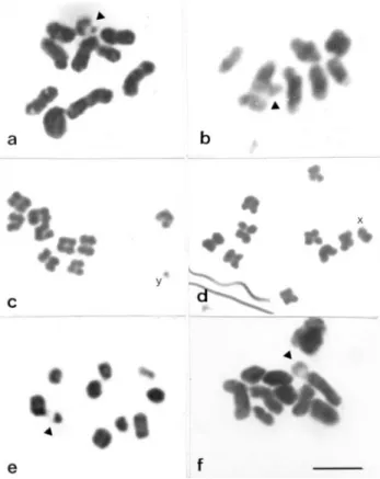
The male karyotypes of most of the species analyzed were 2n = 20 (9II + Xyp) (Figure 1a, c-f), the exception be-ingM. festivawhich had a karyotype of 2n = 18 (8II + Xyp) (Figure 1b). The chromosomes of G. borelli, P. pallidipennisandL. ebenuswere meta-submetacentric and showed a gradual decrease in size. The sex-determining
mechanism of all the species analized was of the parachute type, with a metacêntric X chromosome and a diminutive Y chromosome (Figure 1a, b, e, f).
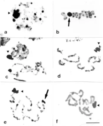
The C-banding method revealed blocks of constitu-tive heterochromatin in the pericentromeric region of all the autosomes of the Rutelinae species (Figure 2a-c) while for the Dynastinae species in addition to the peri-centromeric blocks a terminal block was noted on a small chromosome ofL. ebenus(Figure 2d) and C-banding was absent from oneS. s. hirtus(Figure 2e). The X chromo-somes of the five species studied were all almost completely heterochromatic and no constitutive hetero-chromatin blocks were detected in the y chromosome of any of the species (Figure 2b-d). Heterochromatin associa-tions forming chromocenters between autosomal bivalents were observed in the five species analyzed, these associa-tions being first visible during meiotic prophase and per-sisted until the end of the pachytene phase (Figure 2c).
Fluorochrome staining
For G. borelli CMA3/DA/DAPI staining showed small GC-rich CMA3 positive blocks, coinciding with those visualized by C-banding, in the pericentromeric re-gion of all the chromosomes (Figure 3a), but no DAPI-positive blocks were detected in this species (Figure 3b). In
Figure 1- Meiotic chromosomes stained with acetic orcein. Metaphases I ofG. borelli(a)M. festiva(b),L. ebenus(e) andS. s hirtus(f). Note the
contrast,P. pallidipennispresented DAPI blocks similar in size and location to those detected by C-banding and the heterochromatin of this species was AT-rich except for a small CMA3 -positive block detected in one of the autosomal bivalents and another detected in the sex chro-mosomes (Figure 3e, f), CMA3/DA staining revealed the presence of GC-rich blocks in all chromosomes of the com-plement except for a small bivalent, but no DAPI-positive blocks were detected in this species.
Silver nitrate staining and FISH
Amorphous masses corresponding to nucleolar rem-nants were visualized by silver nitrate staining in the Xyp bivalents ofG. borelli,P. pallidipennis,M. festivaandS. s. hirtus(Figure 4 a, c, d, f), while inL. ebenusthe labeling was detected on an autosomal pair (Figure 4e). These masses were visible until the end of the pachytene phase or the beginning of the diplotene phase. Silver nitrate also la-beled the constitutive heterochromatin blocks (Figure 4a-c, e) and the sex chromosomes showed affinity for silver and continued to be labeled during different phases of meiosis
(Figure 4b). In the three Rutelinae species, FISH of rDNA genes produced results, which coincided with those, ob-tained by silver nitrate staining and permitted the identifi-cation of rDNA genes on the X chromosome (Figure 5a-c).
Discussion
We found thatG. borelli,P. pallidipennis,L. ebenus andS. s. hirtushad the 2n = 20 (9II + Xyp) karyotype typical of the suborder Polyphaga, but M. festiva karyotype of 2n = 18 (8II+ Xyp) which coincided with the karyotype of Macraspis dichroa (Vidal, 1984). Other species of Macraspisare known to have a 2n = 20 karyotype (Martins 1994) but with the Xyp type sex-determining system changed to neoXy system. Karyotypic comparisons M. festivaand other species ofMacraspisof known cytology suggests that karyotype evolution in this genus might have involved different types of chromosome rearrangements. It is possible that the reduction in the chromosome number observed inM. festivamight have been due to a mechanism involving pericentric inversion followed by fusion between autosomes, which would explain the occurrence of karyotypic changes in the absence of alterations in the
Figure 2- Constitutive heterochromatin (CH) distribution pattern in the five species studied. C-bands in a spermatogonial metaphase ofG. borelli
(a). Metaphase I ofP. pallidipennis(b). Pachytene ofM. festiva(c), note the heterochromatic associations. Diplotene and metaphase I ofL. ebenus
(d)and S. s. hirtus(e). The arrowheads point to an almost completely heterochromatic sex bivalent. The arrows indicate the presence of a termi-nal CH block inL. ebenus(d) and the absence of labeling in an autosomal bivalent ofS. s. hirtus(e). Bar = 5mm.
sex-determining system. Changes of this type have been described in the literature and are included in the five types of karyotype evolution proposed for Scarabaeidae by Yadav and Pillai (1979).
Our results show that inG. borelli,P. pallidipennis, M. festiva,L. ebenusandS. s. hirtusthere was some degree of conservation in terms of the size and location of the CH blocks as well as a type of heterochromatic association in which chromocenters formed between some autosomal bi-valents. It is known that the degree of ectopic pairing be-tween heterochromatic segments that promote the formation of chromocenters varies among different coleopteran species and that this type of association seems to play an important role in nuclear organization and the segregation of meiotic chromosomes (Smith and Virkki, 1978; Dretset al., 1983).
The constitutive heterochromatin of the species ana-lyzed by us was located on the pericentromeric region of the chromosomes, similar observations having been re-ported for other Scarabaeidae (Vidal and Giacomozzi, 1978; Vidal and Nocera, 1984). This pattern of distribution has been described for most coleopteran species studied by C-banding (Virkki, 1983; Rozek and Maryanska-Nadachowska, 1991; Rozek and Rudek, 1992), although telomeric blocks in addition to pericentromeric ones have been observed in the tenebrionid Misolampus goudoti (Juan and Petitpierre, 1989) and exclusively telomeric blocks in the carabid Bembidion minimum (Rozek and Rudek, 1992), with extra-heterochromatic segments having been reported in the scarabaeidBubas bison(Colombaet al., 1996).
In our study, CMA3and DAPI staining revealed that qualitative heterogeneity in the constitutive hetero-chromatin of G. borelli andS. s hirtus we found CMA3 positive blocks indicating GC-rich constitutive hetero-chromatin, but in Pelidnota palidipennis we found two types of constitutive heterochromatin, a DAPI positive type
Figure 4- Silver nitrate staining in the five species analyzed. Pachytene and metaphase I ofG. borelli(a, b). Zygotene and initial pachytene ofP. pallidipennis(c). Final pachytene ofM. festiva(d). Pachytenes ofL. ebenus(e) andS. s hirtus(f). Observe the heterochromatic regions and the
strongly silver-labeled sex pair (b). The arrows in (b,e) indicate de Xyp bi-valent. The arrowheads indicate nucleolar organizer regions. Bar = 5mm.
Figure 5- Fluorescentin situhybridization (FISH) in the three Rutelinae species. Zygotenes ofG. borelli(a),P. pallidipennis(b) andM. festiva(c). Note
evenly distributed throughout the karyotype and a CMA3 positive type restricted to one small block located on the sex pair (probably the X chromosome) and another small block located on one of the autosomal bivalents. Reports on the use of base-specific fluorochromes in Scarabaeidae are still scarce, but different patterns have been found in some species. For example, inGymnopleurus sturni(Vitturiet al 1999) and Thorectes intermedius (Colomba et al 2000) GC-rich sequences were detected in all the chromosomes whileLyogenys fuscuspresented AT-rich sequences in ev-ery karyotype complement studied (Mouraet al., 2003).
Data regarding the location of NORs in Coleoptera have suggested that a pair of nucleolar organizer autosomes is widely distributed in this order (Virkki, 1983; Virkkiet al., 1984). In representatives of the family Scarabaeidae rDNA sites are generally found on the X chromosome, al-though sites located on autosomes have been reported for Phyllophaga (Phyllophaga) aff capillata and Gymnopleurus sturni(Mouraet al., 2003; Colombaet al., 2000). InG. borelli,P. pallidipennis,M. festivaandS. s hirtuswe found that the NOR was associated with the sex bivalent, and this confirmed forG. borelli, P. pallidipennis, M. festivaby ourin situhybridization using the 45S rDNA probe.
Studies analyzing the development and segregation of the Xypchromosome in some curculionid species have shown that, even when the NORs are autosomal, the lumen of the sex bivalent is filled from diakinesis to anaphase I with proteinaceous substances which have an affinity for silver and which probably resemble substances present in the synaptonemal complex and chromosome skeleton. It has been proposed by Virkkiet al.(1990; 1991) that these substances function as an adhesive and therefore control the correct disjunction of the sex chromosomes.
In the Scarabaeidae species analyzed we found that the sex bivalent remained silver labeled after the nucleolus disappeared, suggesting that the Xypassociation is not nec-essarily due to the presence of the NOR, but rather to the presence of argyrophilic proteins distributed within the heterochromatin of these species. Argyrophilic proteins have also been observed in the scarabaeid species Thorectes intermedius(Vitturiet al., 1999),Gymnopleurus sturni (Colomba et al., 2000), Phyllophaga (Phytalus) vestita, Phyllophaga (Phyllophaga) aff capillata and Lyogenys fuscus(Mouraet al., 2003) and their presence ap-pears not to depend on the base composition of CH, further studies being needed to establish the real biochemical com-position of scarabeoid beetle heterochromatin and explain the positive reaction to silver staining.
Our results show that in the species studied by us there was a clear relationship between the NOR and the sex chromosomes, with silver staining and FISH demonstrating that NORs are preferentially located on the sex pair.
Al-though not indicating the direct participation of the NOR in the formation of the Xypbivalent, this relationship demon-strates the apparent conservation of the location of the rDNA sites on the X chromosomes in representatives of the family Scarabaeidae.
Acknowledgements
The authors are indebted to Dr. Sérgio Ide, (Insitute of Biology, São Paulo) for the taxonomic identification of the species analyzed in this study; Dr. Marcelo Guerra (De-partment of Botany, UFPE) for providing the infrastructure for performing the FISH experiments; Dr Neide Santos (Department of Genetics, UFPE) for critical reviewing of the manuscript; and Francisca Tavares de Lira for technical assistance. The study was supported by Conselho Nacional de Desenvolvimento Científico e Tecnológico (CNPq) and Fundação de Amparo à Ciência e Tecnologia do Estado de Pernambuco (FACEPE).
References
Angus RB (1983) Separation ofHelophorus grandis, maritimus
and occidentalis sp. n. (Coleoptera, Hydrophilidae) by
banded chromosome analysis. Syst Entomol 8:1-13. Colomba MS, Monteresino E, Vitturi R and Zunino M (1996)
Characterization of mitotic chromosomes of the scarab bee-tlesGlyphoderus sterquilinus(Westwood) andBubas bison
(L.) (Coleoptera, Scarabaeidae) using conventional and banding techniques. Biol Zentbl 115:58-70.
Colomba MS, Vitturi R and Zunino M (2000) Karyotype ana-lyzes, banding, and fluorescentin situhybridization in the Scarab beetleGymnopleurus sturmiMcLeady (Coleoptera,
Scarabaeoidea, Scarabaeidae). The Journal of Heredity 91:260-264.
Drets ME, Corbella E, Panzera F and Folle GA (1983) C-banding and non-homologous association II. The “parachute” Xyp
sex bivalent and the behaviour of heterochromatic segments inEpilachna paenulata. Chromosoma 88:249-255.
Endrödi S (1985) The Dynastinae of the World. Dr. W. Junk, Bu-dapest, 801 pp, xlvi pls.
Juan C and Petitpierre E (1989) C-banding and DNA content in seven species of Tenebrionidae (Coleoptera). Genome 32:834-839.
Maffei EMD, Gasparino E and Pompolo SG (2000) Karyotypic characyerization by mitosis, meiosis and C-banding of
Eriopis connexa Mulsant (Coccinellidae, Coleoptera,
Polyphaga), a predator of insect pests. Hereditas 132:79-85. Martins VG (1994) The chromosomes of five especies of
Scarabaeidae (Polyphaga, Coleoptera). Naturalia 19:89-96. Morón MA, Ratcliffe BC and Deloya. C (1997) Atlas de los escarabajos de Mexico. Coleoptera, Lamelicornia. V. 1 Fa-milia Melolonthidae, SubfaFa-milias Rutelinae, Dynastinae, Cetoniinae, Trichiinae, Valginae y Melolonthinae. Xalapa, Sociedad Mexicana de Entomologia, A.C., xvi + 280 pp. Moscone EA, Matzke MA and Matzke AJM (1996) The use of
Moura RC, Souza M.J, Melo NF and Lira-Neto AC (2003) Karyotypic characterization of representatives from Melolonthinae (Coleoptera, Scarabaeidae): Karyotypic analysis, banding and fluorescent in situ hybridization
(FISH). Hereditas 138:200-206.
Plohl M, Lucijanic-Justic V, Hugarkovic D, Petitpierre E and Juan C (1993) Satellite DNA and heterochromatin of the flour beetleTribolium confusum.Genome 36:467-675.
Rozek M and Lachowska D (2001) C-bands on chromosomes of four beetle species (Coleoptera, Carabidae, Silphidae, Elateridae, Scarabaeidae).Folia Biologica 49:3-4.
Rozek M and Maryanska-Nadachowska A (1991) Studies on C-banding karyotype and meiosis of Harpalus affinis
(Shrank 1781) (Coleoptera, Carabidae). Caryologia 44:317-323.
Rozek M and Rudek Z (1992) Karyotype analysis and C-banding pattern in two species of carabid (Coleoptera, Carabidae). Folia biologica 40:47-52.
Rufas JS, Giménez-Ábian J, Suja JA and Garcia de la Vega C (1987) Chromosome organisation in meiosis revealed by light microscope analysis of silver-stained “cores”. Genome 29:706-712.
Sánchez-Gea JF, Serrano J and Galián J (2000) Variability in rDNA loci in Iberian species of the genus Zabrus
(Coleoptera, Carabidae) detected by fluorescencein situ hy-bridization. Genome 43:22-28.
Schweizer D, Mendelak M, White MJD and Contreras N (1983) Cytogenetics of the parthenogenetic grasshopper
Warramaba virgoand its bisexual relatives. X. Pattern of
fluorescent banding.Chromosoma 88:227-236.
Smith SG and Virkki N (1978) Coleoptera. In: John B (ed) Animal Cytogenetics Borntraeger, Berlin, Stuttgart, 366 pp. Sumner AT (1972) A simple technique form demonstrating
centromeric heterochromatin. Exp Cell Res 75:304-306. Unfried I and Gruendler P (1990) Nucleotide sequence of the 5.8S
and 25S rRNA genes and of the internal transcribed spacers fromArabdopsis thaliana.Nucleic Acids Res 18:4011.
Unfried I, Stocker U and Gruendler P (1989) Nucleotide sequence of the 18S rRNA gene fromArabdopsis thaliana. Nucleic
Acids Res 17:7513.
Vidal OR (1984) Coleoptera from Argentina. Genetica 65:235-239.
Vidal OR and Giacomozzi RO (1978) Los cromosomas de la subfamilia Dynastinae (Coleoptera, Scarabaeidae). Las bandas C enEnema pan(Fabr.). Physis 38:113-119.
Vidal OR and Nocera CP (1984) Citogenética de la tribu Eucranini (Coleoptera, Scarabaeidae). Estudios convencionales y con bandeo C. Physis 42:83-90.
Vidal OR, Giaccomozzi RO and Riva R (1977). Los cromosomas de la subfamilia Dynastinae (Coleoptera, Scarabaeidae). I. Inversión pericéntrica en Diloboderus abderus (Sturm)
1862. Physis 37:303-309.
Virkki N (1983) Banding of Oedionychna (Coleoptera, Alticinae) chromosomes: C- and Ag-bands. J. Agric. Univ. Puerto
Rico 67:221-225.
Virkki N, Flores M and Escudero J (1984) Structure, orientation and segregation of the sex trivalent inPyrophorus luminosus
(Coleoptera, Elateridae). Can J Genet Cytol 26:326-330. Virkki N, Mazzella C and Denton A (1990) Staining of substances
adjacent to the Xyp sex bivalent of some weevils
(Coleoptera, Curculionidae). J Agric Univ Puerto Rico 74:405-418.
Virkki N, Mazzella C and Denton A (1991) Silver staining of the coleopteran Xypsex bivalent. Cytobios 67:45-63.
Vitturi R, Colomba MS, Barbieri R and Zunino M (1999) Ribo-somal DNA location in the scarab beetle Thorectes intermedius(Costa) (Coleoptera, Geotrupidae) using band-ing and fluorescent in-situ hybridization. Chromosome Re-search 7:255-260.
Yadav JS and Pillai RK (1979) Evolution of Karyotypes and phylogenetic relationships in Scarabaeidae (Coleoptera). Zool Anz Jena 202:105-118.