Digital orthodontic radiographic set versus cone-beam computed
tomography: an evaluation of the effective dose
Lillian Atsumi Simabuguro Chinem1, Beatriz de Souza Vilella2, Cláudia Lúcia de Pinho Maurício3, Lucia Viviana Canevaro3, Luiz Fernando Deluiz4, Oswaldo de Vasconcellos Vilella2
1 Specialist in Orthodontics, Universidade Federal Fluminense (UFF), Niterói,
Rio de Janeiro, Brazil.
2 Professor, Graduate program in Orthodontics, Universidade Federal
Fluminense (UFF), Niterói, Rio de Janeiro, Brazil.
3 Professor, Graduate program in Radioprotection and Dosimetry, Instituto de
Radioproteção e Dosimetria (IRD), Rio de Janeiro, Rio de Janeiro, Brazil.
4 Professor, Graduate program in Radiology, Universidade Estácio de Sá
(UNESA), Rio de Janeiro, Rio de Janeiro, Brazil.
Submitted: June 11, 2015 - Revised and accepted: February 21, 2016.
Objective: The aim of this study was to compare the equivalent and effective doses of different digital radiographic methods (panoramic, lateral cephalometric and periapical) with cone-beam computed tomography (CBCT). Methods: Precalibrated thermoluminescent dosim-eters were placed at 24 locations in an anthropomorphic phantom (Alderson Rando Phantom, Alderson Research Laboratories, New York, NY, USA), representing a medium sized adult. The following devices were tested: Heliodent Plus (Sirona Dental Systems, Bernsheim, Germany), Orthophos XG 5 (Sirona Dental Systems, Bernsheim, Germany) and i-CAT (Imaging Sciences International, Hatfield, PA, USA). The equivalent doses and effective doses were calculated considering the recommendations of the International Commission of Ra-diological Protection (ICRP) issued in 1990 and 2007. Results: Although the effective dose of the radiographic set corresponded to 17.5% (ICRP 1990) and 47.2% (ICRP 2007) of the CBCT dose, the equivalent doses of skin, bone surface and muscle obtained by the radiographic set were higher when compared to CBCT. However, in some areas, the radiation produced by the orthodontic set was higher due to the complete periapical examination. Conclusion: Considering the optimization principle of radiation protection, i-CAT tomography should be used only in specific and justified circumstances. Additionally, following the ALARA principle, single periapical radiographies covering restricted areas are more suitable than the complete periapical examination.
Keywords: Cone-beam computed tomography. Digital radiograph. Radiation dose.
DOI: http://dx.doi.org/10.1590/2177-6709.21.4.066-072.oar
How to cite this article: Chinem LAS, Vilella BS, Mauricio CLP, Canevaro LV, Deluiz LF, Vilella OV. Digital orthodontic radiographic set versus cone-beam computed tomography: an evaluation of the effective dose. Dental Press J Orthod. 2016 July-Aug;21(4):66-72.
doi: http://dx.doi.org/10.1590/2177-6709.21.1.066-072.oar
» The authors report no commercial, proprietary or financial interest in the products or companies described in this article.
» Patients displayed in this article previously approved the use of their facial and in-traoral photographs.
Contact address: Lillian Atsumi Simabuguro Chinem
Rua Mário Santos Braga 30, 2o andar, sala 214 - Niterói, Rio de Janeiro, Brazil. E-mail: [email protected]
Objetivo: o objetivo deste estudo foi comparar as doses equivalentes e efetivas absorvidas pelo mesmo receptor, quando exposto a diferentes exames radiográficos digitais (panorâmica, telerradiografia lateral e periapicais) e à tomografia computadorizada de feixe cônico (TCFC). Métodos: dosímetros termoluminescentes pré-calibrados foram colocados em 24 locais em um phantom antropomórfico (Alderson Rando Phantom, Laboratórios de Pesquisas de Alderson, New York, NY, EUA), o que representa um adulto de tamanho mé-dio. Os seguintes equipamentos foram avaliados: Heliodent Plus (Sirona Dental Systems, Bernsheim, Alemanha), Orthophos XG 5 (Sirona Dental Systems, Bernsheim, Alemanha) e i-CAT (Imaging Sciences International, Hatfield, PA, EUA). As doses equivalentes e as doses efetivas foram calculadas tendo-se em conta as recomendações da Comissão Internacional de Proteção Radiológica (International Commission on Radiological Protection, ICRP), publicadas em 1990 e 2007. Resultados: embora a dose eficaz do set radiográfico tenha correspondido a 17,5% (ICRP 1990) e 47,2% (ICRP 2007) da dose da TCFC, as doses equivalentes na pele e superfícies ósseas e musculares desse set foram maiores, quando comparadas às da TCFC. Em algumas áreas, a radiação produzida pelo set de radiografias foi maior, devido à radiação do exame periapical completo. Conclusão: considerando-se o princípio da otimização da proteção radiológica, a tomo-grafia computadorizada adquirida no tomógrafo i-CAT deve ser usada apenas em circunstâncias específicas e justificadas. Além disso, seguindo o princípio ALARA, radiografias periapicais unitárias de áreas pré-selecionadas são mais apropriadas do que o exame periapical completo.
INTRODUCTION
The limitation of radiography due to its two-dimensional representation of tritwo-dimensional
struc-tures is a well-known fact.1,2 In the last decades,
two-dimensional images were gradually replaced by tridi-mensional ones. Cone-beam computed tomography (CBCT) provides a high-resolution image that is
simi-lar to computed tomography,3 but at lower cost and
ra-diation dose.4,5,6 Given these advantages, CBCT use is
widespread in Dentistry nowadays, particularly for
di-agnosis, treatment planning and follow-up.7
On the other hand, the high prevalence of adolescents who seek orthodontic treatment goes against the fact that the radiation emitted by CBCT is greater than the radiation emitted by a radiographic device. The higher frequency of young patients results in a concern regard-ing radiation dose, as children seem to carry the brunt of radiation for a longer period of time than adults, and their
developing organs are more sensitive to radiation efects.8
Furthermore, due to stochastic efects, of which probability of occurring is proportional to the radiation dose without a threshold, limits had to be established.
The ALARA principle is usually applied as a reference.9
In order to control the radiation doses emitted by the devices and to allow evaluations and comparisons of diferent devices, the International Commission of Radiological Protection (ICRP) established values in 1990 and 2007. These values were applied to calcu-late the absorbed dose, the equivalent and the efective dose. Although studies have already compared diferent models and parameters, signiicant diferences between models and between imaging protocols of the same
de-vice were observed.10,11
The aim of this study is to compare the equivalent and efective doses of diferent digital radiographic methods (panoramic, lateral cephalometric and periapical) with cone-beam computed tomography (CBCT) absorbed by the same receptor.
MATERIAL AND METHODS
Calibration and selection of dosimeters
Two types of thermoluminescent dosimeters (TLD) were employed in this study: TLD-100 Chip (Thermo Fisher Scientiic Incorporation, Waltham, MA, USA) and TLD-100 Rod (Thermo Fisher Scientiic Incorpo-ration, Waltham, MA, USA). Due to the attenuation of radiation by the tissues, chip dosimeters were positioned
on skin areas while rods dosimeters, more sensitive, were adapted in the holes inside the phantom. TLDs were prepared, calibrated and evaluated following the routine procedures of the Thermoluminescent Dosim-etry Laboratory of Instituto de Radioproteção e Dosi-metria (IRD), Brazil. Thereater, they were preselected in groups with similar sensitivities (standard deviation of the mean value were lower than 5%) ater three Cs 137 free in air irradiations with electronic equilibrium. The air kerma values were of 1.0 mGy.
The selected dosimeters were wrapped in plastic, so as to protect them from dirt and moisture. Subsequently, each one of these plastic packs was placed in a speciic location inside an Alderson Rando phantom in order to evaluate the organ/tissue equivalent dose. A total of 19 TLDs was chosen to measure the background dose.
The location of dosimeters
The dosimeters were positioned in 24 regions (Table 1) of a phantom that was composed by the skel-eton of a medium sized male adult (1.75 m) covered with equivalent tissue material. The areas were selected
ac-cording to Ludlow’s methods6,12-15 and corresponded to
radiosensitive organs, including eyes and pituitary gland. All dosimeters were placed inside the phantom by the same operator in order to reduce positioning variability.
Protocols and parameters adopted
The devices evaluated were Heliodent Plus (Sirona Dental Systems, Bernsheim, Germany), Orthophos XG 5 (Sirona Dental Systems, Bernsheim, Germany) and CBCT i-CAT (Imaging Sciences International, Hatield, Pa, USA). Protocols for digital radiograph and CBCT parameters for examination of a medium sized male adult were adopted (Table 2). For the periapical examination, exposure time varied according to the re-gion; whereas for the CBCT examination a ield of view (FOV) of 22 cm was necessary in order to obtain the image of all structures.
The phantom was positioned according to the manufacturer’s guidelines without the thyroid
col-lar. Based on the doses obtained in other studies,12,16
The equivalent and the effective doses
The values obtained were divided by the number of repeated irradiations, so as to obtain the value per examination.
For the bone marrow, the equivalent dose was cal-culated based on the distribution of bone throughout the adult body. The mandible contains 1.3%, the cal-varium 11.8% and the cervical spine contains 3.4%. The technique by Underhill et al was adopted to cal-culate the dose for the calvarium. For bone surface, a correction factor was applied:
Bone: muscle attenuation ratio =
-0.0618 X kV peak + 6.9406.17
The proportion of skin area in the head and neck region directly exposed during maxillofacial CBCT
imaging is estimated as 5% of the total body. Muscle and lymphatic nodes are estimated to represent 5%,
esophageal tract 10% and other tissues 100%.12
The salivary glands began to be used in the efective
dose calculation only in ICRP 2007.18 Their equivalent
dose is obtained with the weighted average dose values of parotid, submandibular and sublingual glands.
The equivalent doses (HT) in these organ/tissues
were calculated by the following formula:
HT (µSv): WR DT
WR is the radiation weighting factor and its value is 1
for X-rays. DT is the mean absorbed dose in T.19
The efective dose (E), proposed by ICRP 1990,18 is a
reliable clinical and standardized measure of the biologi-cal efects of radiation, although previous studies have
Organ/Tissue Location TLDs
Bone marrow
Anterior calvarium 1
Posterior calvarium 2
Left calvarium 3
Center cervical spine 12
Right/left ramus 10, 11
Right/left mandibular body 14, 15
Brain Mid brain 7
Pituitary fossa 4
Eyes Right/left orbit 5, 6
Right/left lens of eye 21-24*
Salivary glands
Right/left parotid 8, 9
Right/left submandibular gland 16, 17
Sublingual gland 13
Thyroid Thyroid surface 19*
Midline thyroid 18
Skin Right cheek 25, 26*
Left back of neck 27, 28*
Esophagus Pharyngeal-esophageal space 20
Table 1 - Location of thermoluminescent dosimeters in Alderson Rando phantom.
Table 2 - Parameters adopted.
*Dosimeters positioned on the surface of the phantom. TLDs: thermoluminescent dosimeters.
*Field of view. **Kilovoltage. *** Milliamperes.
FOV* kV** mA*** Exposure time (s)
Heliodent plus Periapical 70 7 0.25 - 0.4
Orthophos XG 5 Panoramic 69 15 14.1
Orthophos XG 5 Cephalometric 80 14 9.4
demonstrated limitations.20 The efective dose deined to estimate an average whole body human radiation risk is calculated with the following formula:
E = ∑wT X HT 18
WT is the weighting factor of the organ or tissue
(T) and is related to its radiation sensitivity. Both tissue
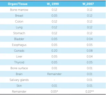
weighting factors of ICRP 60 and ICRP 103 (1990 and 2007) applied in this study are presented in Table 3.
ICRP 103 (2007)18 increased the number of
organs/tissues with wT values, including brain and
salivary glands, and the number of remainder tissues increased to 14. Only lymphatic nodes, muscles, ex-trathoracic airways and oral mucosa were exposed during the tests (Table 4).
The new recommendations stated that brain and salivary glands received factors of 0.01 and 0.1, respec-tively. The oral mucosa equivalent dose was calculated by the salivary glands and mandibular ramus and body
with a conservative estimate of 100%.14
RESULTS
The obtained values of the equivalent and efective doses are listed in Table 5. The lowest equivalent doses were obtained in lateral cephalometric radiograph, fol-lowed by panoramic, periapical and CBCT. Consider-ing thyroid equivalent doses, it was observed that their values were lower in lateral cephalometric and periapical examinations, and higher in CBCT.
By adding salivary glands to the calculations of efective doses, their values increased considerably. The glands and the remainder tissues were the main contributors to the efective dose in lateral cephalo-metric and panoramic radiographs. The efective doses
using values recommended by the ICRP 60 (1990)19
correspond to 48%, 24.7%, 23.8% and 66.6% of the doses calculated with the recommendations of the
ICRP 103 (2007)18 for cephalometric, panoramic,
periapical and CBCT, respectively. These results
cor-roborate those of other studies.6,12
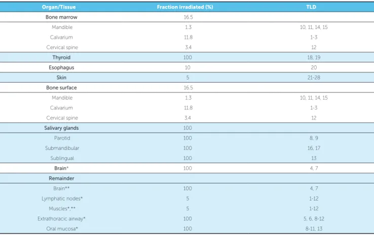
Table 3 - Estimated percentage of tissue irradiated and dosimeters used to calculate mean absorbed dosage.
*ICRP 2007 recommendations , **ICRP 1990 recommendations. TLD: thermoluminescent dosimeter.
Organ/Tissue Fraction irradiated (%) TLD
Bone marrow 16.5
Mandible 1.3 10, 11, 14, 15
Calvarium 11.8 1-3
Cervical spine 3.4 12
Thyroid 100 18, 19
Esophagus 10 20
Skin 5 21-28
Bone surface 16.5
Mandible 1.3 10, 11, 14, 15
Calvarium 11.8 1-3
Cervical spine 3.4 12
Salivary glands 100
Parotid 100 8, 9
Submandibular 100 16, 17
Sublingual 100 13
Brain* 100 4, 7
Remainder
Brain** 100 4, 7
Lymphatic nodes* 5 1-12
Muscles*,** 5 1-12
Extrathoracic airway* 100 5, 6, 8-12
Table 5 - Mean of equivalent doses (µSv) of each organ or tissue, effective doses (µSv) and percentage of equivalent and effective doses of all devices compared to CBCT.
Table 4 - Tissue weighting factors – ICRP 1990 and 2007 recommendations.
*Adrenals, brain, upper large intestine, small intestine, kidney, muscle, pan-creas, spleen, thymus and uterus. **Adrenals, extrathoracic region, gall blad-der, heart, kidneys, lymphatic nodes, muscle, oral mucosa, pancreas, prostate, small intestine, spleen, thymus and uterus.
Organ/Tissue WT 1990 WT2007
Bone marrow 0.12 0.12
Breast 0.05 0.12
Colon 0.12 0.12
Lung 0.12 0.12
Stomach 0.12 0.12
Bladder 0.05 0.04
Esophagus 0.05 0.05
Gonads 0.20 0.08
Liver 0.05 0.04
Thyroid 0.05 0.05
Bone surface 0.01 0.01
Brain Remainder 0.01
Salivary glands - 0.01
Skin 0.01 0.01
Remainder 0.05* 0.10**
Organ/Tissue Cephalometric Panoramic Periapical CBCT Radiographic set Radiographic set/
CBCT (%)
Thyroid 5.1 34.5 1.1 388.5 40.7 10.5
Bone marrow 3.3 21.4 66.3 279.6 91 32.5
Esophagus 0.7 3.4 10 89.7 14.1 15.7
Skin 1 36.2 0.7 0.2 37.9 18950.0
Bone surface 12.1 87.7 3268.4 556.4 3368.2 605.4
Salivary glands 26.4 359.1 932.2 1908.2 1317.7 69.1
Brain** 14 33.9 139.7 2985.3 187.6 6.3
Remainder
Brain* 14 33.9 139.7 2985.3 187.6 6.3
Lymphatic nodes** 1.3 18 46.6 95.4 65.9 69.1
Extrathoracic airways** 26.4 359.1 932.2 1908.2 1317.7 69.1
Muscles*,** 1 5.6 70.4 62.3 77 123.6
Oral mucosa** 23.2 316 839.7 1813.1 1178.9 65.0
Efective dosage ICRP 90 1.2 6.7 16.5 139.2 24.4 17.5
Efective dosage ICRP 07 2.5 27.1 69.1 208.9 98.7 47.2
The equivalent and efective doses obtained by the radiographic set were summed up and the percentages between these values and the CBCT values were cal-culated (Table 5). Although the efective dose of the ra-diographic set corresponded to 17.5% (ICRP 1990) and 47.2% (ICRP 2007) of the CBCT dose, the equivalent doses of skin, bone surface and muscle obtained by the radiographic set were higher when compared to CBCT.
DISCUSSION
In dosimetry, several factors must be considered: the phantom used (made from bones or just equivalent tis-sue material), number and location of dosimeters, type of device tested and its parameters (voltage [kV], amperage
[mA], time of exposure, ield of view [FOV] and voxel).10
Diferent combinations lead to diferent doses. Due to many variables, there are no appropriate parameters to compare these results, especially in relation to efective doses of radiographs and CBCT. Furthermore, in order
to allow comparison between diferent studies, a standard methodology should be established.
A wide variation of efective doses was observed in diferent studies when evaluating CBCT scans. When comparing the same i-CAT model, diferent results were
achieved.6,12,15,21 The high variability of radiation doses
ob-tained compromises comparisons among diferent devices. Studies that evaluated current models, such as i-CAT Next Generation and i-CAT FLX, found efective doses
of 182.1 µSv15 and 69.2 µSv,21 respectively. The reduction
of FOV down to 17 cm and exposure time to 3.7 seconds may have contributed to reduce the efective dose, which reached about 38.9% when the results of these studies were compared with those of i-CAT FLX.
However, it was reported that the average distance
between nasion and menton was 12.28 cm.22 While a
multiethnic population presents much more variation, anterior facial height may reach greater values than these ones. Therefore, to the reduced ield of view, even in the extended ield of view protocol, some essential structures may be cut out of the image obtained.
The efective dose measured in i-CAT in the present study was greater than the sum of the efective doses of all radiographic examinations routinely required for orth-odontic treatment. One reason for this diference might be the radiographic devices used in the study, which pro-duce digital images with lower radiation doses. Addition-ally, i-CAT is a large volume tomographic device with extended FOV. The area exposed during examination is, therefore, increased.
The doses obtained in this study were higher in all devices because a lead apron was not used. The highest equivalent doses found were in the regions of the thyroid, brain and eyes. When a thyroid apron is used, there is a re-duction of 48.7% in the dosage of the thyroid, and 41.7%
in the dosage of the esophagus.23 Examinations with a
large FOV showed a reduction of 61%.24 Therefore, the
use of lead aprons should not be overlooked.
Additionally, the geometrical position of these organs in relation to the X-ray beam may have inluenced the re-sults. In i-CAT, due to the largest FOV, the organ is closer to the X-ray center of the beam.
The efective dose of the radiographic set corresponded to less than a half of the dose calculated for CBCT. On the other hand, the equivalent doses of skin, bone surface and muscle were higher (Table 5). The periapical examination
was the most responsible for the highest dose. It could be due to proximity of dosimeters to the molars area.
There-fore, to follow the ALARA9 principle (as low as possible
radiation), the orthodontist should not request full peri-apical examination. Instead, single periperi-apical radiographs covering restricted areas are more suitable.
Although the equivalent and efective doses of CBCT scans are high when compared to X-rays, the doses of mul-tidetector CT scanners, used routinely for medical
exami-nations, are dozens of times higher.13,25 Furthermore, it is
estimated that the population is exposed to an average dose
of natural radiation of 2400 µSv per year,26 and that the risk
of developing cancer from exposure during CBCT
exami-nation is between 1:100.000 and 1:350.000 for adults.19
Moreover, tomography accepts the capture of a range of images otherwise inaccessible to radiography, whenever more in-depth information is needed about the patient. Nevertheless, based on the results of the current and other studies, CBCT examination with the i-CAT device should be indicated only in special cases and should not be used routinely.
CONCLUSION
The efective doses produced by i-CAT were higher than the doses generated by the digital radiographs of the orthodontic set. However, in some areas, the radiation produced by the orthodontic set was higher due to com-plete periapical examination. Replacing radiographs with tomographic images generated by this device goes against the principle of ALARA and should be carried out only in speciic cases. Furthermore, single periapical radiographs covering restricted areas are more suitable than complete periapical examination.
Acknowledgements
We would like to thank Instituto de Radioproteção e Dosimetria (IRD) and Centro de Ensino e Diagnóstico em Tomograia (CEDT) for all the support.
Author contributions
1. Adams GL, Gansky SA, Miller AJ, Harrell WE Jr, Hatcher DC. Comparison between traditional 2-dimensional cephalometry and a 3-dimensional approach on human dry skulls. Am J Orthod Dentofacial Orthop. 2004 Oct;126(4):397-409.
2. Tsao DH, Kazanoglu A, McCasland JP. Measurability of radiographic images. Am J Orthod. 1983 Sept;84(3):212-6.
3. Swennen GR, Schutyser F. Three-dimensional cephalometry: spiral multi-slice vs cone-beam computed tomography. Am J Orthod Dentofacial Orthop. 2006 Sept;130(3):410-6.
4. White SC, Pharoah MJ. The evolution and application of dental maxillofacial imaging modalities. Dent Clin North Am. 2008 Oct;52(4):689-705, v. 5. Yamamoto K, Ueno K, Seo K, Shinohara D. Development of dento-maxillofacial
cone beam X-ray computed tomography system. Orthod Craniofac Res. 2003;6 Suppl 1:160-2.
6. Roberts JA, Drage NA, Davies J, Thomas DW. Efective dose from cone beam CT examinations in dentistry. Br J Radiol. 2009 Jan;82(973):35-40. 7. Scarfe WC, Farman AG, Sukovic P. Clinical applications of cone-beam computed
tomography in dental practice. J Can Dent Assoc. 2006 Feb;72(1):75-80. 8. Halazonetis DJ. From 2-dimensional cephalograms to 3-dimensional computed
tomography scans. Am J Orthod Dentofacial Orthop. 2005 May;127(5):627-37. 9. Farman AG. ALARA still applies. Oral Surg Oral Med Oral Pathol Oral Radiol
Endod. 2005 Oct;100(4):395-7.
10. Helmrot E, Thilander-Klang A. Methods for monitoring patient dose in dental radiology. Radiat Prot Dosimetry. 2010 Apr-May;139(1-3):303-5.
11. Qu XM, Li G, Ludlow JB, Zhang ZY, Ma XC. Efective radiation dose of ProMax 3D cone-beam computerized tomography scanner with diferent dental protocols. Oral Surg Oral Med Oral Pathol Oral Radiol Endod. 2010 Dec;110(6):770-6. 12. Ludlow JB, Davies-Ludlow LE, Brooks SL, Howerton WB. Dosimetry of 3 CBCT
devices for oral and maxillofacial radiology: CB Mercuray, NewTom 3G and i-CAT. Dentomaxillofac Radiol. 2006 July;35(4):219-26.
13. Ludlow JB, Ivanovic M. Comparative dosimetry of dental CBCT devices and 64-slice CT for oral and maxillofacial radiology. Oral Surg Oral Med Oral Pathol Oral Radiol Endod. 2008 July;106(1):106-14.
14. Ludlow JB. A manufacturer’s role in reducing the dose of cone beam computed tomography examinations: efect of beam iltration. Dentomaxillofac Radiol. 2011 Feb;40(2):115-22.
REFERENCES
15. Grünheid T, Kolbeck Schieck JR, Pliska BT, Ahmad M, Larson BE. Dosimetry of a cone-beam computed tomography machine compared with a digital X-ray machine in orthodontic imaging. Am J Orthod Dentofacial Orthop. 2012 Apr;141(4):436-43.
16. Garcia Silva MA, Wolf U, Heinicke F, Gründler K, Visser H, Hirsch E. Efective dosages for recording Veraviewepocs dental panoramic images: analog ilm, digital, and panoramic scout for CBCT. Oral Surg Oral Med Oral Pathol Oral Radiol Endod. 2008 Oct;106(4):571-7.
17. Statistics NBO. Physical aspects of irradiation. Washington, DC: US Government Printing Oice; 1964.
18. International Commission on Radiological Protection. The 2007
Recommendations of the International Commission on Radiological Protection. ICRP publication 103. Ann ICRP. 2007;37(2-4):1-332.
19. International Commission on Radiological Protection.
1990 Recommendations of the International Commission on Radiological Protection. Ann ICRP. 1991;21(1-3):1-201.
20. Loubele M, Bogaerts R, Van Dijck E, Pauwels R, Vanheusden S, Suetens P, et al. Comparison between efective radiation dose of CBCT and MSCT scanners for dentomaxillofacial applications. Eur J Radiol. 2009 Sept;71(3):461-8. 21. Ludlow JB, Walker C. Assessment of phantom dosimetry and image quality of
i-CAT FLX cone-beam computed tomography. Am J Orthod Dentofacial Orthop. 2013 Dec;144(6):802-17.
22. Silva MBG, Gois BC, Sant’Anna EF. Evaluation of the reliability of measurements in cephalograms generated from cone beam computed tomography. Dental Press J Orthod. 2013 July-Aug;18(4):53-60.
23. Qu XM, Li G, Sanderink GC, Zhang ZY, Ma XC. Dose reduction of cone beam CT scanning for the entire oral and maxillofacial regions with thyroid collars. Dentomaxillofac Radiol. 2012 July;41(5):373-8.
24. Qu X, Li G, Zhang Z, Ma X. Thyroid shields for radiation dose reduction during cone beam computed tomography scanning for diferent oral and maxillofacial regions. Eur J Radiol. 2012 Mar;81(3):e376-80.
25. Silva MA, Wolf U, Heinicke F, Bumann A, Visser H, Hirsch E. Cone-beam computed tomography for routine orthodontic treatment planning: a radiation dose evaluation. Am J Orthod Dentofacial Orthop. 2008 May;133(5):640.e1-5. 26. UNSCEAR 2000. The United Nations Scientiic Committee on the Efects of