Shoulder and Elbow Group and Arthroscopy Laboratory of Department of Orthopedics, Faculty of Medicine, University of Sao Paulo,Sao Paulo, SP, Brazil
Email: [email protected]
Received for publication on April 24, 2006. Accepted for publication on June 27, 2006.
BASIC RESEARCH
AN ANATOMICAL STUDY OF THE SUBCORACOID
SPACE
Arnaldo Amado Ferreira Neto, Adriano Marques de Almeida, Renzo Maiorino, Américo Zoppi Filho, Eduardo Benegas
Ferreira Neto AA, de Almeida AM, Maiorino R, Zoppi Filho A, Benegas E. An anatomical study of the subcoracoid space. CLINICS. 2006;61(5):467-72.
PURPOSE: To evaluate the amplitude of the subcoracoid space under maximum internal and external rotations of the humeral head and measure the distance between the apex of the coracoid process and the following anatomical structures: (a) point of entry of the musculocutaneous nerve and its branches into the coracobrachial muscles and into the short head of the biceps brachii muscle; (b) acromial artery; (c) lesser tubercle of the humerus.
METHOD: Thirty shoulders of fresh cadavers, without any kind of shoulder pathology, (9 males and 6 females) were dissected, and the distances (in mm) were measured between the anatomical structures defined above and the apex of the coracoid process.
RESULTS: The mean distance between the apex of the coracoid process and the musculocutaneous nerve was 49.2 mm (in all
specimens a proximal branch of the nerve was identified 34.2 mm away from the apex of the coracoid process), which was not significantly different between the sexes or body sides; the mean distance between the apex of the coracoid process and the acromial artery was 12.4 mm, which was not significantly different between the sexes or body sides; the mean distance between the apex of the coracoid process and the lesser tubercle of the humerus, with the humeral head under internal rotation, was 10.6 mm in men and 8.6 mm in women, values that were significantly different between the sexes.
DISCUSSION: In women, the smaller distance between the apex of the coracoid process and the lesser tubercle of the humerus in the arm internal rotation suggests a higher chance of impingement between those bone structures among the female sex.
KEYWORDS: Coracoid process. Musculocutaneous nerve. Shoulder surgery. Shoulder arthroscopy. Neurovascular shoulder
injury.
INTRODUCTION
Pain in the anterior region of the shoulder is a frequent complaint among patients with scapular girdle injuries. The most common cause of this pain is the progressive lesion of the supraspinatus tendon caused by the impact of its in-sertion into the greater tubercle of the humerus against the coracoacromial arch. This condition was well defined by Neer1 who, in a landmark article, showed the etiopathogeny, the natural history, and the way to manage the process that
he called the impingement syndrome.
The pain in the anterior region of the shoulder may have different origins, one of them being the impingement of the lesser tubercle of the humerus (LTH) against the cora-coid process (CP).
Gerber2 reported a few cases of pain in that site where classical subacromial decompression was not successful, and observed, in agreement with the anatomical study by Goldthwait,3 that the mechanical shock of the LTH against the posterolateral face of the CP can also cause a similar condition. He then described what he called the coracoid impingement syndrome (CIS), with the specific clinical pic-ture and management.
in the horizontal plane, associated with internal rotation.4,5 The apex of the coracoid process is painful, but the pain may be suppressed by local anaesthesia.
An increase in size and a change in the shape of the corachoid process that makes it more prominent or protu-berant, all of which may be difficult to evaluate by simple X-ray examination can be the cause of CIS. Some authors have tried to show the etiology and the consequences of CIS by axial computerized tomography, which shows the reduction in the distance between the LTH and the apex of the coracoid process (ACP), reduction in the coracoid index, and the reduction of the coracoglenoidal angle.6 Other changes such as edema located at the ACP level, a subscapular tendon injury, changes in the rotator interval7,8 thickening of the coracoacromial ligament and the clavipectoral fascia, as well as the reduction in the cora-coid index, may be shown by magnetic resonance.
The initial treatment indicated for CIS is conservative and includes anti-inflammatory medication, local anaesthe-sia, and physiotherapy. Coracoplasty (excision of the pos-terolateral border of the coracoid process) is indicated in patients who are refractory to those treatments. However, there are few cases described in literature, and there is no consensus on how to perform it.
Dines4 described the 1.5-cm excision of the apex of the CP by the deltopectoral approach, which is possible to per-form by detachment of the joint tendon and its immediate reinsertion.
Karnaugh3 and Lo9 reported satisfactory results obtained by arthroscopic excision of the posterolateral border of the coracoid process through different portals (portal for the scope) to visualize the CP. The first author used an antero-lateral portal through the subacromial space, while the latter used a transarticular portal that reaches the subcoracoid space through the rotator interval.
The complexity of the anatomy in the anterior region of the shoulder, meaning the proximity of nerves and blood vessels, the relation between musculoskeletal structures, and possible anatomical changes10 could make surgical ac-cess difficult, primarily if the arthroscopic approach is used. The purpose study was to explore the anatomy of sub-coracoid space in cadavers to establish the relationship be-tween the ACP and other local anatomical structures, such as the musculocutaneous nerve (MCN) and its branches, adjacent vascular structures, and the coracohumeral inter-val at different degrees of rotation, for enabling a safe sur-gical approach.
METHODS
Thirty shoulders from 15 fresh adult cadavers, 9 males
and 6 females, without any kind of shoulder pathology were dissected.
After the specimens had been placed in the horizontal dorsal decubitus position, the dissection was started by the deltopectoral approach. After separating the cephalic vein laterally and opening the deltopectoral space, we identi-fied the following structures: 1) the coracoid process and its tendinous insertions; (it was determined that the ACP is the most laterally located point in relation to its long lon-gitudinal axis); 2) the acromial artery (AA); 3) the muscu-locutaneous nerve (MCN), its branches, and respective points of entry into the coracobrachial muscle (CBM); and 4) the lesser tubercle of the humerus (LTH).
A hand caliper graded in millimeters was used to meas-ure the following distances: 1) distance between the ACP and the point of entry of the MCN into the CBM (dMC); 2) distance between the ACP and the point of entry of the proximal motor branch of the MCN into the CBM; 3) smallest distance between the ACP and the MCN (mini-mum dMC); 4) smallest distance between the ACP and the acromial artery (dAA); and 5) distances between the LTH and the ACP as measured at maximum internal rotation (dIR), neutral position (dNP), and maximum external ro-tation (dER) of the humerus.
Statistical analysis of the obtained data was performed using the following tests: paired Student’s t test, to analyze
the distances measured along the right and left shoulders; nonpaired Student’s t test, to analyze the distances
meas-ured in the male and female specimens.
RESULTS
The descriptive statistics for the measurements are listed in Tables 1.
The distance between the ACP and the LTH in the in-ternal rotation position in male and female specimens was significantly smaller in women (P < 0.05) (Table 2).
There were no significant differences in the other meas-urements analyzed either by sex or by side of body (Table 3).
these branches were located less than 30 mm away from the ACP. The smallest distance between the ACP and the coracoid muscle (CM) varied from 15.1 to 39.2 mm (aver-age: 30.28 mm), medially to the ACP. There were no sig-nificant differences in these parameters with regard to sex or side of body.
Figure 3 shows the vascular structures that are located medially to the CP and emit branches laterally, all of them close to it.
The distance of the acromial artery from the apex of the CP, as measured proximally, varied from 9.1 to 17.8 mm (average: 12.47 mm), with no significant differences
with regard to sex or side of body (Figure 4).
DISCUSSION
Coracoid impingement syndrome has been increasingly diagnosed, and its management requires a proper surgical approach.4,5,9,11,12 Detailed knowledge of the relationship between the relevant anatomical structures located near the coracoid process is required to enable a safe surgical ap-proach.13
The point of entry of the MCN into the CBM is often considered to be located between 50 and 80 mm away from Table 1 - Distances (mm) between the apex of coracoid process and the anatomical structures analyzed
dMC minimum dMC dAA dIR dNP maximum dER
Average 49.28 30.28 12.47 9.88 28.04 37.89
Standard deviation 9.72 6.54 2.15 2.45 5.47 6.29
dMC = distance between the apex of coracoid process and the point of entry of the musculocutaneous nerve into the coracobrachial muscle minimum dMC = smallest distance between the apex of coracoid process and the musculocutaneous nerve
dAA = smallest distance between the apex of coracoid process and the acromial artery
dIR =distance between the lesser tubercle of the humerus and the apex of coracoid process as measured at maximum internal rotation of the humerus dNP = distance between the lesser tubercle of the humerus and the apex of coracoid process as measured at neutral position of the humerus
dER = distance between the lesser tubercle of the humerus and the apex of coracoid process as measured at maximum external rotation of the humerus.
Table 3 - Distances (mm) between the apex of coracoid process and the anatomical structures analyzed, stratified by side of body, average (standard deviation)
Side dMC minimum dMC dAA dIR dNP dER
Right 49.07 29.82 12.73 10.06 29.13 37.97
(11.2) (7.10) (2.18) (2.86) (5.52) (6.56)
Left 49.48 30.73 12.21 9.71 26.97 37.83
(8.61) (6.14) (2.17) (2.05) (5.40) 6.24
dMC = distance between the apex of coracoid process and the point of entry of the musculocutaneous nerve into the coracobrachial muscle minimum dMC = smallest distance between the apex of coracoid process and the musculocutaneous nerve
dAA = smallest distance between the apex of coracoid process and the acromial artery
dIR =distance between the lesser tubercle of the humerus and the apex of coracoid process as measured at maximum internal rotation of the humerus dNP = distance between the lesser tubercle of the humerus and the apex of coracoid process as measured at neutral position of the humerus
dER = distance between the lesser tubercle of the humerus and the apex of coracoid process as measured at maximum external rotation of the humerus Table 2 - Distances (mm) between the apex of coracoid process and the anatomical structures analyzed, stratified by sex, average (standard deviation)
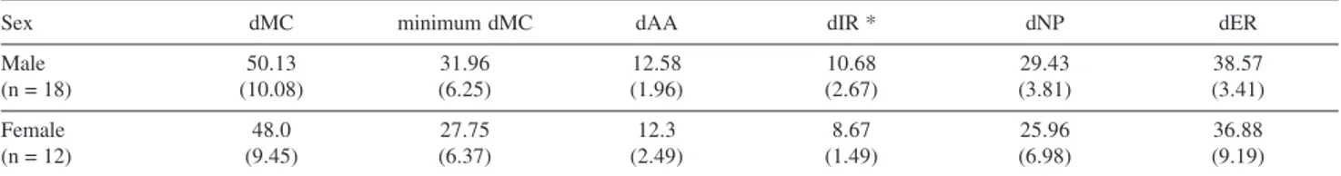
Sex dMC minimum dMC dAA dIR * dNP dER
Male 50.13 31.96 12.58 10.68 29.43 38.57
(n = 18) (10.08) (6.25) (1.96) (2.67) (3.81) (3.41)
Female 48.0 27.75 12.3 8.67 25.96 36.88
(n = 12) (9.45) (6.37) (2.49) (1.49) (6.98) (9.19)
*P < 0.05.
dMC = distance between the apex of coracoid process and the point of entry of the musculocutaneous nerve into the coracobrachial muscle minimum dMC = smallest distance between the apex of coracoid process and the musculocutaneous nerve
dAA = smallest distance between the apex of coracoid process and the acromial artery
dIR =distance between the lesser tubercle of the humerus and the apex of coracoid process as measured at maximum internal rotation of the humerus dNP = distance between the lesser tubercle of the humerus and the apex of coracoid process as measured at neutral position of the humerus
Figure 1 - Distribution of the distances (mm) of the site of entry of the musculocutaneous nerve into the coracobrachial muscle, in relation to the apex of the coracoid process (CP)
Figure 2 - Musculocutaneous nerve (wide arrow) and its proximal motor branch (narrow arrow), as well as their points of entry into the medially separated coracobrachial muscle
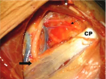
Figure 3 - Relationship between the coracoid process (CP), joint tendon, and neurovascular structures: musculocutaneous nerve (wide arrow) and acromial artery (narrow arrow)
Figure 4 - Left shoulder: the musculocutaneous nerve (wide arrow) laterally to the median nerve, and origin of the acromial artery (narrow arrow), a branch of the deltoid artery
the CP. However, Flatow and Bigliani10 showed that in 29% of cases, it is located less than 50 mm away from the CP and that it is possible for the coracobrachial muscle to have 1 or even more motor branches even more proximal, lo-cated between 17 mm and 31 mm away from the CP. In our study, the point of entry of the MCN into the CBM varied from 35 to 75.1 mm, and in 60% of cases this point was located more proximally than about 50 mm. We no-ticed the presence of motor branches located more proxi-mally in relation to the main stem in all cases, where the point of entry of the CBM varied from 14.8 to 56.5 mm in relation to the ACP. In 30% of the cases, these branches were located less than 30 mm away from the ACP. The dis-tance between the ACP and the coracoid muscle (CM) var-ied from 15.1 to 39.2 mm, medially to the ACP.
The vascular structures are located medially to the CP and emit branches laterally; all of them are close to it, hence the importance of this study. The acromial artery (AA) is 1 of the 4 branches of the thoracoacromial artery. It is directed towards the acromioclavicular joint, accom-panying the CP,14 and sends anastomotic branches to the suprascapular and posterior humeral circumflex arteries, in addition to sending branches to the deltoid muscle15 and to an important cutaneous branch.16 Detailed knowledge of the location of the acromial artery is important for the arthroscopic approach to the CP, since its injury may cause a large hematoma, in addition to significantly impairing the surgical view. We found that the acromial artery is located on the CP and that its distance as measured proximally from its apex varied from 9.1 to 17.8 mm with no signifi-cant differences for sex or side.
all techniques address the excision of the ACP. Dines4 rec-ommends the disinsertion of the joint tendon for a 1.5 cm-excision from the ACP, with reinsertion later.4 However, according to published anatomical studies describing the relationship between the CP, the CBM, and the MCN and corresponding branches, there is a risk for injury of those nerve structures during the joint tendon disinsertion, which is the reason why it should be avoided.11,12 Also, with re-gard to arthroscopic treatment, some authors describe pro-cedures for CP excision that preserve the origin of the joint tendon. We agree with Lo9 in the sense that the transarticular posterior portal for the access with the op-tics through the rotator interval allows proper access to the subcoracoid space, so that one can have a direct view of the posterolateral part of the CP. The excision is performed using a shaver through the anterior portal laterally to the CP (Figure 5), with good safety margin as regards the neu-rovascular structures studied in this paper.
The small distance between the LTH and the CP with the arm in internal rotation position4,5 has been held respon-sible for the pathogenesis of the CIS. The reduction of this distance during internal rotation of the arm using radio-graphic measurements has been sought. Gerber measured
it by computerized tomography in normal subjects, hav-ing obtained, with the arm in internal rotation position, mean values of 9.1 mm for men and 8.2 mm for women.6 Although reported epidemiological studies do not show that CIS is more frequent in women, our study revealed signifi-cantly smaller values for the distance between the LTH and the CP in women, with the arm in internal rotation. This apparent disagreement has led us to believe that a larger number of properly studied CIS cases are required to prove the diagnostic value of that parameter.17
In spite of the complex anatomy of the subcoracoid space, we believe that a safe surgical approach of the CP is possible, provided that disinsertion of the joint tendon is avoided—a recommendation that we make—to prevent the risk of damaging the important neurovascular structures described above.
Although it was not possible to prove a correlation be-tween the reduction of the distance from the ACP to the LTH and the etiology of CIS, it is necessary to insist on improving the new imaging diagnostic techniques to be able to establish consistent parameters of sensitivity and specificity to confirm its clinical diagnosis.
The apparent rarity of this syndrome in daily clinical practice—perhaps due to the fact that it is hard to diag-nose, or even due to lack of knowledge of its existence— makes it difficult to perform prospective studies to com-pare the efficacy of the different treatment methods.
Still, we believe that it is important to understand the anatomical details involved, especially when considering the benefits of the arthroscopic approach for decompres-sion in cases of coracoid impingement syndrome.
CONCLUSIONS
1. The location of neurovascular structures in relation to the apex of the coracoid process is variable; however, their proximity represents a risk in surgical approaches.
2. The distance between the involved neurovascular structures and the apex of the coracoid process is not re-lated to sex or side.
3. The distance between the lesser humeral tubercle and the apex of the coracoid process with the arm under maxi-mum internal rotation is smaller in women.
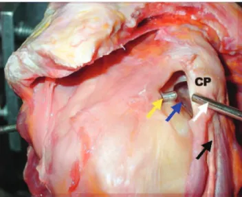
Figure 5 - Right shoulder: CP (coracoid process); transarticular access for the optics through the posterior portal (yellow arrow); subcoracoid space (blue arrow), anterior portal for shaver access (white arrow); musculocutaneous nerve (black arrow)
RESUMO
Ferreira Neto AA, de Almeida AM, Maiorino R, Zoppi Fi-lho A, Benegas E. Um estudo anatômico do espaço subcoracóide. CLINICS. 2006;61(5):467-72.
as seguintes estruturas anatômicas: (a) ponto de entrada do nervo musculocutâneo e suas ramificações nos músculos coracobraquiais e na cabeça curta do músculo bíceps do braço; (b) artéria acromial; (c) tubérculo menor do úmero. MÉTODO: Foram dissecados trinta ombros de cadáveres novos (nove do sexo masculino e seis do sexo feminino) sem nenhum tipo de patologia de ombro, tendo sido feita a medida (em mm) entre as estruturas anatômicas mencio-nadas acima e o ápice do processo coracóide.
RESULTADOS: A distância média entre o ápice do pro-cesso coracóide e o nervo musculocutâneo foi 49,2 mm (em todas as amostras foi identificado um ramo proximal do nervo a 34,2 mm de distância do ápice do processo coracóide) sem significado estatístico em termos de sexo e lado do corpo; a distância média entre o ápice do
pro-cesso coracóide e a artéria acromial foi 12,4 mm, sem sig-nificado estatístico em termos de sexo e lado do corpo; a distância média entre o ápice do processo coracóide e o tubérculo menor do úmero com a cabeça do úmero em ro-tação interna foi 10,6 mm nos homens e 8,6 mm nas mu-lheres, valores esses significativos em termos de sexo. DISCUSSÃO: Nas mulheres, a distância menor entre o ápice do processo coracóide e o tubérculo menor do úmero em rotação interna do braço sugere uma probabilidade mai-or de invasão entre aquelas estruturas ósseas no sexo fe-minino.
UNITERMOS: Processo coracóide. Nervo musculo
cutâneo. Cirurgia de ombro. Artroscopia de ombro. Lesão vasculonervosa de ombro.
REFERENCES
1. Neer CS. Anterior acromioplasty for the chronic impingement syndrome in the shoulder: a preliminary report. J Bone Joint Surg Am. 1972;54:41-50.
2. Gerber C, Terrier F, Ganz R. The role of the coracoid process in the chronic impingement syndrome. J Bone Joint Surg Br. 1985;67:703-8. 3. Goldthwait JE. An anatomic and mechanical study of the shoulder joint, explaining many of the cases of painful shoulder, many of the recurrent dislocations and many of the cases of brachial neuralgias or neuritis. Am J Orthop Surg. 1909;6:579-606.
4. Dines DM, Warren RF, Inglis AE, Pavlov H. The coracoid impingement syndrome. J Bone Joint Surg Br. 1990;72:314-6.
5. Ferrick MR. Coracoid impingement: a case report and review of the literature. Am J Sports Med 2000; 28: 117-9.
6. Gerber C, Terrier F, Zender R, Ganz R. The subcoracoid space: an anatomic study. Clin Orthop. 1987;215:132-8.
7. Dumontier C, Sautet A, Gagey O, Apoil A. Rotator interval lesions and their relation to coracoid impingement syndrome. J Shoulder Elbow Surg. 1999;8:130-5.
8. Neer CS, Satterlee CC, Dalsey RM, Flatow EL. The anatomy and potential effects of contracture of the coracohumeral ligament. Clin Orthop. 1992:280:182-5.
9. Lo KY, Burkhart SS. Arthroscopic coracoplasty through the rotator interval. Arthroscopy. 2003;19:667-71.
10. Flatow EL, Bigliani LU, April EW. An anatomic study of the musculocutaneous nerve and its relationship to the coracoid process. Clin Orthop. 1989;244:166-71.
11. Karnaugh RD, Sperling JW, Warren RF. Arthroscopic treatment of coracoid impingement. Arthroscopy. 2001;17:784-7.
12. Patte D. The subcoracoid impingement. Clin Orthop. 1990;254:55-9. 13. Lo KY, Burkhart SS, Parten PM. Surgery about the coracoid:
neurovascular structures at risk. Arthroscopy. 2004; 20:591-5. 14. Gray H, Lewis W. Tratado de anatomia humana. Rio de Janeiro:
Guanabara; 1946. p. 654-6.
15. Testut L, Latarjet A. Tratado de anatomia humana. Barcelona: Salvat; 1944. p. 292.
16. Rockwood CA, Matsen FA 3rd. The shoulder. Philadelphia: Saunders; 1990. p. 79-80.