Sterilizing elastomeric chains without losing
mechanical properties. Is it possible?
Matheus Melo Pithon1, Caio Souza Ferraz2, Francine Cristina Silva Rosa3, Luciano Pereira Rosa4
How to cite this article: Pithon MM, Ferraz CS, Rosa FCS, Rosa LP. Steriliz-ing elastomeric chains without losSteriliz-ing mechanical properties. Is it possible? Dental Press J Orthod. 2015 May-June;20(3):96-100.
DOI: http://dx.doi.org/10.1590/2176-9451.20.3.096-100.oar
Submitted: August 11, 2014 - Revised and accepted: September 26, 2014
Contact address: Matheus Melo Pithon
Av. Otávio Santos, 395, sala 705, Centro Odontomédico Dr. Altamirando da Costa Lima, Bairro Recreio, CEP 45020-750 - Vitória da Conquista - Bahia - Brazil - E-mail: [email protected]
1 Professor of Orthodontics, Universidade Estadual do Sudoeste da Bahia
(UESB), Jequié, Bahia, Brazil.
2 Undergraduate student in Dentistry, Universidade Estadual do Sudoeste da
Bahia (UESB), Jequié, Bahia, Brazil.
3 Professor of Microbiology, Universidade Federal da Bahia (UFBA), Vitória da
Conquista, Bahia, Brazil.
4 Professor of Radiology, Universidade Federal da Bahia (UFBA), Vitória da
Conquista, Bahia, Brazil.
» The authors report no commercial, proprietary or financial interest in the products or companies described in this article.
Objective: To investigate the effects of different sterilization/disinfection methods on the mechanical properties of orth-odontic elastomeric chains. Methods: Segments of elastomeric chains with 5 links each were sent for sterilization by cobalt 60 (Co60) (20 KGy) gamma ray technology. After the procedure, the elastomeric chains were contaminated with clinical samples of Streptococcus mutans. Subsequently, the elastomeric chains were submitted to sterilization/disinfection tests carried out by means of different methods, forming six study groups, as follows: Group 1 (control - without con-tamination), Group 2 (70°GL alcohol), Group 3 (autoclave), Group 4 (ultraviolet), Group 5 (peracetic acid) and Group 6 (glutaraldehyde). After sterilization/disinfection, the effectiveness of these methods, by Colony forming units per mL (CFU/mL), and the mechanical properties of the material were assessed. Student’s t-test was used to assess the number of CFUs while ANOVA and Tukey’s test were used to assess elastic strength. Results: Ultraviolet treatment was not completely effective for sterilization. No loss of mechanical properties occurred with the use of the different sterilization methods (p > 0.05). Conclusion: Biological control of elastomeric chains does not affect their mechanical properties.
Keywords:Orthodontics. Disinfection. Elastomers.
DOI: http://dx.doi.org/10.1590/2176-9451.20.3.096-100.oar
Objetivo: verificar os efeitos de diferentes métodos de esterilização/desinfecção nas propriedades mecânicas de elásticos ortodônticos em cadeia. Métodos: segmentos de elástico em cadeia com 5 elos cada foram enviados para esterilização em radiação gama com cobalto 60 (20 KGy). Após esterilização, esses foram contaminados com amostras clínicas de
Streptococcus mutans. Passado esse período, foram submetidos aos testes de esterilização/desinfecção por diferentes métodos, formando seis grupos de estudo, assim denominados: Grupo 1 (controle - sem ter sido contaminado), Grupo 2 (álcool 70°GL), Grupo 3 (autoclave), Grupo 4 (ultravioleta), Grupo 5 (ácido peracético) e Grupo 6 (glutaraldeído). Após esteri-lização/desinfecção, avaliou-se a efetividade desses métodos, por meio de contagem de unidades formadoras de colônias por mL (UFC/mL), e as propriedades mecânicas desses materiais. Utilizou-se o teste t de Student para avaliar o número de UFC, além do ANOVA e, posteriormente, do teste de Tukey para avaliação da força. Resultados: verificou-se que o ultravioleta não obteve eficácia total quanto à esterilização. E não ocorreu perda das propriedades mecânicas dos elásticos, com os diferentes métodos de esterilização utilizados (p > 0,05). Conclusão: o controle biológico de elásticos em cadeia não interfere nas suas propriedades mecânicas.
INTRODUCTION
Fighting infections in dental oices has been a daunting challenge to dentists, researchers and immu-nologists. Most of times, germs have been able to dodge contemporary safety measures, thereby exposing profes-sionals and patients to risk. On the other hand, lack of care by some professionals with regard to biosafety has
favored the intensiication of infection.1,2
Of the dental specialties, Orthodontics is outstanding among those with a higher number of predisposing
fac-tors for cross-infection.3,4 Orthodontics is characterized
by a high turnover of patients and multiplicity of ve-hicles for disease transmission (equipment, instruments, operators’ hands, etc.), thus exposing clinicians,
assis-tants and patients to serious risks of infection.5,6
In Orthodontics, elastomeric chains are among the diferent types of material that highly favor the occur-rence of cross-infection, given that this type of mate-rial is commercially presented in reels ranging from
1 to 4.5 meters, which hinders its individual use.7,8
Despite wide acceptance and use of elastomeric chains, doubt is cast on their mechanical and biological properties ater they have been submitted to sterilization
procedures.9 Considering that elastics and elastomeric
chains are amorphous polymers made of polyurethane material, presenting characteristics of both rubber and plastic, their characteristics may be altered in contact
with physical and or chemical agents.10
Thus, the present study aimed at assessing which method would be most indicated to sterilize elas-tomeric chains without causing them to lose their mechanical properties.
MATERIAL AND METHODS
Elastomeric chains (Morelli, Sorocaba, Brazil) of the short spacing type were carefully removed from the reel without being elongated/stretched, and cut into segments with 5 links each. Subsequently, they were wrapped in surgical grade paper (n = 15) and sent to sterilization by gamma radiation with co-balt 60 (20 KGy) (Empresa Brasileira de Radiação - EMBRARAD, Cotia-SP, Brazil) without alterations in their physical properties.
Assessing effectiveness of different methods After specimens were sterilized, they were con-taminated in test tubes containing 10 mL of TODD
liquid culture medium with 100 microliters of stan-dardized suspension for assessment by spectropho-tometry (optical density = 0.620; wavelength = 398)
of 1 X 10⁶ cells/mL of ten different randomly selected
clinical samples of Streptococcus mutans. Specimens
were then incubated at 37 oC for 48 h.
Ater the incubation period and Streptococcus
mu-tans monospecies bioilm formation adherent to the
specimens, the latter were introduced into polypro-pylene tubes, containing 2 mL of sterile saline solution (0.85% NaCl), for 10 seconds, so as to remove excess bioilm. Specimens were then introduced into appro-priate and sterile receptacles so as to be subjected to sterilization tests, as follows:
» Group 1: Elastomeric chains which were not submitted to any sterilization method (control group).
» Group 2: Elastomeric chains immersed in poly-propylene tubes containing 2 mL of 70° GL al-cohol for 1 minute.
» Group 3: Elastomeric chains autoclaved for a cycle of 15 minutes.
» Group 4: Elastomeric chains sterilized in ul-traviolet light (SPLabor, Presidente Prudente, São Paulo, Brazil) for 30 minutes, divided by 15 minutes on each side of the elastic.
» Group 5: Elastomeric chains immersed in polypropylene tubes containing 2 mL of per-acetic acid for 30 minutes.
» Group 6: Elastomeric chains immersed in polypropylene tubes containing 2 mL of 2% glutaraldehyde solution for 30 minutes.
Ater sterilization/disinfection procedures were car-ried out by the diferent methods, the specimens were removed in a sterile environment inside a laminar low chamber and introduced into polypropylene tubes con-taining 2 mL of sterile saline solution (0.85% NaCl), agitated in a vortex appliance for 1 minute. From the
suspension obtained, decimal dilutions of 10-1, 10-2 were
made. Aliquots of 100 microliters of initial suspension and the other dilutions were seeded on Petri dishes
con-taining Todd Hewitt broth at 37 oC for 48 h.
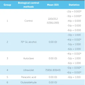
Table 1 - Methods with description of respective groups.
Table 2 - Mean, standard deviation and statistical analysis of the number of colony forming units for the different groups evaluated.
SD = standard deviation; *= statistical differences (p < 0.05).
Sterilization method Time Pressure Volume Temperature
Group 1 - - - -
-Group 2 70° GL alcohol 1 min - 1 mL Room
Group 3 Autoclave 15 min 1 atm - 121 °C
Group 4 Ultraviolet 15 min p/surface - - Room
Group 5 Peracetic acid 30 min - 2 mL Room
Group 6 Glutaraldehyde 30 min - 2 mL Room
Group Biological control
methods Mean (SD) Statistics
1 Control 220133.2 (53911.093)
-2/p = 0.000* -3/p = 0.000* -4/p = 0.000 -5/p = 0.000 -6/p = 0.000
2 70° GL alcohol 0.00 (0)
-3/p = 1.000 -4/p = 0.010* -5/p = 1.000 -6/p = 1.000
3 Autoclave 0.00 (0)
-4/p = 0.010* -5/p = 1.000 -6/p = 1.000
4 Ultraviolet 75956 (83643) -5/p = 0.010* -6/p = 0.010* 5 Peracetic acid 0.00 (0) -6/p = 1.010 6 Glutaraldehyde 0.00 (0)
Assessing mechanical properties
Ater being submitted to diferent biological con-trol methods, the strength generated by the elastomeric chains was measured (n = 15)according to the previously established sequence of groups.
Elastomeric chains were taken to a digital dynamometer (Instrutherm DD-300, São Paulo, Brazil) mounted on a platform speciically set up for this investigation. Elasto-meric chains were distended for 23.5 cm.
Statistical analysis
Ater assessing the number of colonies formed and the maximum values obtained by the elastomeric chains, sta-tistical analyses were carried out. To this end, SPSS 13.0 sotware (SPSS Inc., Chicago, Illinois, USA) was used. Descriptive statistical analysis including mean and stan-dard deviation was carried out for all groups. The values referring to the number of colonies formed were submit-ted to Student’s t-test with a signiicance level set at 5%. The values referring to the amount of strength released were submitted to analysis of variance (ANOVA) so as to determine whether there were statistical diferences among groups. Tukey’s test was later performed.
RESULTS
Results referring to the mean number of colony forming units (CFU/mL) reveal that the control group obtained the highest mean of around 220,000 CFU/mL, whereas the group in which ultraviolet light (UV) was used as the method for microorganism control obtained an approximate mean of 80,000 CFU/mL.
When UV was compared to the other biological control methods, it proved to be the least efective in reducing microorganisms (p = 0.010) (Table 1). There were statistical diferences between the control group and the other groups (p < 0.05) (Fig 1 and Table 2).
With regard to the percentage of decontamination of elastomeric chains, the UV group obtained the lowest percentage of around 65%, whereas the other methods obtained 100%.
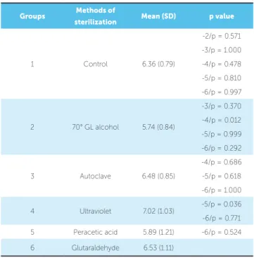
In terms of mechanical properties, no diferences were found among the diferent sterilization methods (p > 0.005) (Table 3).
DISCUSSION
Table 3 - Statistics of different biological control methods in terms of evalua-tion of the mechanical properties of elastomeric chains.
SD = standard deviation. *= statistical differences (p < 0.05).
Groups Methods of
sterilization Mean (SD) p value
1 Control 6.36 (0.79)
-2/p = 0.571 -3/p = 1.000 -4/p = 0.478 -5/p = 0.810 -6/p = 0.997
2 70° GL alcohol 5.74 (0.84)
-3/p = 0.370 -4/p = 0.012 -5/p = 0.999 -6/p = 0.292
3 Autoclave 6.48 (0.85)
-4/p = 0.686 -5/p = 0.618 -6/p = 1.000
4 Ultraviolet 7.02 (1.03) -5/p = 0.036 -6/p = 0.771 5 Peracetic acid 5.89 (1.21) -6/p = 0.524 6 Glutaraldehyde 6.53 (1.11)
Cross-infection is deined as the transmission of infectious agents among patients and health personnel within a clinical environment. Transmission occurs from person to person or by contact with contaminated objects. Transmission may occur through blood, saliva droplets, or instruments contaminated with blood, sa-liva and tissue debris. Transmission pathway is either by
contact, inhalation or inoculation.1
According to Silva et al,11 there is a high incidence of
cross-infection in the dental oice. Thus, the use of de-contaminating agents is relevant in clinical practice. A number of methods is used in the dental oice with a view to dodging cross-infection, namely: autoclave, alcohol, glutaraldehyde, peracetic acid and ultraviolet radiation.
According to Berger,12 ultraviolet radiation (UV) is
used in Dentistry as a disinfectant agent for toothbrush surfaces; however, its efectiveness is greatly related to the time of exposure. In the present study, ultraviolet radia-tion obtained the lowest percentage (65%) in the reduc-tion of colony forming units (CFU/mL) in comparison to the other groups in which disinfectant agents were used. The latter reduced CFUs/mL in 100%. When the mechanical properties of elastomeric chains were com-pared, UV obtained the best mean, around 7.02; how-ever, without signiicant diferences among groups.
In the present study, glutaraldehyde proved an ei-cient disinfectant agent, in addition to not afecting the mechanical properties of elastomeric chains, given that there was no signiicant diference between this group (6.53) and the control group (6.36). These data
cor-roborate the indings by Suprono et al13 who reported
that glutaraldehyde does not cause deterioration of the elastomeric chain surface.
Peracetic acid has been used in food and water indus-tries, sewage treatment companies and for decontami-nation and sterilization of heat-sensitive
medical-hos-pital devices and equipment.14-17 Peracetic-acid proved
an eicient decontaminating agent and completely re-duced the number of colony forming units (CFU/mL). Furthermore, peracetic acid does not leave residues and does not produce harmful products, as its mechanism of action involves the release of free oxygen and hydroxyl radicals in decomposition in water, oxygen and acetic
acid.14-17 This was proved in the present study, since
all elastomeric chains evaluated kept their mechanical properties, in addition to being completely sterilized.
The method most used for sterilization of medical and dental instruments worldwide is damp steam sterilization
(autoclave).18 It proved eicient in reducing the number
of colony forming units (CFU/mL), thereby completely reducing the existent bacteria. Moreover, it yielded sur-prising results in terms of the mechanical properties of elastomeric chains, since even in contact with heat, the mechanical properties remained the same, without statis-tical diferences in comparison to the control group.
Figure 1 - Mean CFU/mL of S. Mutans on orthodontic elastomeric chains after applying the different methods of microorganisms control.
Mean CFU/ml of S. mutans in orthodontic elastomeric chains after applying the different methods of microorganisms control
70% alcohol control
autoclave ultraviolet peracetic acid glutaraldehyde 240000
200000
140000
60000
20000 220000
160000
80000 180000
100000 120000
40000
Based on the results obtained in this study, the simplest method of promoting sterilization/disinfec-tion of orthodontic elastomeric chains was alcohol. After 1 minute, it was possible to eliminate the mi-croorganisms adhered to the elastics without losing their mechanical characteristics. Nevertheless, the fact that only S. Mutans was used in the experi-ment must be considered. In spite of being the most prevalent and most important infectious agent in the oral cavity, this bacterium is not the most resistant; therefore, further studies are warranted to investi-gate other microorganisms.
Importantly, orthodontic clinic success not only in-volves mastery of corrective techniques to achieve the ideal dental occlusion, but also requires the application of biosafety rules and concerns about the local and systemic consequences of dental material used for this purpose.
CONCLUSION
Based on the results of this study it is reasonable to conclude that except for the ultraviolet method, all other methods promoted sterilization of elastomeric chains; no sterilization methods led to loss of elasto-meric chains mechanical properties.
1. Barghout N, Habashneh RA, Ryalat ST, Asa’ad FA, Marashdeh M. Patients’ perception of cross-infection prevention in dentistry in Jordan. Oral Health Prev Dent. 2012;10(1):9-16.
2. Sofola OO, Uti OG, Onigbinde OO. Public perception of cross-infection control in dentistry in Nigeria. Int Dent J. 2005;55(6):383-7.
3. Davies C. Orthodontic products update. Cross infection control and elastomeric module delivery systems. Br J Orthod. 1998;25(4):301-3.
4. Shcherbakov AS, Ivanova SB, Nikonorov VI. [The organizational problems of preventing cross infection in orthodontic departments and oices]. Stomatologiia (Mosk). 1996;Spec No:94-5.
5. Casaccia GR, Gomes JC, Alviano DS, Ruellas ACO, Sant’Anna EF. Microbiological evaluation of elastomeric chains. Angle Orthod. 2007;77(5):890-3.
6. Tebbett PM. Gloves: a recommended aid in cross infection control in orthodontics. A comparison of gloves available from UK supply houses. Br J Orthod. 1993;20:367-371.
7. Dittmer MP, Demling AP, Borchers L, Stiesch M, Kohorst P, Schwestka-Polly R. The inluence of simulated aging on the mechanical properties of orthodontic elastomeric chains without an intermodular link. J Orofac Orthop. 2012;73(4):289-97.
8. Takla GS, Cunningham SJ, Horrocks EN, Wilson M. The efectiveness of an elastomeric module dispenser in cross-infection control. J Clin Orthod. 1998;32(12):721-6.
9. Jefries CL, von Fraunhofer JA. The efects of 2% alkaline gluteraldehyde solution on the elastic properties of elastomeric chain. Angle Orthod. 1991;61(1):25-30. 10. Josell SD, Leiss JB, Rekow D. Force degradation in elastomeric chain. Semin
Orthod. 1997;3(3):189-97.
REFERENCES
11. Silva FC, Kimpara ET, Mancini MN, Balducci I, Jorge AO, Koga-Ito CY. Efectiveness of six diferent disinfectants on removing ive microbial species and efects on the topographic characteristics of acrylic resin. J Prosthodont. 2008;17(8):627-33.
12. Berger JR, Drukartz MJ, Tenenbaum MD. The eicacy of two UV toothbrush sanitization devices. A pilot study. NY State Dent J. 2008;74(1):50-2. 13. Suprono MS, Kattadiyil MT, Goodacre CJ, Winer MS. Efect of disinfection on
irreversible hydrocolloid and alternative impression materials and the resultant gypsum casts. J Prosthet Dent. 2012;108(4):250-8.
14. Kauppinen A, Ikonen J, Pursiainen A, Pitkanen T, Miettinen IT. Decontamination of a drinking water pipeline system contaminated with adenovirus and Escherichia coli utilizing peracetic acid and chlorine. J Water Health. 2012;10(3):406-18.
15. Sagsen B, Ustun Y, Aslan T, Canakci BC. The efect of peracetic acid on removing calcium hydroxide from the root canals. J Endod. 2012;38(9):1197-201. 16. Gonzalez A, Gehr R, Vaca M, Lopez R. Disinfection of an advanced primary
eluent with peracetic acid and ultraviolet combined treatment: a continuous-low pilot plant study. Water Environ Res. 2012;84(3):247-53.
17. Fernandes FH, Orsi IA, Villabona CA. Efects of the peracetic acid and sodium hypochlorite on the colour stability and surface roughness of the denture base acrylic resins polymerised by microwave and water bath methods. Gerodontology. 2013;30(1):18-25.