CLINICAL SCIENCE
Skeletal muscle major histocompatibility complex
class I and II expression differences in adult and
juvenile dermatomyositis
Samuel Katsuyuki Shinjo,IAdriana Maluf Elias Sallum,IIClovis Artur Silva,IISuely Kazue Nagahashi MarieIII IFaculdade de Medicina da Universidade de Sa˜o Paulo, Division of Rheumatology, Sa˜o Paulo/SP, Brazil.IIFaculdade de Medicina da Universidade de Sa˜o Paulo,
Pediatric Rheumatology Unit, Sa˜o Paulo/SP, Brazil.IIIFaculdade de Medicina da Universidade de Sa˜o Paulo, Department of Neurology, Sa˜o Paulo/SP, Brazil.
OBJECTIVE: To analyze major histocompatibility complex expression in the muscle fibers of juvenile and adult dermatomyositis.
METHOD: In total, 28 untreated adult dermatomyositis patients, 28 juvenile dermatomyositis patients (Bohan and Peter’s criteria) and a control group consisting of four dystrophic and five Pompe’s disease patients were analyzed. Routine histological and immunohistochemical (major histocompatibility complex I and II, StreptoABComplex/HRP, Dakopatts) analyses were performed on serial frozen muscle sections. Inflammatory cells, fiber damage, perifascicular atrophy and increased connective tissue were analyzed relative to the expression of major histocompatibility complexes I and II, which were assessed as negatively or positively stained fibers in 10 fields (200X).
RESULTS:The mean ages at disease onset were 42.0¡15.9 and 7.3¡3.4 years in adult and juvenile dermatomyositis, respectively, and the symptom durations before muscle biopsy were similar in both groups. No significant differences were observed regarding gender, ethnicity and frequency of organ involvement, except for higher creatine kinase and lactate dehydrogenase levels in adult dermatomyositis (p,0.050). Moreover, a significantly
higher frequency of major histocompatibility complex I (96.4% vs. 50.0%, p,0.001) compared with major
histocompatibility complex II expression (14.3% vs. 53.6%,p =0.004) was observed in juvenile dermatomyositis.
Fiber damage (p =0.006) and increased connective tissue (p,0.001) were significantly higher in adult
dermatomyositis compared with the presence of perifascicular atrophy (p,0.001). The results of the histochemical
and histological data did not correlate with the demographic data or with the clinical and laboratory features.
CONCLUSION:The overexpression of major histocompatibility complex I was an important finding for the diagnosis of both groups, particularly for juvenile dermatomyositis, whereas there was lower levels of expression of major histocompatibility complex II than major histocompatibility complex I. This finding was particularly apparent in juvenile dermatomyositis.
KEYWORDS: Adult Dermatomyositis; Idiopathic Inflammatory Myopathies; Juvenile Dermatomyositis; Major
Histocompatibility Complex; Muscle Biopsy.
Shinjo SK, Sallum AM, Silva CA, Marie SK. Skeletal muscle major histocompatibility complex class I and II expression differences in adult and juvenile dermatomyositis. Clinics. 2012;67(8):885-890.
Received for publication onDecember 29, 2011;First review completed onFebruary 7, 2012;Accepted for publication onApril 2, 2012
E-mail: [email protected]
Tel.: 55 11 3061-7176
INTRODUCTION
Idiopathic inflammatory myopathies are a heterogeneous group of autoimmune diseases and include dermatomyosi-tis (DM), polymyosidermatomyosi-tis, and inclusion body myosidermatomyosi-tis. These myopathies differ in regard to their clinical, histological, and immunopathological features (1-3).
The incidence of DM is 5-10 cases per million per year, with the adult form of the disease primarily affecting individuals between 45 and 55 years old, whereas the pediatric form affects children and adolescents between 5 and 10 years old. DM is twice as common in women as in men, with no ethnic predilection (4).
With respect to the immunopathological analysis, the expression of major histocompatibility complex (MHC) I on target cells is a prerequisite for antigen-specific T cell-mediated cytotoxicity (5). Normal muscle fibers do not express this complex (6,7), but it is overexpressed in inflammatory myopathies (8-11).
MHC I overexpression in muscle fibers in early juvenile DM (JDM) without therapy and even in the absence of lymphocytic infiltration and muscle damage has been
Copyrightß2012CLINICS– This is an Open Access article distributed under
the terms of the Creative Commons Attribution Non-Commercial License (http:// creativecommons.org/licenses/by-nc/3.0/) which permits unrestricted non-commercial use, distribution, and reproduction in any medium, provided the original work is properly cited.
reported (12). Moreover, we have recently shown that MHC I overexpression in muscle fibers is both a premature and late marker of JDM regardless of the evolution time of disease, glucocorticosteroid therapy and the grading of inflammation by clinical, laboratory and histological para-meters (13). Similarly, MHC I overexpression, mainly in the perifascicular area, has been described in adult DM (ADM), even in muscle biopsies without typical features, as inflammatory infiltrates and perifascicular atrophy.
In contrast, MHC II is expressed on antigen-presenting cells and is necessary to activate T helper lymphocytes and to initiate an immune response. It is not usually expressed on normal myofibers (7) but is expressed on myoblasts in culture (14) that behave as antigen-presenting cells (15). However, the actual function of MHC II in inflammatory myopathies remains controversial in the literature.
MHC I and II have been studied in ADM and JDM patients, although not systematically and simultaneously. Therefore, the aim of this study was to assess the expression of this class of proteins comparatively in ADM and JDM muscle biopsies and to describe the possible MHC expres-sion mechanism(s) involved in both groups.
MATERIALS AND METHODS
In total, 28 JDM and 28 ADM consecutive patients fulfilling Bohan & Peter’s criteria (3) were included in this study. The patients were followed between 1990 and 2010 in the Pediatric Rheumatology Unit of the Children’ Institute and the Inflammatory Myopathies Unit of the Hospital das Clı´nicas da Faculdade de Medicina da Universidade de Sa˜o Paulo. As a control group, we included nine muscle biopsies of the following patients: two with limb-girdle muscular dystrophy (27 and 40 years-old, with immunohistochemical studies), two with facio-scapular-humeral dystrophy (20 and 51 years-old, with molecular confirmations) and five with Pompe’s disease (ages of 2 years 1 month, 2 years 11 months, 11 years 10 months, 19 years 6 months and 43 years, with molecular diagnoses). No patients or controls had evidence of an associated malignancy.
The study was approved by the local research ethics committee of our university hospital.
None of the ADM and JDM patients had a malignancy. The demographics, clinical manifestations at disease onset (cuta-neous involvement: heliotrope and Gottron’s papule; heart involvement: myocarditis and acute myocardial infarct; gastrointestinal tract involvement: dysphagia; articular invol-vement: arthralgia and/or arthritis; and pulmonary involve-ment: pulmonary alterations in computer tomography and symptoms such as dyspnea) and laboratory data were obtain-ed through a systematic review of the patient records. Limb muscle strength was graded according to the Medical Research Council as the following: grade 0, absence of muscle contraction; grade I, slight signs of contractility; grade II, movements of normal amplitude but not against the action of gravity; grade III, normal range of motion against gravity; grade IV, full mobility against gravity and degree of resis-tance; and grade V, complete mobility and strong resistance against the action of gravity (16).
The laboratory data corresponded to information col-lected at disease onset. Creatine kinase (normal range: 24-173 IU/L), lactate dehydrogenase (20-350 IU/L), amino-transferase alanine (10-36 IU/L), aminoamino-transferase aspartate (10-36 IU/L) and aldolase (1.0-7.5 IU/L) were determined by the automated kinetic method.
Muscle biopsies of the brachial biceps were routinely performed on the same day as the laboratory exams.
Sequential frozen sections were first stained for hematox-ylin-eosin (HE) and then by immunohistochemistry. Each muscle biopsy specimen was coded and analyzed separately by two investigators (SKS and AMES), who were blinded to the diagnosis and clinical status. When any discrepancy was noted, the sample was reviewed concomitantly. Fiber damage, inflammatory infiltrate, perifascicular atrophy and increased connective tissue were assessed semi-quantitatively as absent, minimal, moderate or intense. A visual ana-log scale (VAS) was also included to score the global degree of abnormality from 0 (no abnormality) to 10 (most abnormal). Monoclonal antibodies (Dakopatts, Glostrup, Denmark) against MHC I and II were used at dilutions of 1:100. The StreptABComplex/HRP immunohistochemical procedure was performed as follows. Serial frozen sections of 5mm
Endogenous peroxidase was blocked with 1% H2O2 in absolute methanol three times for 10 minutes. After a rinse in phosphate-buffered saline (PBS 0.01 M, pH 7.4) for 5 minutes, the specimen was incubated in fetal bovine serum in a wet chamber for 1 hour at 37˚C. The primary antibody, diluted in PBS and 1% BSA, was applied in a wet chamber at 37˚C overnight. The slides were then washed in PBS, and the prepared secondary mouse biotinylated antibody (StreptAB Complex/HRP) was applied for 30 minutes at 37˚C, followed by rinsing in PBS. Subsequently, the prepared StreptAB Complex/HRP complex at a 1:100 dilution was applied for 30 minutes at 37˚C, and after rinsing in PBS, the slides were incubated with a chromogenic substrate solution for perox-idase 3,39-diaminobenzidine tetrahydrochloride solution, a chromogenic substrate for peroxidase. After a final rinse, hematoxylin counterstaining was performed. The slides were mounted and cover-slipped with an aqueous-based mount-ing medium. The preparations of all muscle specimens were performed at the same time as a batch. Under 200X magnification, 10 random fields, representing almost the entire area of the specimen and including an average of 500-1000 muscle fibers, were analyzed.
The immunohistochemical preparations were analyzed for the expression levels of MHC I and II using a semi-quantitative method: (-) = positively stained muscle fibers were absent; (+) = 1 to 10% positively stained fibers; (++) = 11 to 25% positively stained fibers; (+++) = 26 to 50% positively stained fibers; and (++++) = 51 to 100% positively stained fibers.
The results were expressed as means¡standard devia-tions (SD) or percentages. The chi-square and Student‘s
t-tests were employed for non-parametric and parametric values, respectively. The 95% confidence interval (95% CI)
of the percentages was calculated using a binomial distribution. The age- and disease duration-adjusted odds ratios (OR) and 95% CI were calculated based on an unconditional logistic model. These calculations were performed using the STATA version 7.0 (STATA, College Station, TX, USA) computer program. A p-value,0.05 was considered to be statistically significant.
RESULTS
The demographic, clinical and laboratory features are shown in Table 1. The ages at disease onset were 42.0¡15.9 and 7.3¡3.4 years of age in ADM and JDM, respectively, and the disease times before the muscle biopsy were similar in both groups (9.4¡11.1vs. 11.3¡16.0 months,p =0.616). There were no differences between the groups with respect to the clinical and laboratory data, except for the creatine kinase and lactate dehydrogenase levels, which were higher in ADM than in JDM (Table 1).
In general, the immunohistochemistry analysis revealed significantly higher MHC I-positive expression in the JDM group compared with the ADM group (96.4% vs. 50.0%,
p,0.001). Analyzed individually, MHC I expression was observed in more than 50% (4+) of the fibers in four (14.3%)
JDM cases, in up to 25% (2+) of the fibers in five (17.8%)
cases, and in up to 50% (3+) of fibers in three (10.7%) cases.
In 15 (53.6%) cases, MHC I was expressed in less than 10% (1+) of the fibers (Table 2). Of note, negative MHC I expression was observed in only one patient. In contrast, MHC I was positive in only half of the ADM cases, with 10 (35.7%) having more than 50% (4+) positive fibers. Scores of
1+and 3+were observed in one (3.6%) case each, whereas a
Table 1 -Clinical and laboratory features of adult and juvenile dermatomyositis.
Features ADM (n = 28) JDM (n = 28) p-value
Age at disease onset¡SD (years) 42.0¡15.9 7.3¡3.4
Disease duration¡SD (months) 9.4¡11.1 11.3¡16.0 0.616
Gender: female (%) 24 (85.7) 20 (71.4) 0.193
Constitutional symptoms (%) 14 (50.0) 11 (39.3) 0.420
Cutaneous involvement
Heliotrope (%) 26 (92.9) 21 (75.0) 0.127
Gottron‘s papule (%) 28 (100.0) 22 (78.6) 0.313
Heart involvement (%) 0 3(10.7) 0.075
Gastrointestinal tract involvement (%) 0 2 (7.1) 0.150
Articular involvement (%) 11 (39.3) 8 (28.6) 0.397
Pulmonary involvement (%) 7 (25.0) 3 (10.7) 0.163
Muscle strength Upper limbs
Grade I (%) 0 0 0.945
Grade II (%) 3 (10.7) 3 (10.7)
Grade III (%) 11 (39.3) 12 (42.9)
Grade IV (%) 12 (42.9) 12 (42.9)
Grade V (%) 2 (7.1) 1 (3.5)
Lower limbs
Grade I (%) 0 0 0.774
Grade II (%) 4 (14.3) 5 (17.8)
Grade III (%) 11 (39.3) 11 (29.3)
Grade IV (%) 12 (42.9) 12 (42.9)
Grade V (%) 1 (3.5) 0
Laboratory alterations
Creatine kinase¡SD (IU/L) 3820.9¡5332.3 0.030 171.4¡400.8
Lactate dehydrogenase¡SD (IU/L) 1105.7¡1321.6 200.7¡254.4 0.863
Aspartate aminotransferase¡SD (IU/L) 0.020 174.0¡366.1 56.5¡103.6
Alanine aminotransferase¡SD (IU/L) 1665.0¡1311.2 0.807 16.7¡7.7
Aldolase¡SD (IU/L) 937.8¡552.1 192.5¡305.1 0.329
score of 2+was detected in two cases (7.1%) (Table 2). Figure
1 shows comparative representatives of the DMJ and DM biopsies. In the control group, only one patient with facio-scapular humeral dystrophy was positive for MHC I, with less than 10% expression (1+).
The percentage of MHC II expression was significantly higher in ADM than in JDM (50.0%vs. 14.3%,p =0.004). In contrast, all patients in the control group were negative for MHC II.
Inflammatory cell infiltration was detected in 26 out of 28 (92.9%) JDM muscle biopsies and in the majority of perifascicular atrophy patients (27, 96.4%) (Table 2). Additionally, minimal alterations in muscle fiber damage were observed in 15 (53.6%) patients, and only a slight increase in connective tissue was observed in two (7.1%) patients. Rimmed vacuoles were not detected in any biopsy.
Compared with JDM, inflammatory cell infiltration was observed in 22 (78.6%) ADM muscle specimens (p =0.384). In contrast, fiber damage and increased connective tissue was observed in 23 (82.1%) ADM cases for both parameters, with significant differences between JDM and ADM (p =0.006 and p,0.001, respectively). Interestingly, perifas-cicular atrophy was observed in only 14 (50.0%) ADM patients (p,0.001).
The overview of the histopathological findings was graded using a VAS and was similar between the two groups (p =0.748). Following the result pattern of the analyses of the individual histological parameters, the VAS was not correlated with the clinical parameters measuring disability, including limb muscle strength.
Neither MHC I nor MHC II expression correlated with the interval of disease evolution to the biopsy, the age at disease onset, the grading of muscle strength, the creatine kinase level or any other clinical manifestation in either ADM and JDM (Table 3). Moreover, these proteins were not correlated with any of the histological parameters (inflammatory cell infiltration, fiber damage, perifascicular atrophy and con-nective tissue proliferation) in either group (p.0.050).
DISCUSSION
Our study showed that MHC I and II were expressed in the muscle fibers of JDM and ADM, two idiopathic inflammatory myopathies, with a distinct pattern: there was greater MHC I expression in JDM and greater MHC II expression in ADM. These findings were observed regard-less of the clinical, laboratory or histological features.
The major strengths of this study were the stringent inclusion of untreated patients who fulfilled Bohan & Peter‘s criteria (3) for DM, the analysis of only one type of skeletal muscle, the use of standardized histological, histochemical and immunohistochemical processing meth-ods, and the double-blind analyses by two independent investigators, which assured the homogeneity of the studied parameters and consistency of the results.
The presence of inflammatory cells in the skeletal muscle biopsy in conjunction with the clinical and laboratory features resulted in the diagnosis of an inflammatory myopathy. However, the muscle biopsy findings may be non-specific or even normal, and in such cases, the diagnosis of inflammatory myopathy and, consequently,
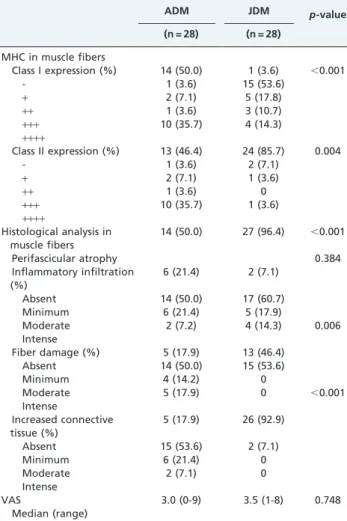
Table 2 -Immunohistological and histological analyses of muscle biopsies from untreated adult and juvenile dermatomyositis patients.
ADM JDM p-value
(n = 28) (n = 28)
MHC in muscle fibers
Class I expression (%) 14 (50.0) 1 (3.6) ,0.001
- 1 (3.6) 15 (53.6)
+ 2 (7.1) 5 (17.8)
++ 1 (3.6) 3 (10.7)
+++ 10 (35.7) 4 (14.3)
++++
Class II expression (%) 13 (46.4) 24 (85.7) 0.004
- 1 (3.6) 2 (7.1)
+ 2 (7.1) 1 (3.6)
++ 1 (3.6) 0
+++ 10 (35.7) 1 (3.6)
++++
Histological analysis in muscle fibers
14 (50.0) 27 (96.4) ,0.001
Perifascicular atrophy 0.384
Inflammatory infiltration (%)
6 (21.4) 2 (7.1)
Absent 14 (50.0) 17 (60.7)
Minimum 6 (21.4) 5 (17.9)
Moderate 2 (7.2) 4 (14.3) 0.006
Intense
Fiber damage (%) 5 (17.9) 13 (46.4)
Absent 14 (50.0) 15 (53.6)
Minimum 4 (14.2) 0
Moderate 5 (17.9) 0 ,0.001
Intense
Increased connective tissue (%)
5 (17.9) 26 (92.9)
Absent 15 (53.6) 2 (7.1)
Minimum 6 (21.4) 0
Moderate 2 (7.1) 0
Intense
VAS 3.0 (0-9) 3.5 (1-8) 0.748
Median (range)
ADM: adult dermatomyositis; JDM: juvenile dermatomyositis; MHC: major histocompatibility complex; VAS: visual analog score.
Fiber damage, inflammatory infiltrate and increased connective tissue were assessed semi-quantitatively as absent, minimal, moderate or intense.
The expression levels of MHC I and II were assessed by a semi-quantitative method: (-) = positively stained endothelial cells only; (+) = 1 to 10% positively stained fibers; (++) = 11 to 25% positively stained fibers; (+++) = 26 to 50% positively stained fibers; and (++++) = 51 to 100% positively stained fibers.
Table 3 -Relationship among various myositis parameters and the MHC I / II presence in dermatomyositis.
Parameters ADM (n = 28) JDM (n = 28)
MHC I MHC II MHC I MHC II
Interval of disease to biopsy
0.807 0.807 0.787 0.515
Age at onset 0.086 0.086 0.296 0.767
Degree of muscle strength 0.251 0.0251 0.179 0.484 Creatine kinase level 0.362 0.362 1.000 0.141 Other clinical
manifestations
.0.050 .0.050 .0.050 .0.050
Perifascicular atrophy 0.257 0.257 1.000 1.000
Fiber damage 0.045 0.045 0.464 1.000
Increased connective tissue 0.537 0.537 1.000 1.000 Inflammatory cell
infiltration
0.512 0.512 1.000 0.253
the therapeutic approach might be delayed. MHC protein expression analysis by immunohistochemistry has proven to be extremely helpful in these cases as an additional diagnostic tool (2,11,17).
In fact, in this study, MHC I expression was present in all JDM cases except for in one patient, who had a muscle biopsy showing perifascicular atrophy and minimal inflam-matory cell infiltration without fiber damage. The over-expression of MHC I has been previously described by others (11,12,18-20), even in muscle biopsies reported as normal by conventional histology. In our study, 66.7% of the MHC I-positive JDM cases presented no (7.4%) or very low (59.3%) inflammatory cell infiltration in the analyzed muscle specimen. In contrast, MHC I overexpression was identified in only half of the ADM patients in our series. The previously reported frequency of MHC I positivity has been quite variable, ranging from 53.3% to 100% (11,17-19,21,22). The smaller number and heterogeneity of pre-viously studied cases may partially explain these discre-pancies. Similarly, MHC I expression was detected in half of the ADM cases, including cases presenting no or only scattered inflammatory cell infiltration in the muscle biopsy (14.3% and 42.9% of the cases, respectively). Therefore, the positive MHC I expression in such histologically near-normal cases proved to be a powerful tool to establish the diagnosis of inflammatory myopathy in combination with the compatible clinical and laboratorial findings, mainly in the JDM cases. The MHC II expression was present in fewer of our cases; it was observed in 50.0% and 14.3% of the ADM and JDM cases, respectively. Variable frequencies, ranging from 0% to 28.6%, have been reported in other studies (17,19,21).
Whether the MHC complex up-regulation in DM results from a nonspecific response to muscle degeneration and regeneration or from a specific disease process is not fully understood. Highly expressed pro-inflammatory cytokines in DM (21,23), such as INFcand TNFa, have been reported
to induce MHC I expression inin vitroassays (24,25). The overexpression of the MHC complex might also be second-ary to the diffusion of secreted cytokines by inflammatory cells near the biopsied muscle area, as up-regulation of interleukin-1 might even reflect a stress response secondary to the early ischemic injury of the muscle fibers (18,21,26).
The overexpression of MHC I and II in the muscle biopsies proved to be an independent factor of not only the duration of the disease and the degree of muscle damage (as determined by either the degree of muscle strength recorded during clinical examination or the muscle enzy-matic measurements) but also the muscle histological findings. In fact, despite the absence of inflammatory cell infiltrates and/or perifascicular atrophy, MHC I and/or II overexpression was clearly detected in the muscle biopsies from patients with clinical features of DM, as demonstrated in this study and in other studies (11,18,20-22,27). Moreover, in our previous study, MHC I expression was independent of even corticosteroid therapy administered prior to the muscle biopsy (13).
Our results indicated that MHC I is overexpressed in the muscle biopsies of JDM and ADM patients. MHC I- and II-dependent processes require the expression of ICAM-1. The ICAM-1 molecule is necessary to stabilize the interactions between the cell receptor and the MHC-peptide complex in antigen recognition (28). The ICAM-1 molecule, which is essential for cell adhesion, also triggers the cytotoxic
mechanism. The results of this study combined with our previous observation of differential positive ICAM-1 expres-sion in the muscle fibers of ADM, in contrast to the increased expression of ICAM-1 in the muscle vessels of JDM (29), corroborates the myocytotoxic mechanism in adult inflam-matory myopathies, in contrast to muscle lesions through the involvement of microvasculature in JDM.
The limitation of this study is inherent to the applied immunohistochemistry method, in which absolute quanti-tative data are difficult to obtain. However, the semiquanti-tative analysis performed by two independent investigators and based on microscopic fields chosen with uniform criteria, along with the staining of specimens performed in a single batch, were the strategies adopted to minimize bias. The analyses of these expression profiles in the context of the clinical evolution, therapy response, and the results of other complementary exams (such as capillaroscopy), along with comparisons to other inflammatory myopathies (such as polymyositis and inclusion body myositis), may improve our understanding of the pathophysiology of these diseases. In summary, JDM patients presented more MHC I positivity and perifascicular atrophy and less fiber damage, connective tissue proliferation and increased creatine kinase / lactate dehydrogenase levels compared with ADM patients. Immunohistochemical analysis of MHC I and II in muscle biopsies is confirmed as a complementary diagnostic tool for inflammatory myopathy, particularly with respect to the overexpression of MHC I in JDM cases.
ACKNOWLEDGMENTS
Sponsored by Conselho Nacional de Desenvolvimento Cientı´fico e Tecnolo´gico - CNPQ (grant 300248/2008-3 to CAS) and the Federico Foundation to SKS and CAS.
AUTHOR CONTRIBUTIONS
Shinjo SK participated in the collection of the data, performed the statistical analysis and wrote the manuscript. Sallum AM participated in the collection of the data and performed the data analysis. Silva CA revised the manuscript. Marie SK contributed to the study design, wrote the manuscript and revised the manuscript.
REFERENCES
1. Engel AG, Hohlfeld R, Banker BQ. Inflammatory myopathies: the polymyositis and dermatomyositis syndromes. In: Engel AG, Franzini-Armstrong C, eds. Myology. New York, NY: McGraw Hill Publishing; 2004:1335-83.
2. Dalakas MC, Hohlfeld R. Polymyositis and dermatomyositis. Lancet. 2003;362(9388):971-82, http://dx.doi.org/10.1016/S0140-6736(03)14368-1.
3. Bohan A, Peter JB. Polymyositis and dermatomyositis. N Engl J Med. 1975;292(7):344-7, http://dx.doi.org/10.1056/NEJM197502132920706. 4. Drake LA, Dinehart SM, Farmer ER, Goltz RW, Graham GF, Hordinsky
MK, et al. Guidelines of care for dermatomyositis. Am Acad Dermatol. 1996;34(5 Pt 1):824-9.
5. McMichael AJ. HLA restriction of human cytotoxic T cell. Springer Semin Immunopathol. 1980;3(1):3-22.
6. Appleyard ST, Dunn MZ, Dubowitz V, Rose ML. Increased expression of HLA-ABC class I antigens by muscle fibers in Duchenne mus-cular dystrophy, inflammatory myopathy and other neuromusmus-cular disorders. Lancet. 1985;1(8425):361-3, http://dx.doi.org/10.1016/S0140-6736(85)91384-4.
7. Daar AS, Fuggle SV, Fabre JW, Ting A, Morris PJ. The detailed distribution of HLA-A,B,C antigens in normal human organs. Transplantation. 1984;38(3):287-92, http://dx.doi.org/10.1097/00007890-198409000-00018.
Proc Natl Acad Sci USA. 2000;97(16):9209-14, http://dx.doi.org/ 10.1073/pnas.97.16.9209.
9. Figarella-Branger D, Lacroix C, Coquet M, Gherardi R, Pellissier JF. Idiopathic inflammatory myopathies. Ann Pathol. 2001;21(3):279-84. 10. Pavlath GK. Regulation of class I MHC expression in skeletal muscle:
deleterious effect of aberrant expression on myogenesis. J Neuroimmunol. 2002;125(1-2):42-50, http://dx.doi.org/10.1016/S0165-5728(02)00026-7. 11. Civatte M, Schleinitz N, Krammer P, Fernandez C, Guis S, Veit V, et al.
Class I MHC detection as a diagnostic tool in noninformative muscle biopsies of patients suffering from dermatomyositis (DM). Neuropathol Appl Neurobiol. 2003;29(6):546-52, http://dx.doi.org/10.1046/j.1365-2990.2003.00471.x.
12. Li CK, Varsani H, Holton JL, Gao B, Woo P, Wedderburn LR. Juvenile dermatomyositis research group. MHC class I overexpression on muscles in early juvenile dermatomyositis. J Rheumatol. 2004;31(3):605-9. 13. Sallum AME, Kiss MHB, Silva CAA, Wakamatsu A, Sachetti S, Lotufo S, et al. MHC class I and II expression in juvenile dermatomyositis skeletal muscle. Clin Exp Rheumatol. 2009;27(3):519-526.
14. Cifuentes-Diaz C, Delaporteg C, Dautreaux B, Charron D, Fardeau M. Class II MHC antigens in normal human skeletal muscle. Muscle Nerve. 1992;15(3):295-302, http://dx.doi.org/10.1002/mus.880150307. 15. Goebels N, Michaelis D, Wekerle H, Hohlfeld R. Human myoblasts as
antigen-presenting cells. J Immunol. 1992;149(2):661-7.
16. Hanissian AS, Masi AT, Pitner SE, Cape CC, Medsger TA Jr. Polymyositis and dermatomyositis in children: an epidemiologic and clinical comparative analysis. J Rheumatol. 1982;9(3):390-4.
17. Jain A, Sharma MC, Sarkar C, Bhatia R, Singh S, Handa R. Major histocompatibility complex class I and II detection as a diagnostic tool in idiopathic inflammatory myopathies. Pathol Lab Med. 2007;131(7):1070-6. 18. Emslie-Smith AM, Arahata K, Engel AG. Major histocompatibility complex class I antigen expression, immunolocalization of interferon subtypes, and T-cell mediated cytotoxicity in myopathies. Hum Pathol. 1989;20(3):224-31, http://dx.doi.org/10.1016/0046-8177(89)90128-7. 19. Bartoccioni E, Gallucci S, Scuderi F, Ricci E, Servidei S, Broccolini A, et al.
MHC Class I, II and Intercellular adhesion molecule-I (ICAM-I) expression in inflammatory myopathies. Clin Exp Immunol. 1994;95(1):166-72.
20. Topaloglu H, Muntoni F, Dubowitz V, Sewry C. Expression of HLA class I antigens in skeletal muscle is a diagnostic marker in juvenile dermatomyositis. Child Neurol. 1997;12(1):60-3, http://dx.doi.org/ 10.1177/088307389701200111.
21. Karpati G, Pouliot Y, Carpenter S. Expression of immunoreactive major histocompatibility complex products in human skeletal muscles. Ann Neurol. 1998;23(1):64-72.
22. Van der Pas J, Hengstman JD, Ter Laak HJ, Borm GF, Van Engelen BGM. Diagnostic value of MHC class I staining in idiopathic inflammatory myopathies. J Neurol Neurosurg Psychiatry. 2004;75(1):136-9. 23. Isenberg DA, Rowe DJ, Shearer M, Novick D, Beverley PC. Localization
of interferons and interleukin 2 in polymyositis and muscular dystrophy. Clin Exp Immunol. 1986;63(2):450-8.
24. Bao SS, King NJ, Dos Remedios CG. Elevated MHC class I and II antigens in cultured human embryonic myoblasts following stimulation with IFNc. Immunol Cell Biol. 1990;68(Pt 4):235-41, http://dx.doi.org/ 10.1038/icb.1990.33.
25. Michaelis D, Goebels N, Hohlfeld R. Constitutive and cytokine-induced expression of human leukocyte antigens and cell adhesion molecules by human myotubes. Am J Pathol. 1993;143(4):1142-9.
26. Nyberg P, Wilkman AL, Nennesmo I, Lundberg I. Increased expression of interleukin 1aand MHC class I in muscle tissue of patients with chronic, inactive polymyositis and dermatomyositis. J Rheumatol. 2000;27(4):940-8.
27. Englund F, Lindroos E, Nennesmo I, Klareskog L, Lundberg IE. Skeletal muscle fibers express major histocompatibility complex class II antigens independently of inflammatory infiltrates in inflammatory myopathies. Am J Pathol. 2001;159(4):1263-73, http://dx.doi.org/10.1016/S0002-9440(10)62513-8.
28. Kumamoto T, Abe T, Ueyama H, Sugihara R, Shigenaga T, Tsuda T. Elevated soluble intercellular adhesion molecules-1 in inflammatory myopathy. Acta Neurol Scand. 1997;95(1):34-7, http://dx.doi.org/ 10.1111/j.1600-0404.1997.tb00065.x.