The expression of gene
ANKRD1
correlates with hypoxia
status in muscle biopsies of treatment-näive adult
dermatomyositis
Samuel Katsuyuki Shinjo,I Sueli Mieko Oba-Shinjo,II Miyuki Uno,II Suely Kazue Nagahashi MarieII
I Laboratory of Inlammatory Myopathies, Division of Rheumatology, Faculdade de Medicina FMUSP, Universidade de Sao Paulo, Sao Paulo, Brazil (BR). II Laboratory of Molecular and Cellular Biology, Departament of Neurology, Faculdade de Medicina FMUSP, Universidade de Sao Paulo, Sao Paulo, Brazil (BR).
OBJECTIVES. The ANKRD1 gene codes for the ankyrin repeat domain containing protein 1 and has an important role in myogenesis and possibly also in angiogenesis. Microvasculopathy is a cornerstone and an early pathological marker of change in dermatomyositis, leading to hypoxia and muscle perifascicular atrophy. These alterations could upregulate genes involved in myogenesis and angiogenesis such as ANKRD1. Therefore, we analyzed ANKRD1 expression in muscle biopsies of dermatomyositis and correlated with other hypoxia parameters and with histological changes.
METHODS. Total RNA was extracted from frozen muscle biopsies samples of 29 dermatomyositis patients. A control group consisted of 20 muscle biopsies from adult patients with non-inlammatory myopathy diseases. The gene coding for hypoxia-inducible factor 1, alpha subunit (HIF1A), was analyzed to estimate the degree of hypoxia. ANKRD1 and HIF1A transcript expression levels were determined by quantitative real time PCR.
RESULTS. Signiicantly higher ANKRD1 and HIF1A expression levels were observed in dermatomyositis relative to the control group (P<0.001, both genes). In addition, ANKRD1 and HIF1A were coexpressed (r=0.703, P=0.001) and their expression levels correlated positively to perifascicular atrophy (r=0.420, P=0.023 and r=0.404, P=0.030, respectively).
CONCLUSIONS. Our results demonstrate ANKRD1 overexpression in dermatomyositis correlated to HIF1A expression and perifascicular atrophy. ANKRD1 involvement in myogenesis and angiogenesis mechanisms indicates that further investigation is worthwhile.
KEYWORDS: Dermatomyositis; hypoxia; myogenesis; perifascicular atrophy; RNA expression.
Shinjo SK, Oba-Shinjo SM, Uno M, Marie SKN. The expression of gene ANKRD1 correlates with hypoxia status in muscle biopsies of treatment-näive adult dermatomyositis. MedicalExpress (São Paulo, online). 2017;4(4):M170402
Received for Publication on May 24, 2017; First review on July 1, 2017; Accepted for publication on July 4, 2017; Online on July 18, 2017
E-mail: [email protected]
■
INTRODUCTIONThe ankyrin repeat domain 1 (ANKRD1) is a transcription cofactor that translocates to the nucleus in response to mechanical stretch.1 It is present not
only in the nucleus but also in the I-band region of the sarcomere as a member of the titin-N2A mechanosensory unit1 and interacts with myopalladin.2 Moreover, the
interaction of ANKRD1 and myopalladin is required for the maintenance of sarcomeric integrity.2
ANKRD1 is present in fetal skeletal muscle fibers3 and
its expression decreases after birth, being scarcely expressed in the normal skeletal muscle of adults.3-5 This expression
profile in early development suggests potential functional
roles of ANKRD1, as in cardiogenesis and myogenesis. The up-regulated production of ANKRD1 in adult muscles has been associated with pathological conditions such as denervated skeletal muscle,4-6 myopathies,5,7-9 muscle hypoxia;10 it has
also been associated to exercise.11-15 ANKRD1 overexpression
was also observed during experimental wound healing, with neovascularization and increased blood perfusion, also
Total RNA extraction and cDNA Synthesis
Total RNA was extracted from muscle tissues using the RNeasy Fibrous Tissue Mini Kit (Qiagen Inc, Hilden, Germany). Evaluation of RNA quantification and
purification was carried out by measuring absorbance and
A260/A280 ratios, and ranges of 1.8 - 2.0 were considered satisfactory for purity standards. Denaturing agarose gel electrophoresis was used to assess the quality of the samples. Synthesis of cDNA was performed by reverse transcription from 1 µg total RNA, previously treated with 1 unit of DNase I (FPLC-pure, GE Healthcare, Piscataway, NJ), using random and oligo(dT) primers, RNase inhibitor and SuperScript III reverse transcriptase (Thermo Fisher
Scientific, Carlsbad, CA), all according to the manufacturer’s recommendations. The resulting cDNA was then treated with 1 unit of RNase H (GE Healthcare). For 200-500 ng of total RNA, the High Capacity cDNA Reverse Transcription Kit (Thermo Fisher Scientific) was utilized. Ten μL of total RNA was reverse transcribed using MultiScribe™ Reverse Transcriptase according to the manufacturer’s instructions. All cDNA samples were diluted with TE buffer, and stored
at - 20 ˚C until later use.
Quantitative real time (qRT-PCR)
The expression levels of ANKRD1 and HIF1A (hypoxia-inducible factors 1-alpha) were determined by qRT-PCR using the SYBR Green approach, in duplicate. Quantitative data were normalized relative to the geometric mean expression of three internal housekeeping control genes: hypoxanthine guanine phosphoribosyl transferase 1 (HPRT), ribosomal protein L41 (RPL41) and heterogeneous nuclear
ribonucleoprotein K (HNRPK). Primer sequences were
as follows (5´-3´): ANKRD1 F: GAGTGCGCGGAGCATCTTA,
A N K R D 1 R : G T C T C A C C G C A T C A T G C A A, H I F 1 A F : C A T C C A A G A A G C C C T A A C G T G T , H I F 1 A R : C A T T T T T C G C T T T C T C T G A G C A T , H P R T F : T G A G G A T T T G G A A A G G G T G T , H P R T R : G A G C A C A C A G A G G G C T A C A A , R P L 4 1 F : G C G C C A T G A G A G C C A A G T, R P L 4 1 R : C T C C A C G G T G C A A C A A G C T A , H N R P K F : G A G C C C A T C A G A A T G G C A G A T, H N R P K R : AAGATCACCATATGAGCCACGA; these were synthesized by IDT (Integrated DNA Technologies, Coralville, IA). Sybr
Green I amplification mixtures (12 µL) contained 3 µL of
cDNA, 6 µL of 2x Maxima SYBR Green/ROX qPCR Master
Mix (Thermo Fisher Scientific), and forward and reverse
primers. Reactions were run on an ABI 7500 Real-Time PCR System (Applied Biosystems, Foster City, CA). The
cycle conditions comprised incubation at 50 ˚C for 2 min to activate UNG, initial denaturation at 95 ˚C for 10 min, and 40 cycles at 95 ˚C for 15 s and 60 ˚C for 1 min. The
Dermatomyositis (DM) is a systemic idiopathic
inflammatory myopathy characterized by a subacute onset
and proximal symmetric muscle weakness. The disease is also associated with cutaneous manifestations, including heliotrope and Gottron’s papules.17 The cornerstone of the
DM pathogenesis involves vascular disturbances that lead to hypoxia, capillary necrosis and muscle perifascicular atrophy.17,18
Therefore, the hypoxic condition in muscle tissue leading to perifascicular atrophy found in DM may also upregulate ANKRD1 expression, which has been related to myogenesis and/or angiogenesis. To test this hypothesis, we analyzed ANKRD1 expression in treatment-naïve skeletal muscle biopsies of adult DM and correlated this expression to skeletal muscle alterations due to hypoxia.
■
MATERIALS AND METHODSStudy design
The present retrospective study was performed at one center and included 29 drug therapy naïve DM patients
and 20 adult controls (free of evidence of inflammatory
myopathies), who were submitted to routine diagnostic skeletal muscle biopsies of the brachial biceps. The patients
fulfilled at least four of the five Bohan and Peter criteria
items,19 and they were initially seen at the Myopathies
Unit of our tertiary service. We excluded patients with (i) clinically amyopathic DM, (ii) myositis associated to overlap syndromes or to neoplasia, and (iii) necrotizing myopathies. The study was approved by our University Hospital Research Ethnics Committee (case # 01445312.3.0000.0068).
Patient data
All the participants underwent a clinical evaluation and all available information was extensively reviewed. The following data were collected at the time of the muscle biopsy procedure: demographics (age, gender and ethnicity), clinical (disease duration) and laboratory data (serum level of creatine phosphokinase and aldolase). Serum level of creatine phosphokinase (normal range: 24 - 173 IU/L) and aldolase (normal range: 1.0 - 7.5 IU/L) levels were determined by the automated kinetic method. Limb muscle strength was graded according to the Medical Research Council Scale as grade 0: absence of muscle contraction; grade I: slight signs of contractility; grade II: movements of normal amplitude but not against gravity; grade III: normal range of motion against gravity;
minimum concentration of primers was determined by the
lowest threshold cycle (Ct) and maximum amplification efficiency while minimizing non-specific amplification (at final concentration of 200 nM for ANKRD1, HIF1A, HPRT
and HNRPK, and 100 nM for RPL41). Analysis of DNA melting curves demonstrated a single peak for the whole set of primers. Standard curves were analyzed to check the
efficiency of amplification of each gene. Additionally, the size of the amplified PCR products was checked by agarose
gel electrophoresis. The equation 2-ΔΔCT was applied to
calculate the relative expression of DM samples, where ∆Ct
= mean Ct ANKRD1 - Ct geometric mean of housekeeping
genes and ∆∆Ct = ∆Ct ANKRD1 - mean ∆Ct controls.21
Histological muscle biopsy analysis
S e q u e n t i a l f ro z e n s e c t i o n s we re f i r s t
stained for hematoxylin-eosine (HE), and then by immunohistochemistry. Each muscle biopsy specimen was coded and analyzed separately. The presence of perifascicular atrophy was assessed semi-quantitatively. A visual analogue scale (VAS) was also included to score
global degree of muscle inflammatory and abnormality from 0 (no inflammation or no abnormality) to 10 (most inflammation or most abnormal).
Immunoluorescence analysis
Immunofluorescence staining was performed
to analyze ANKRD1 expression in muscle biopsies. The
mouse polyclonal antibody against human ANKRD1 (ab88661, Abcam, Cambridge, MA, 1:13) was used. Serial
frozen sections of 5 µm - thickness were fixed for 10
min with methanol and acetone (1:1). Membranes were
permeabilized with 0.1 % Triton X-100 and unspecific
sites were blocked with 2 % bovine serum albumin (BSA). The primary antibody was incubated overnight at 4˚C; the secondary antibody - goat anti-mouse IgG-Alexa Fluor
(Thermo Fisher Scientific) was incubated for one hour. Nuclei were stained with DAPI (Thermo Fisher Scientific).
Staining structures were scanned using a Zeiss LSM 510 Meta Confocal microscope (Carl Zeiss Inc, Thornwood, NY). Human cardiac muscle was used as a positive control for the reactions.
Statistical analysis
The Kolmogorov-Smirnov test was used to evaluate the distribution of each parameter. The demographic, clinical and laboratory features are expressed as mean and standard deviation (SD) for continuous variables or as frequencies and percentages for categorical variables. The median (25th - 75th percentile) was calculated for continuous
variables not normally distributed. Comparisons between
ANKRD1 and HIF1A relative expression levels of patients
were analyzed using Student’s t-test or the Mann-Whitney test. Chi-squared or Fisher’s exact test was used to evaluate the categorical variables. The correlations between the parameters were analysed by Spearman correlation. All of the analyses were performed using the SPSS 15.0 statistics software (Chicago, , IL,USA). Values of P < 0.05 were
considered statistically significant.
■
RESULTSThe present study included 29 dermatomyositis patients (6 males and 23 females, mean age 51.2 ± 17.4 years) and 20 adult control individuals (5 males and 15 females, mean age 50.4 ± 15.5 years). The patients’ demographic, clinical, laboratory and muscle biopsy features are presented in Table 1.
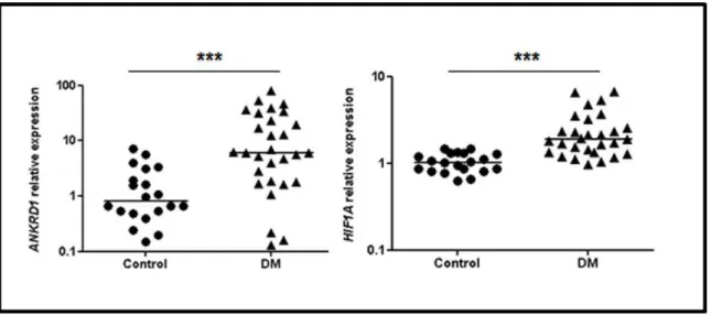
Higher ANKRD1 and HIF1A expression levels were observed in samples of DM patients vs. controls (P = 0.0002 and P < 0.0001, respectively), as shown in Figure 1. ANKRD1
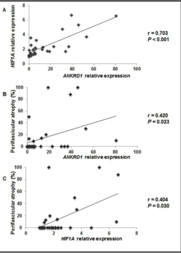
relativeexpression correlated positively to HIF1A relative expression (r = 0.703, P < 0.001), as shown in Figure 2A. Additionally, the relative expression levels of both genes correlated positively to perifascicular atrophy (r = 0.420,
P = 0.023 and r = 0.404, P = 0.003, for ANKRD1 and HIF1A, respectively), as shown in Figures 2B and 2C.
Nevertheless, both ANKRD1 and HIF1A relative expression levels did not correlate to demographics (age at biopsy muscle procedure, gender and ethnicity), to clinical data (disease duration, muscle strength) and to laboratory features (creatine kinase and aldolase) as shown in Table 1. Moreover, these genes did not correlate to muscle biopsy
characteristics as VAS inflammatory infiltration and VAS
abnormality.
The ANKRD1 was localized in the perifascicular area
of muscle fibers of DM patients.
■
DISCUSSIONTo our knowledge, this is the first study that
addressed ANKRD1 and HIF1A expression in muscle
biopsies of DM patients not previously submitted to drug therapy. ANKRD1 was overexpressed in DM muscle biopsies, and the expression levels were positively correlated to
HIF1A expression and perifascicular atrophy.
In adult muscles, ANKRD1 overexpression is generally associated with pathological and stress conditions, as in denervated skeletal muscle,4,6 muscle pathologies5,7-9 and
inherited myopathies, such as muscular dystrophies,22,23
congenital myopathies,24 motor neuron diseases (spinal
muscular atrophy)24 and amyotrophic lateral sclerosis.25 It is
Table 1 - Demographic, clinical, laboratory and biopsy features of patients with dermatomyositis, and correlation with ANKRD1 and HIF1A relative expression.
Features Dermatomyositis ANKRD1 HIF1A
(n=29) P value P value
Age at biopsy muscle procedure (years), mean 44.2±18.1 0.100 0.080
Disease duration (months), interquartile 6 (5-12) 0.123 0.236
Gender (female), % 23 (79.3) 1.000 0.173
Ethnicity (white), % 35 (86.2) 0.711 0.711
Muscle strength
Upper limbs
Grade III, % 3 (10.4) 0.070 0.155
Grade IV, % 21 (72.4)
Grade V, % 5 (17.2)
Lower limbs
Grade III, % 3 (10.4) 0.183 0.951
Grade IV, % 21 (72.4)
Grade V, % 5 (17.2)
Laboratory alterations
Creatine phosphokinase (IU/L), interquartile 2200 (1805-3335) 0.879 0.517
Aldolase (IU/L), interquartile 43 (17-167) 0.809 0.605
Muscle biopsy
Number of fascicule / sample, interquartile 10 (4-20) -
Perifascicular atrophy, % 10 (34.5) -
VAS inlammatory iniltration (0-10mm), interquartile 0.8 (0.4-2.0) 0.106 0.156
VAS abnormality (0-10mm), interquartile 0.8 (0.6-1.8) 0.106 0.339
Results are expressed as mean ± standard deviation, median (25th – 75th interquartile) or percentage (%).
ANKRD1: ankyrin repeat domain 1; HIF1A: hypoxia-inducible factor 1 - alpha subunit; VAS: visual analogue score.
Figure 1. Relative expression proiles of ANKRD1 and HIF1A in dermatomyositis (DM) and control group muscle biopsies. The horizontal bars indicate
Figure 2. (A) Correlation between ANKRD1 and HIF1A relative expression levels; (B) ANKRD1 relative expression levels and perifascicular atrophy; and (C) HIF1A relative expression levels and perifascicular atrophy for DM cases.
is an important finding in DM muscle biopsy, which may
be related to high expression of ANKDR1. Upregulation of ANKRD1 has been demonstrated in the early stage of muscle regeneration in animal model of muscle damage,16 as well as
in experimental denervation-induced atrophy and muscular dystrophy models.9 Therefore, ANKRD1 has been pointed as a
general marker of muscle damage. ANKRD1 is also suggested
to play an anti-inflammatory role through inhibition of NF-κB
transcriptional activity by modulating the balance between
physiological and pathological inflammatory responses in
skeletal muscle.26
ANKRD1 high expression was also demonstrated in skeletal muscle under hypoxic conditions in in vivo
models.10 The latter situation hints at a putative link to the
adaptation of specific mechanisms in order to maintain
adequate oxygenation of critical tissues under hypoxic stress.10 Moreover, the hypoxic status could activate Hypoxia
inducible factors, which act as a master switch to induce expression of several angiogenic factors including vascular endothelial growth factor, nitric oxide synthase, platelet-derived growth factor and others.27,28
HIFs are regulated through hydroxylation by oxygen sensitive hydroxylases, notably by factor-inhibiting HIF-1 (FIH-1).29 HIF hydroxylation directly regulates
transcriptional activities of the HIF-induced targets. It
should be noted that FIH-1 appears to have high affinity
to many substrates containing the ankyrin repeat motif. In fact, FIH-1 hydroxylates well conserved asparagine residues within the ankyrin repeats.29 The affinity of FIH-1 for many ankyrin containing proteins is higher than its affinity
for HIF-1; therefore, oxygen sensitive hydroxylases may directly regulate activity of proteins with ankyrin repeat proteins implicating an active role in the hypoxia response mechanisms.
The atrophic muscle fibers from perifascicular areas
correlated with ANKDR1 expression. Moreover, ANKRD1
expression correlated significantly to HIF1A expression and to the degree of perifascicular atrophy. This perifascicular
muscle fiber atrophy is associated to microvascular capillary
pathology, including a reduction in the number as well as in the enlargement of some remaining endomysial capillaries.30
Apparent capillary loss is most prominent within regions
of muscle fiber atrophy at the edge of fascicles. Deposition
of C5b9 complement on endomysial capillaries has been reported as further evidence that microvasculopathy may play a pathogenic role in DM.31-33 This condition could
promote a hypoxic tissue status that generates a sequence of gene expressions involved in hypoxia, including HIF1A and consequently ANKRD1 gene. Furthermore, in the present study,
the immunohistochemical analysis showed that ANKRD1
and HIF1A presented higher expression in muscle fibers in
perifascicular area, corroborating our present hypothesis. Nevertheless, ANKRD1 and HIF1A expression did not correlate to any demographic, clinical or laboratory parameters.
■
SUMMARYANKRD1 was overexpressed in muscle biopsies of patients with DM and correlated positively to perifascicular atrophy and HIF1A gene expression. Further investigation about ANKRD1 involvement in myogenesis and angiogenesis mechanisms will better clarify the pathomechanism in DM.
■
AUTHOR CONTRIBUTIONS K Shinjo: planning, reviewing literature, executing and writing the present article.
S M Oba-Shinjo: planning, reviewing literature and writing the present article.
M Uno: planning, executing and writing the present article.
■
CONFLICT OF INTERESTAll authors declare no conflict of interest.
■
ACKNOWLEDGEMENTSThis work was supported by: (a) Federico Foundation, (b) Fundação de Amparo à Pesquisa do Estado de São Paulo (FAPESP) [#2014/09079-1, #2012/09633-3], and (c) Fundação Faculdade de Medicina to S.K.S, S.M.O.S and S.K.N.M.
A EXPRESSÃO DO GENE ANKRD1 CORRELACIONA-SE COM O STATUS DE BIÓPSIAS MUSCULARES EM ADULTOS PORTADORES DE DERMATOMIOSITE SEM EXPOSIÇÃO PRÉVIA A TRATAMENTO
OBJETIVOS: ANKRD1 codifica “ankyrin repeat
domain containing protein 1” e tem um papel importante na miogênese e possivelmente também na angiogênese. Microvasculopatia é considerada como um ponto central e uma alteração patológica precoce na dermatomiosite
(DM), levando à hipóxia e à atrofia perifascicular muscular.
Estas alterações poderiam estimular genes envolvidos
na miogênese e angiogênese como ANKRD1. Portanto,
analisamos a expressão de ANKRD1 em biópsias musculares de DM e correlacionamos com outros parâmetros de hipóxia e alterações histológicas.
MÉTODOS: O RNA total foi extraído de biópsias de músculos congelados de 29 pacientes com DM. Como grupo controle, foram usadas 20 biópsias de músculo de pacientes adultos com miopatia não-inflamatória. O gene que codifica a subunidade alfa do fator 1 induzido por hipóxia (HIF1A) foi analisado para estimar o grau de hipóxia. Os níveis
de expressão dos transcritos ANKRD1 e HIF1A foram
determinados por PCR quantitativa em tempo real.
RESULTADOS: Níveis aumentados de expressões de ANKRD1 e HIF1A foram observados em DM quando
comparados ao grupo controle (P<0,001, ambos os
genes). Além disso, ANKRD1 e HIF1A apresentaram
coexpressão (r=0,703, P=0,001) e seus níveis de expressão
correlacionaram-se também positivamente com atrofia
perifascicular (r=0,420, P=0,023 e r=0,404, P=0,030, respectivamente).
CONCLUSÕES: Nossos resultados demonstraram aumento de expressão de ANKRD1 na DM, que correlacionou com a expressão de HIF1A e atrofia perifascicular.
Investigações adicionais do envolvimento de ANKRD1
no mecanismo de miogênese e angiogênese devem ser realizadas.
PALAVRAS-CHAVE: Dermatomiosite, hipóxia,
miogênese, atrofia parafascicular, expressão de RNA.
■
REFERENCES1. Miller MK, Bang ML, Witt CC, Labeit D, Trombitas C, Watanabe K, et al. The muscle ankyrin repeat proteins: CARP, ankrd2/Arpp and DARP as
a family of titin filament-based stress response molecules. J Mol Biol.
2003;333(5):951-64. DOI: 10.1016/j.jmb.2003.09.012.
2. Bang ML, Mudry RE, MCelhinny AS, Trombitas K, Geach AJ, Yamasaki R, et al. Myopalladin, a novel 145-kilodalton sarcomeric protein with multiple roles in Z-disc and I-band protein assemblies. J Cell Biol. 2001;153(2):413-27. DOI: 10.1083/jcb.153.2.413.
3. Ishiguro N, Baba T, Ishida T, Takeuchi K, OSAKI M, Araki N, et al. CARP, a cardiac ankyrin-repeated protein, and its new homologue, Arpp, are differentially expressed in heart, skeletal muscle, and rhabdomyosarcomas. Am J Pathol. 2002;160(5):1767-78. DOI: 10.1016/S0002-9440(10)61123-6.
4. Tsukamoto Y, Senda T, Nakano T, Nakada C, Hida T, Ishiguro N, et al. Arpp, a new homolog of CARP, is preferentially expressed in type 1
skeletal muscle fibers and is preferentially expressed in type 1 skeletal muscle fibers and is markedly induced by denervation. Lab Invest.
2002;82(5):645-55. DOI: 10.1038/labinvest.3780459.
5. Nakamura K, Nakada C, Takeuchi K, Osaki M, Shomori K, Kato S, et al. Altered expression of cardiac ankyrin repeat protein and its homologue, ankyrin repeat protein with PEST and proline-rich region, in atrophic muscles in amyotrophic lateral sclerosis. Pathobiology. 2002;70(4):197-203. DOI: 10.1159/000069329.
6. Baumeister A, Arber S, Caroni P. Accumulation of muscle ankyrin repeat protein transcript reveals local activation of primary myotube end compartments during muscle morphogenesis. J Cell Biol. 1997;139(5):1231-42. DOI: 10.1083/jcb.139.5.1231.
7. Bakay M, Zhao P, Chen J, Hoffman EP. A web-accessible complete transcriptome of normal human and DMD muscle. Neuromuscul Disord. 2002;12(suppl 1):S125-41. DOI: 10.1016/S0960-8966(02)00093-7.
8. Nakada C, Oka A, Nonaka I, Sato K, Mori S, Ito H, et al. Cardiac-restricted ankyrin-repeated protein is differentially induced in Duchenne and spinal muscular atrophy. Pathol Int. 2003;53(10):653-8. DOI: 10.1046/j.1440-1827.2003.01541.x.
9. Laure L, Suel L, Roudaut C, Bourg N, Ouali A, Bartoli M, et al. Cardiac ankyrin repeat protein is a marker of skeletal muscle pathological remodeling. FEBS J. 2009;276(3):669-84. DOI: 10.1111/j.1742-4658.2008.06814.x.
10. Band M, Joel A, Avivi A. The muscle ankyrin repeat proteins are hypoxia-sensitive: in vivo mRNA expression in the hypoxia-tolerant blind subterranean mole rat, Spalax ehrenbergi. J Mol Evol. 2010;70(1):1-12. DOI: 10.1007/s00239-009-9306-6.
11. Carson JA, Nettleton D, Reecy JM. Differential gene expression in the rat soleus muscle during early work overload-induced hypertrophy.
FASEB. J 2002;16(14):207-9. DOI: 10.1096/fj.01-0544fje.
12. Chen YW, Nader GA, Baar KR, Fedele MJ, Hoffman EP, Esser KA.
Response of rat muscle to acute resistance exercise defined by transcriptional and translational profiling. J Physiol.
2002;545(1):27-41. DOI: 10.1113/jphysiol.2002.021220.
13. Barash IA, Mathew L, Ryan AF, Chen J, Lieber RL. Rapid muscle-specific
gene expression changes after a single bout of eccentric contractions in the mouse. Am J Physiol Cell Physiol. 2004;286(2):C355-64. DOI: 10.1152/ajpcell.00211.2003.
14. Hentzen ER, Lahey M, Peters D, Mathew L, Barash IA, Friden J, et al. Stress-dependent and -independent expression of the myogenic regulatory factors and the MARP genes after eccentric contractions in rats. J Physiol. 2006;570(1):157-67. DOI: 10.1113/ jphysiol.2005.093005.
16. Shi Y, Reitmaier B, Regenbogen J, Slowey RM, Opalenik SR, Wolf E, et al. CARP, a cardiac ankyrin repeat protein, is up-regulated during wound healing and induces angiogenesis in experimental granulation tissue. Am J Pathol. 2005;166(1):303-12. DOI: 10.1016/S0002-9440(10)62254-7
17. Dalakas MC, HohlfedL R. Polymyositis and dermatomyositis. Lancet. 2003;362(9388):971-82. DOI: 10.1016/S0140-6736(03)14368-1
18. Dalakas MC. Inflammatory muscle diseases: a critical review on
pathogenesis and therapies. Curr Opin Pharmacol. 2010;10(3):346-52. DOI: 10.1016/j.coph.2010.03.001.
19. Bohan A, Peter JB. Polymyositis and dermatomyositis.I. N Engl J Med. 1975;292(7);344-7. DOI: 10.1056/NEJM197502132920706. 20. Medical Research Council. Aids to the examination of the peripheral
nervous system, Memorandum no. 45. Her Majesty’s Stationery Office,
London, 1981.
21. Livak KJ, Schmittgen TD. Analysis of relative gene expression data using real-time quantitative PCR and the 2(-Delta Delta C(T)) Method. Methods. 2001;25(4):402-8. DOI: 10.1006/meth.2001.1262. 22. Furukawa Y, Hashimoto N, Yamakuni T, Ishida Y, Kato C, Ogashiwa M,
et al. Down-regulation of an ankyrin repeat-containing protein, V-1, during skeletal muscle differentiation and its re-expression in the regenerative process of muscular dystrophy. Neuromuscul Disord. 2003;13(1):32-41. DOI: 10.1016/S0960-8966(02)00185-2. 23. Ojima K, Kawabata Y, Nakao H, Nakao K, Doi N, Kitamura F, et al.
Dynamic distribution of muscle-specific calpain in mice has a key role
in physical-stress adaptation and is impaired in muscular dystrophy. J Clin Invest. 2010;120(8):2672-83. DOI: 10.1172/JCI40658. 24. Nakada C, Oka A, Nonaka I, Sato K, Mori S, Ito H, et al. Cardiac ankyrin
repeat protein is preferentially induced in atrophic myofibers
of congenital myopathy and spinal muscular atrophy. Pathol Int. 2003;53(10):653-8. DOI: 10.1046/j.1440-1827.2003.01541.x.
25. Nakamura K, Nakada C, Takeuchi K, Osaki M, Shomori K, Kato S, et al. Altered expression of cardiac ankyrin repeat protein and its homologue, ankyrin repeat protein with PEST and proline-rich region, in atrophic muscles in amyotrophic lateral sclerosis. Pathobiology. 2003;70(4):197-203. DOI: 10.1159/000069329
26. Liu XH, Bauman WA, Cardozo C. ANKRD1 modulates inflammatory
responses in C2C12 myoblasts through feedback inhibition of NF-κB signaling activity. Biochem Biophys Res Commun. 2015;464(1):208-13. DOI: 10.1016/j.bbrc.2015.06.118.
27. Semenza GL. Hypoxia-inducible factor 1: master regulator of O2
homeostasis. Curr Opin Genet Dev. 1998;8(5):588-94. DOI: 10.1016/ S0959-437X(98)80016-6.
28. Loges S, Roncal C, Carmeliet P. Development of targeted angiogenic medicine. J Thromb Haemost. 2008;7:21-33. DOI: 10.1111/j.1538-7836.2008.03203.x.
29. Linke S, Hampton-Smith RJ, Peet DJ. Characterization of ankyrin repeat-containing proteins as substrates of the asparaginyl hydroxylase factor inhibiting hypoxia-inducible transcription factor. Methods Enzymol. 2007;435:61-85. DOI: 10.1016/S0076-6879(07)35004-0.
30. Emslie-Smith AM, Engel AG. Microvascular changes in early and advanced dermatomyositis: a quantitative study. Ann Neurol. 1990;27(4):343-56. DOI: 10.1002/ana.410270402.
32. Kissel JT, Mendell JR, Rammohan KW. Microvascular deposition of complement membrane attack complex in dermatomyositis. N Engl J Med. 1986;314(6):329-34. DOI: 10.1056/NEJM198602063140601. 33. Miles L, Bove KE, Lovell D, Wargula JC, Bukulmez H, Shao M, et al.
Predictability of the clinical course of juvenile dermatomyositis based on initial muscle biopsy: a retrospective study of 72 patients. Arthritis Rheum. 2007;57(7):1183-91. DOI: 10.1002/art.22993.