Functional and Morphological Correlates in
the Drosophila LRRK2
loss-of-function
Model
of Parkinson
’
s Disease: Drug Effects of
Withania somnifera
(Dunal) Administration
Francescaelena De Rose1☯, Roberto Marotta2☯, Simone Poddighe3☯, Giuseppe Talani4☯,
Tiziano Catelani2, Maria Dolores Setzu3, Paolo Solla5, Francesco Marrosu5, Enrico Sanna1, Sanjay Kasture6, Elio Acquas1, Anna Liscia1☯*
1Department of Life and Environmental Sciences, University of Cagliari, Cagliari, Italy,2Nanochemistry Department, Fondazione Istituto Italiano di Tecnologia, Genova, Italy,3Department of Biomedical Sciences, University of Cagliari, Cagliari, Italy,4Institute of Neuroscience, National Research Council (CNR), Monserrato, Cagliari, Italy,5Department of Public Health, Clinical and Molecular Medicine, University of Cagliari, Cagliari, Italy,6Pinnacle Biomedical Research Institute, Bhopal, India
☯These authors contributed equally to this work.
Abstract
The common fruit flyDrosophila melanogaster(Dm) is a simple animal species that contrib-uted significantly to the development of neurobiology whose leucine-rich repeat kinase 2 mutants (LRRK2)loss-of-functionin the WD40 domain represent a very interesting tool to look into physiopathology of Parkinson’s disease (PD). Accordingly, LRRK2Dmhave also the potential to contribute to reveal innovative therapeutic approaches to its treatment. With-ania somniferaDunal, a plant that grows spontaneously also in Mediterranean regions, is known in folk medicine for its anti-inflammatory and protective properties against neurode-generation. The aim of this study was to evaluate the neuroprotective effects of its standard-ized root methanolic extract (Wse) on the LRRK2loss-of-function Dmmodel of PD. To this end mutant and wild type (WT) flies were administeredWse, through diet, at different con-centrations as larvae and adults (L+/A+) or as adults (L-/A+) only. LRRK2 mutants have a
significantly reduced lifespan and compromised motor function and mitochondrial morphol-ogy compared to WT flies 1%Wse-enriched diet, administered toDmLRRK2 as L-/A+and
improved a) locomotor activity b) muscle electrophysiological response to stimuli and also c) protected against mitochondria degeneration. In contrast, the administration ofWseto
DmLRRK2 as L+/A+, no matter at which concentration, worsened lifespan and determined
the appearance of increased endosomal activity in the thoracic ganglia. These results, while confirming that the LRRK2loss-of-functionin the WD40 domain represents a valid model of PD, reveal that under appropriate concentrationsWsecan be usefully employed to counteract some deficits associated with the disease. However, a careful assessment of the risks, likely related to the impaired endosomal activity, is required.
a11111
OPEN ACCESS
Citation:De Rose F, Marotta R, Poddighe S, Talani G, Catelani T, Setzu MD, et al. (2016) Functional and Morphological Correlates in the Drosophila LRRK2 loss-of-functionModel of Parkinson’s Disease: Drug Effects ofWithania somnifera(Dunal) Administration. PLoS ONE 11(1): e0146140. doi:10.1371/journal. pone.0146140
Editor:Véronique Sgambato-Faure, INSERM / CNRS, FRANCE
Received:September 25, 2015
Accepted:December 14, 2015
Published:January 4, 2016
Copyright:© 2016 De Rose et al. This is an open access article distributed under the terms of the
Creative Commons Attribution License, which permits unrestricted use, distribution, and reproduction in any medium, provided the original author and source are credited.
Data Availability Statement:All relevant data are within the paper and its Supporting Information files.
Funding:This work was supported by Fondazione Banco di Sardegna (ITA) no.0172 /2014. The funders had no role in study design, data collection and analysis, decision to publish, or preparation of the manuscript.
Introduction
Parkinson’s disease (PD) is the second most common neurodegenerative disorder[1] affecting 2% of the population over 60 years with an increasing incidence over age 85 [2]. The progres-sive loss of dopaminergic neurons in the substantia nigra of the midbrain leads to a deficiency of dopamine causing the typical motor symptoms such as tremor, bradykinesia and rigidity [3] [4]. Although the etiopathogenesis is not fully understood and most cases seem sporadic, genetic variables play a key role in the predisposition to PD onset with at least 5 to 10% of PD patients clearly associated with genetic factors[5]. Indeed, since the seminal paper of Polymero-poulos et al. [6], which identified the first mutation related to PD in the alpha-synuclein gene, other genes involved in the etiology of familial forms of parkinsonism have been discovered[7–
15]. Among them, the identification of several leucine-rich repeat kinase 2 (LRRK2) gene mutations has opened a novel scenario in Parkinson’s disease genetics[16]. In fact, the G2019S LRRK2 mutation is the most common in Caucasian patients occurring in 1–2% of sporadic cases of PD [17][18], while other mutations, such as the G2385R variants contribute to the sus-ceptibility to develop PD especially in Chinese patients[19]. LRRK2 encodes for a protein with a number of independent domains that is expressed, although at a low level, in all tissues. In the brain it is found in the cortex, striatum, hippocampus, cerebellum, and at the level of the dopaminergic neurons in the substantia nigra [20–23]. Most mutations of this gene are associ-ated with a late onset Parkinsonism [15]. Mutations of the gene LRRK2 that elicit the disease occur at the level of the functional domain Roc (R1441C and G), at the level of the COR (Y1699C and R1628P) and of MAPKKK domains (G2019S and I2020T) and in only one of the WD40 domains (G2385R)[11][15][24]. This latter is known to be crucial in several basic cell functions such as vesicle sorting during endocytosis and exocytosis of synaptic vesicles as well as vesicle-mediated transport and cytoskeleton assembly [25][26]. The role of the WD40 domain is suggested to be crucial in controlling the LRRK2-regulated kinase activity having a critical role in the self-interaction and autophosphorylation-mediated mechanisms of neuronal toxicity [27]. Accordingly, deletion of this domain has been shownin-vitroto cause the reduc-tion of the kinase activity that is restored over-expressing the gain of funcreduc-tion mutareduc-tion of the gene[28].
Translational animal models are particularly useful in studying neuronal dysfunction and investigating the etiology and molecular aspects of neurodegenerative diseases. Among the ani-mal species that significantly contributed to the development of these studies, theDrosophila melanogaster(Dm) represents a simple, yet experimentally and translationally powerful, organ-ism that contributed significantly not only to the development of neurobiology but also to the progress of knowledge on neurodegenerative diseases. Notably, most of the genes implicated in familial forms of PD have a counterpart in this insect [29], andDmmutants of PD have been genetically engineered to model key features of the human condition and have been success-fully used in studying PD pathogenesis and in exploring new strategies of disease treatment [30–33]. Previous studies on LRRK2 PD form usingDmmutants (dLRRK2) did not clarify the role of LRRK2 in Drosophila, both in mutantsgain-of-functionfor the kinase domain[15][34] andloss-of-function(LRRKex1mutant) [35–37].
Fully effective medications to treat neurodegenerative diseases are currently lacking and the discovery of novel drug targets for long-sought therapeutics is a great challenge for researchers and clinicians. The use of plant extracts is largely employed worldwide in traditional medicine, constituting the basis of health care in many societies, to treat disparate pathologies [38]. The well-known therapeutic properties of the medicinal plants have been investigated in various animal models and the observations of such investigations have served in many instances as the basis of new drugs development [39][40][33]. A common plant of the Indian flora, also
Abbreviations:Dm,Drosophila melanogaster; LRRK2, leucine-rich repeat kinase 2; LRRK2WD40, LRRK2loss-of-functionin the WD40 domain; PD, Parkinson’s disease;Wse,Withania somnifera extract; WT, wild type; L-Dopa,
found in Southern Europe, including Sardinia (Italy), isWithania somnifera(Ws) Dunal. Its roots, used in Ayurvedic medicine for many central nervous system disorders [41][42], are a valuable herbal medication and the recognized pharmacological effects ofWs, such as anti-oxi-dant, neuroprotection and functional recovery made it of prime interest also in the treatment of PD [43][44].
The aim of this paper was twofold: on one hand to confirm the validity of the LRRKex1 mu-tant [35][37], from now on namedLRRK2 WD40 loss-of-function(LRRK2WD40), as animal model of parkinsonism inDm; on the other hand, to investigate the antiparkinsonian potential of the standardized methanolic extract ofWseroots on this mutant, as compared toDmwild type (WT, Canton-S). To this end we tested lifespan, climbing activity, electrophysiological muscle parameters and subcellular ultrastructure (mitochondria and lysosomes) of the neurons involved in the motor circuitry, as those present in theDmthoracic ganglia.
Materials and Methods
For these experiments we used adult wild type (WT; Canton -S) and LRRK2WD40mutant (LRRKex1, #34750, from Bloomington Stock Center)Drosophila melanogaster(Dm) males. After emergence from pupae, WT and LRRK2 mutant males were separated. WT and mutant flies were reared on a standard cornmeal-yeast-agar medium in controlled environmental con-ditions (24–25°C; 60% relative humidity; light/dark = 12/12 hours). In addition, groups of mutant and WT flies were reared on a standard medium supplemented with the standardized methanolic extract ofWithania somniferaroot (Wse) (gift of Natural Remedies Ltd, Bangalore, India) at three different concentrations (0.1, 1 and 10% w/w) whereas other independent groups of WT and mutant flies were reared with 0.01% (0.5 mM) L-3,4-dihydroxyphenylala-nine (L-Dopa).Wseand L-Dopa were added once the mixture was stirred for 10 min and had cooled down sufficiently[45].All treatments were performed in two combinations concerning their life cycle: as adults (L-/A+) or from larvae and adults (L+/A+). Standard genetic procedures were used during the study.
Survival curves
With the aim of selecting the optimalWse’s concentration to perform the whole study,Dm
were grown on standard diet supplemented with different concentrations ofWseat 25°C. Cohorts of 60 flies (6 flies/tube) from each experimental group (i.e.Wse-untreated and
Wse-treated WT,Wse-untreated andWse-treated LRRK2WD40) were monitored every 2 days for their survival. Mortality was analyzed using Kaplan-Meier survival curves and the statistical comparisons were made with a Gehan-Breslow-Wilcoxon test. All experiments were done in triplicate.
Climbing assay
Wse-untreated andWse-treated LRRK2WD40were indicated. The statistical evaluation was made with a one-way analysis of variance (ANOVA) followed by LSD post-hoc test.
Electrophysiological recordings
At the time of the experiments, flies from groupIIwere anesthetized by using CO2and
care-fully anchored to a wax support ventral side down, as previously reported [47][48]and placed underneath a stereomicroscope. In details, two tungsten stimulating electrodes, connected to a stimulator (Master 8, A.M.P.I, Jerusalem, IL) and a stimulus isolation unit (DS2A, Digitimer Ltd., Hertfordshire, UK) were placed into both eyes of the fly in order to activate the Giant Fiber System (GFS). Stimulus intensity and duration were adjusted in every single experiment until the muscle response was detected; maximal stimulation intensity was not greater than 10 V, and stimulus duration was not greater than 0.5 ms. A ground tungsten wire was placed into the fly abdomen. A borosilicate recording electrode, shaped by a puller (P97, Sutter Instru-ments, Novato, CA) with a resistance of 40-50MOwhen filled with 3M KCl, was placed into the right or left backside of the fly in order to record Post Synaptic Potentials (PSPs) from the Dorsal Longitudinal Muscle fibers (DLMs). PSPs were recorded with an Axopatch 2-B ampli-fier (Axon Instruments, Foster City, CA), filtered at 0.5 kHz and digitized at 1 kHz. PSPs were recorded in bridge mode, measured using peak and event detection software pCLAMP 8.2 (Axon Instruments, Foster City, CA) and analyzed off-line by pCLAMP fit software (Axon Instruments, Foster City, CA). All recordings were obtained from at least 10 different flies belonging to each experimental group (i.e. WT,Wse-untreated andWse-treated LRRK2WD40). Experimenters were blind to the treatment.
Additional electrophysiological experiments were performed by applying a protocol consist-ing in a sconsist-ingle GFS stimulation, delivered every 20 s, followed by PSPs recordconsist-ing. In this differ-ent set of experimdiffer-ents, the“frequency of following”was determined by delivering trains of 10 stimuli at frequencies of 100 Hz (with 10 ms between stimuli) or 200 Hz (with 5 ms between stimuli). Data are expressed as mean + SEM and one or two-way ANOVA followed by Tukey’s or Bonferroni’s post-hoc test (p<0.05), were used in order to determine significant differences
between groups.
Electron microscopy analysis
DrosophilaeWT,Wse-untreated andWse-treated at 1% (L-/A+) and 10% (L+/A+) LRRK2WD40 from groupIIwere anesthetized with CO2before brains and thoracic ganglia being rapidly
Results
Effects of
Wse
on the lifespan of LRRK2
WD40Fig 1Ashows that LRRK2WD40mutants exhibit a significantly shorter life span than WT con-trols. To evaluate a possible toxic effect,Wsewas tested at different concentrations (0.1, 1 and 10% w/w in their standard diet) as L-/A+onto WT insects. In this respect, no significant effects were detected at anyWseconcentration but 10% which significantly reduces the duration of life (Fig 1B) as compared to untreated WT controls. To evaluate the influence of the extract of
Wseon the duration of life of the LRRK2WD40mutants that, as reported above, demonstrated a reduced life span in respect to untreated- WT, they were treated withWseat the same concen-trations as L-/A+(Fig 1C) or as L+/A+(Fig 1D). As shown by the Kaplan-Meier survival curves, administration ofWseinduces a statistically significant increase, even if by a different extent, in the lifespan of mutants LRRK2WD40, when the insects were fed in the adult stage only at 0.1% and especially 1% concentrations (p<0.05 Breslow-Gehan-Wilcoxon test). This restoring
effect was lost when insects were treated at10%WseL-/A+(Fig 1C), and at any concentration when administrated to larvae and adults (L+/A+) LRRK2WD40(Fig 1D).The overall results are in accordance with the hypothesis thatWseaccumulation, due to high concentration and/or long period administration, can induce a possible toxic effect.
Fig 1. Effects ofWseon lifespan.(A) Lifespan, expressed as % survival rates, of wild type (WT) and LRRK2 flies. (B) Lifespan of untreated WT compared to treated WT, only when adults (L-/A+), withWse, 0.1%, 1% and 10%. (C)Lifespan of untreated LRRK2 mutants compared to treated LRRK2 mutants, only
when adults (L-/A+), withWithania somniferaextract (Wse), 0.1%, 1% and 10%. (D) Lifespan of untreated LRRK2 mutants compared to treated LRRK2
mutants, from their larval stage to the end of their life-cycle (L+/A+), withWse, 0.1%, 1% and 10%.*indicates p<0.05 at Kaplan-Meier survival curves
(Gehan-Breslow–Wilcoxon—Graph Pad Prism 5.01), (A) untreated LRRK2 compared to untreated WT, (B) untreated WT compared to treated WT and (C-D) untreated LRRK2 compared to treated LRRK2.
Effect of
Wse
on the locomotor ability of LRRK2
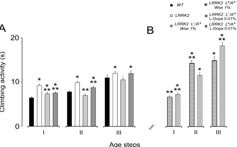
WD40According to results obtained followingWseadministration paralleled with life span we decided to testWseat 1% w/w effects on the climbing activity (negative geotaxis) of mutants.
Fig 2Ashows a significant increase in the climbing time in the threeage groups tested (I: 3–6;
II: 10–15;III: 20–25 days old) of LRRK2WD40as compared to subjects of the WT group (p<0.001) with a tendency to deterioration of the motor performance with aging. The exposure
of LRRK2WD40to 1% w/wWseas L-/A+, induces, in groupsIandII, the recovery of motor dis-ability showing a significant decrease of time to climb compared to untreated mutants; a simi-lar result was also found in insects of groupsI-IIthat were fed 1%Wsefrom larvae and adults (L+/A+). On the other hand,Wseadministration both to L-/A+and L+/A+failed to significantly ameliorate motor behavior in groupIIIaged flies with respect to untreated mutants. L-/A+flies treated withWseshowed a clear tendency toward rescue.
Moreover, as in zebrafish LRRK2loss-of-function-WD40, another PD model in which a sig-nificant rescue of motor impairment after L-Dopa treatment was obtained [49] we also tested L-Dopa at 0.01% (0.5 mM) concentration in the feeding diet of both L-/A+and L+/A+mutant flies. The results presented inFig 2Bshow that inDmmutants the administration of L-Dopa rescued the impairment of climbing activity only in insects of groupI, while worsening the per-formance in groupsII-III.
Fig 2. Effects ofWseon climbing activity.(A-B) Climbing activity of LRRK2 adult males treated withWse1% as compared with WT and untreated LRRK2 (A) and climbing activity of LRRK2 adult males treated with L-Dopa 0.01% (0.5mM) as compared with WT and untreated LRRK2 (B). Values are
average±SEM.*indicates p<0.05 at one-way ANOVA followed by LSD post hoc test as compared to WT;**indicates p<0.05 at one-way ANOVA followed
by LSD post hoc test as compared to LRRK2.
We also considered the percentages of flies that were able to complete the test and the results are shown inS1 Fig. In this respect, results confirm the rescue of insects of groupsI-II,treated withWseboth as L-/A+and L+/A+, increase with respect to untreated ones. It is noteworthy that the percentage of insects of groupIIthat completed the test was 75.2% in WT, 55.6% in untreated mutants, 80.6% in L-/A+and 69.5% in L+/A+Wse-treated mutants. In groupIII, the percentage of mutant insects achieving the target was the same no matter the treatment (being 40.9%, 43.4% and 37.9%, respectively) while more than 52% of WT insects accomplished the task, according to the evaluation criterion (10 sec).
The percentages of flies that were able to complete the test after L-dopa administration are shown inS1B Figand demonstrate that the worsening was positively correlated to age and treatment duration. Thus, the effects ofWseas well as those of L-Dopa administration decrease with age but that of L-Dopa was drammatic. In fact, groupIIIof L-Dopa-treated flies as L+/A+ the percentage achieving the target was only 15%.
Effects of
Wse
on the kinetic properties of evoked PSPs recorded from
DLM in LRRK2
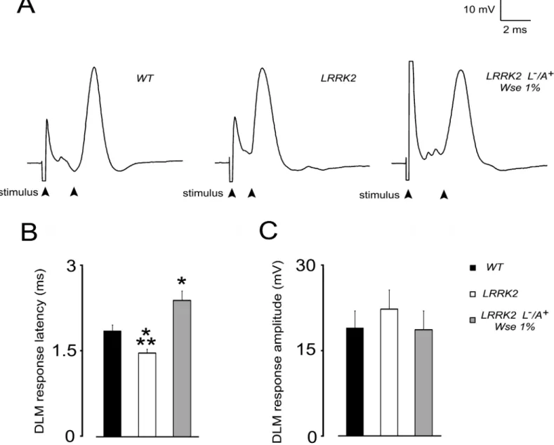
WD40In order to detect potential changes in the function of the DLM neuromuscular junction of LRRK2WD40flies, from groupII, we first evaluated the basal kinetic properties of evoked PSPs (ePSPs) recorded from the DLM after GFS electrical stimulation. More precisely, we evaluated the response latency, that is the time between stimulation of the GFS and subsequent muscle PSP peak, and PSP peak amplitude, that is the maximal muscle depolarization from baseline value.Fig 3shows that the basal properties of ePSPs recorded from DLM muscle of WT ani-mals results in a latency of 1.84 ± 0.1 ms and in an averaged amplitude of 19 ± 3 mV. Notably, LRRK2WD40mutation results in a significant decrease (21%, p<0.05) of ePSPs latency when
compared to WT animals (Fig 3A and 3B). Such effect was no longer apparent in LRRK2 (L-/ A+) flies that were treated withWse1%. Surprisingly, latency in LRRK2 treated flies was signifi-cantly higher with respect to both WT as well as untreated LRRK2 animals. No significant change was detected in PSP peak amplitude among flies from the different experimental groups (Fig 3A and 3C).
Effects of
Wse
on the PSP responses to high frequency stimulation of
GFS of LRRK2
WD40Effects of
Wse
on the subcellular morphology of LRRK2
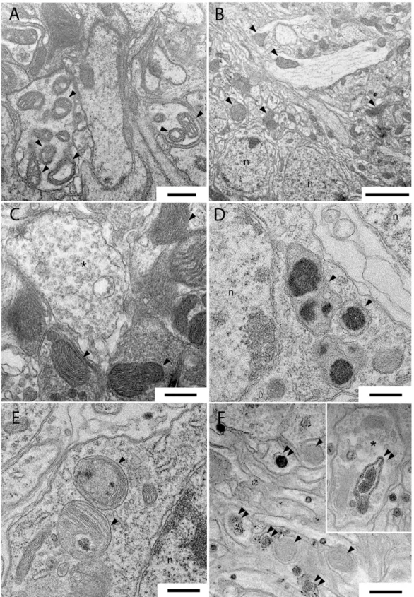
WD40Fig 5shows representative transmission electron microscopy images of thoracic ganglia and antennal lobes (ALs) of untreatedDmLRRK2 mutants (A) and of 1% and 10%Wse-treated, as L-/A+(B and C) and as L+/A+(D-F), insects. In mitochondria of the thoracic ganglia of LRRK2 mutants, we observed regions with several damaged, swollen, and with clearly fragmented cris-tae, that we failed to find in the corresponding regions after treatment with 1%Wse(inFig 5
compare A with B and C). However, after treatment with 10%WseL+/A+, we observed, in the
Fig 3. Effect of LRRK2 gene mutation and treatment withWse1% (L-/A+) on PSP latency and amplitude recorded fromDrosophilaDLM.(A) Representative traces obtained from three different flies in which PSP latency is calculated as the time (ms) from stimulus application to the peak of PSP (black arrows). (B, C) Bar graphs represent the mean±SEM of PSP latency (ms) and amplitude (mV) recorded from flies of the indicated experimental groups.*indicates p<0.05 compared to WT,**indicate p<0.05 compared to treated LRRK2; one-way ANOVA, followed by Bonferroni post-hoc test.
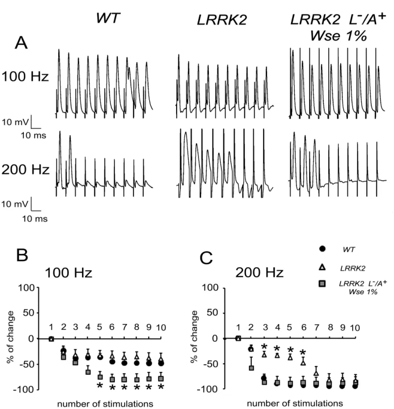
Fig 4. Effect of LRRK2 gene mutation and treatment withWseon the“frequency of following”recorded inDrosophilaDLM.(A) Representative traces obtained from three different flies in which PSPs were evoked in response to 10 stimulations at 100 (top) or 200 Hz (bottom). (B,C) Scatter plot graphs showing the changes in PSP amplitude following stimulation at 100 (B) or 200 Hz (C). All values are expressed as the mean±SEM of the % relative to the amplitude of the first PSP.*indicates p<0.05 compared to WT andWse-untreated LRRK2 (B) and compared to WT andWse-treated LRRK2 (C), two-way ANOVA.
Fig 5. Samples of transmission electron microscopy images of thoracic ganglia and antennal lobes inDrosophilaLRRK2 mutant (A) and after treatment with 1% in L-/A+insects (B, C) and 10% L+/A+(D-F) extract ofWse.(A) abnormal mitochondria in the thoracic ganglia neuropil ofDrosophila
LRRK2. (B, C) conventional mitochondria in thoracic ganglia ofDrosophilaLRRK2 after treatment with 1%WseL-/A+imaged at low (B) and higher magnification (C). (D, E) abnormal mitochondria inDrosophilaLRRK2 thoracic ganglia cell bodies after treatment with 10%WseL+/A+. Note the irregular
electron-dense substance clearly recognizable inside the mitochondria. (F and Inset) numerous endosomes are present inside the antennal lobes neurites of
DrosophilaLRRK2 after treatment with 10%Wse. Scale bars are 0.5μm except in B that is 2.5μm.
corresponding regions of the thoracic ganglia, numerous altered mitochondria with a granular, irregularly shaped electron-dense material in their matrix (Fig 5D and 5E). Moreover after the same treatment we observed, inDrosophilaLRRK2WD40ALs numerous late endosomes/ phago-somes vacuoles inside presynaptic terminals and dendrites (Fig 5F).
Discussion and Conclusions
One of the aims of the present study was to validate the use of LRRK2WD40as a model of PD. In this respect, these mutant flies show reduced lifespan, and motor impairments (face validity) and mitochondrial dysfunctions (construct validity) that characterize Parkinsonism. Further-more, this study was aimed at evaluating the action of the standardized extract of the roots of
Withania somnifera (Wse) and its possible neuroprotective effects on the Parkinson’s genetic model ofDrosophila melanogasterLRRK2WD40. Although almost all of the mutations in LRRK2 have a number of related features, these mutants object of the present study lack, in particular, the WD40 domain responsible for coding a protein chaperone known to be involved in a number of cellular functions such as cytoskeletal, neurotransmitter vesicular pathway and lyso-endosomal activities [25] The results presented here show that the addition of 1%Wseto standard diet of only LRRK2WD40adults (L-/A+), but not of L+/A+, significantlya)increases their lifespan compared to untreated controls andb)improves their locomotor abilities andc)
affects evoked electrophysiological parameters. Furthermore, in thoracic ganglia, under elec-tron microscopy observation, we found thatWseadministration dramatically rescued the mutation-related loss of mitochondrial structural integrity. Interestingly,Wsechronic adminis-tration to flies as L+/A+, no matter the concentration, induces a worsening of symptoms associ-ated with parkinsonism and a further decrease of lifespan as compared to WT controls as well as to untreated LRRK2WD40(Fig 1B)
The flight muscle degeneration accompanied by defects in motor activity [50–52] detected in our study is probably related to dysfunction of dopaminergic neurons. Accordingly, in a zeb-rafish model LRRK2loss-of-functionin the WD40 domain, it was previously reported a rescue of motor impairment following L-Dopa administration in the early larval stage from days post fecundation (DPF) 5 to 6 [49]. Notably, although this and our model of LRRK2loss-of-function
differ in a number of factors such as animal species, life period and duration of L-Dopa admin-istration, the present results also demonstrate an improvement of motor deficit (climbing activ-ity) in the mutants of the groupItreated as L-/A+. However, extension of the treatment to flies of groupIIandIIIdid not rescue the mutation-dependent impairment but elicited a worsening in both L-/A+and L+/A+treated flies (Fig 2B).
The observed rescue of impaired motor ability byWseadministration to LRRK2WD40Dm
while confirming the condition of mutation-dependent impaired motility, as shown in tests of climbing (Fig 2), also supports the suggestion thatWse’s effects might be attributable to increased neurotransmission [53][54]that would result in a better locomotion. Electrophysio-logical data showed that mutation of the LRRK2 gene was associated with a significant decrease in PSP latency when compared to WT animals, an effect that was no longer apparent in LRRK2 (L-/A+) flies that were treated withWse1%. However, no significant change of PSP peak amplitude was detected among flies from the different experimental groups suggesting that in LRRK2WD40mutants there is a higher probability of (but not necessarily an optimally coordinated) muscle contraction compared with WT without changes in muscle contraction
to well correlate with the motility impairment observed in these flies. As for the possible mech-anism, Augustin and colleagues [48] reported that recording the“frequency of following”, a GFS train stimulation at 200 Hz induced in WT a significant decrement of PSP amplitude rela-tive to the first PSP because the intermediary synapses do not have sufficient time to recover between stimuli. Conversely, a stimulation train at 200 Hz performed in untreated LRRK2WD40 flies showed that, relative to the first PSP, the amplitude of PSPs was only slightly diminished, starting from the second response, and treatment with 1%Wsemade the responses similar to those observed in WT. Thus, the effect ofWseon the functional changes associated with the mutation clearly discloses a beneficial aspect of this treatment. At this time, we cannot explain in deep details the abnormal effect ofWsetreatment in LRRK2 flies (i.e. increased PSP latency and exacerbated effect on 100 Hz response vs WT), and this might at least in part be justified recalling the complexity of the projection pathway from the brain to the thoracic ganglion, where axons form electrical synapses with interneurons and the latter form chemical synapses on each motor neuron innervating the DLMs [55][56]. However, mutation of LRRK2WD40 may be correlated with a significant impairment in neurotransmitter release from presynaptic terminals [25][57].
The impaired motility shown by the LRRK2 mutants is paralleled by the presence of scat-tered abnormal mitochondria in their thoracic ganglia, an observation corroborated by other studies that suggest the involvement of LRRK2WD40in mitochondrial homeostasis, responsible of mitochondrial degradation[58][59]. Intriguingly, the conventional mitochondrial morphol-ogy of LRRK2WD40flies observed after treatment with 1%Withaniaextract, and paralleled by an improvement in their motor capacity, suggests thatWsemay also act suppressing mito-chondrial dysfunction, as has been recently demonstrated for a green tea-derived catechin, epi-gallocatechin gallate (EGCG) [59] and as well as already demonstrated in the case of the mutant PINK1B9treated with the standardized seeds extract of another plant,Mucuna pruriens
[33].
In conclusion, based on our results we can infer that the LRRK2loss-of-functionin the WD40domain is a plausible model that recapitulates some of the essential features of Parkin-sonism and that the extract ofWscan be usefully employed to counteract some deficits associ-ated with this condition. However, as demonstrassoci-ated by Poddighe et al., [33] afterMucuna pruriensadministration toDmPINK1B9mutant model of PD, the use of a whole herbal extract requires careful assessment. In fact, the effects ofWseon LRRK2WD40might also be related to age (group IvsIII), length of exposure (L-/A+vsL+/A+) andWse(0.1%vs1%vs10%) concen-trations as suggested by the observation of its effects on climbing (Fig 2A and 2C) as well as on life duration (Fig 1C and 1D). Indeed, the negative effect of 10%Wseboth on WT (Fig 1B) and on theloss-of-functionLRRK2WD40mutant indicates that one or more components of the extract, when administered chronically and at a concentration higher than optimal, may have toxic effects. This conclusion is supported by the observation that chronic administration of
Wseto flies as L+/A+, no matter the concentration, and also at 10% to L-/A+, induces a worsen-ing of symptoms associated with parkinsonism and a further reduction of lifespan as compared to WT controls and untreated LRRK2WD40. This observation also indicates thatWseshows a concentration threshold, below which it does not work; b) has an optimal value for its effects; but c) whose effects at higher concentrations and/or after longer exposures became toxic. As discussed above, this suggests thatWseexerts its effects -as a drug- following a hormesis-like dose-response curve [60] and further highlights the need to assess the proper concentration of
Supporting Information
S1 Fig. Percentages of insects able to achieve the test.(A-B) Percentages of adult males WT, LRRK2,Wse1% treated LRRK2 (A) and L-Dopa 0.01% (0.5mM) treated LRRK2 (B), that could climb unto, or above, the line drawn at 6 cm from the bottom of the tube within 10 sec-onds.Treatments were administered to flies both only when adults (L−/A+) and from their
lar-val stage to the end of their life-cycle (L+/A+), and their effects were assayed at three different age steps (I: 3–6;II: 10–15;III: 20–25 days) of flies’life-span. Values are average ± SEM.
indi-cates p<0.05 at one-way ANOVA followed by LSD post hoc test as compared to WT;
indi-cates p<0.05 at one-way ANOVA followed by LSD post hoc test as compared to LRRK2.
(TIF)
Acknowledgments
We are indebted to Natural Remedies Ltd., Bangalore, India for generous gift ofWithania som-niferaextract. The Authors would like to thank Dr. Ignazio Collu, Dr. Giuliana Colella (Uni-versity of Cagliari) for taking care of flies and Dr. Valentina Corda for technical support.
Author Contributions
Conceived and designed the experiments: FDR SP RM GT EA AL. Performed the experiments: FDR SP GT TC RM. Analyzed the data: FDR SP RM GT TC MDS PS AL. Contributed
reagents/materials/analysis tools: SK ES EA FM AL. Wrote the paper: EA ES SK PS FM AL.
References
1. Lang AE, Lozano AM. Parkinson’s disease. First of two parts. N Engl J Med. 1998; 339: 1044–1053. doi:10.1056/NEJM199810083391506PMID:9761807
2. Gasser T. Genetics of Parkinson’s disease 52. CurrOpinNeurol. 2005; 18: 363–369.
3. Jankovic J. Parkinson’s disease: clinical features and diagnosis. J Neurol Neurosurg Psychiatry. 2008; 79: 368–376. doi:10.1136/jnnp.2007.131045PMID:18344392
4. Gelb DJ, Oliver E, Gilman S. Diagnostic criteria for Parkinson disease. Arch Neurol. 1999; 56: 33–39. doi:10.1001/archneur.56.1.33PMID:9923759
5. Lees AJ, Hardy J, Revesz T. Parkinson’s disease. Lancet. Elsevier Ltd; 2009; 373: 2055–2066. doi:10. 1016/S0140-6736(09)60492-X
6. Polymeropoulos MH, Lavedan C, Leroy E, Ide SE, Dehejia A, Dutra A, et al. Mutation in theα-Synuclein Gene Identified in Families with Parkinson‘s Disease. Science (80-). 1997; 276: 2045–2048.
7. Bonifati V, Rizzu P, van Baren MJ, Schaap O, Breedveld GJ, Krieger E, et al. Mutations in the DJ-1 gene associated with autosomal recessive early-onset parkinsonism. Science. 2003; 299: 256–259. doi:10.1126/science.1077209PMID:12446870
8. Di Fonzo A, Dekker MCJ, Montagna P, Baruzzi A, Yonova EH, Correia Guedes L, et al. FBXO7 muta-tions cause autosomal recessive, early-onset parkinsonian-pyramidal syndrome. Neurology. 2009; 72: 240–245. doi:10.1212/01.wnl.0000338144.10967.2bPMID:19038853
9. Kitada T, Asakawa S, Hattori N, Matsumine H, Yamamura Y, Minoshima S, et al. Mutations in the parkin gene cause autosomal recessive juvenile parkinsonism. Nature. 1998. pp. 605–608. doi:10.1038/ 33416
10. Lautier C, Goldwurm S, Dürr A, Giovannone B, Tsiaras WG, Pezzoli G, et al. Mutations in the GIGYF2 (TNRC15) Gene at the PARK11 Locus in Familial Parkinson Disease. Am J Hum Genet. 2008; 82: 822–833. doi:10.1016/j.ajhg.2008.01.015PMID:18358451
11. Paisán-Ruíz C, Jain S, Evans EW, Gilks WP, Simón J, van der Brug M, et al. Cloning of the gene con-taining mutations that cause PARK8-linked Parkinson’s disease. Neuron. 2004; 44: 595–600. doi:10. 1016/j.neuron.2004.10.023PMID:15541308
13. Ramirez A, Heimbach A, Gründemann J, Stiller B, Hampshire D, Cid LP, et al. Hereditary parkinsonism with dementia is caused by mutations in ATP13A2, encoding a lysosomal type 5 P-type ATPase. Nat Genet. 2006; 38: 1184–1191. doi:10.1038/ng1884PMID:16964263
14. Valente EM, Abou-Sleiman PM, Caputo V, Muqit MMK, Harvey K, Gispert S, et al. Hereditary early-onset Parkinson’s disease caused by mutations in PINK1. Science. 2004; 304: 1158–1160. doi:10. 1126/science.1096284PMID:15087508
15. Zimprich A, Biskup S, Leitner P, Lichtner P, Farrer M, Lincoln S, et al. Mutations in LRRK2 cause auto-somal-dominant parkinsonism with pleomorphic pathology. Neuron. 2004; 44: 601–7. doi:10.1016/j. neuron.2004.11.005PMID:15541309
16. Bonifati V. Parkinson’s disease: the LRRK2-G2019S mutation: opening a novel era in Parkinson's dis-ease genetics. Eur J Hum Genet. 2006; 14: 1061–1062. doi:10.1038/sj.ejhg.5201695PMID: 16835587
17. Berg D, Schweitzer KJ, Leitner P, Zimprich A, Lichtner P, Belcredi P, et al. Type and frequency of muta-tions in the LRRK2 gene in familial and sporadic Parkinson’s disease*. Brain. 2005; 128: 3000–3011. doi:10.1093/brain/awh666PMID:16251215
18. Schrag A, Schott JM. Epidemiological, clinical, and genetic characteristics of early-onset parkinsonism. Lancet Neurol. 2006; 5: 355–363. doi:10.1016/S1474-4422(06)70411-2PMID:16545752
19. Xie CL, Pan JL, Wang WW, Zhang Y, Zhang SF, Gan J, et al. The association between the LRRK2 G2385R variant and the risk of Parkinson’s disease: a meta-analysis based on 23 case-control studies. Neurol Sci. 2014; 2: 1495–1504. doi:10.1007/s10072-014-1878-2
20. Biskup S, Moore DJ, Celsi F, Higashi S, West AB, Andrabi S, et al. Localization of LRRK2 to membra-nous and vesicular structures in mammalian brain. Ann Neurol. 2006; 60: 557–569. doi:10.1002/ana. 21019PMID:17120249
21. Higashi S, Biskup S, West AB, Trinkaus D, Dawson VL, Faull RLM, et al. Localization of Parkinson’s disease-associated LRRK2 in normal and pathological human brain. Brain Res. 2007; 1155: 208–219. doi:10.1016/j.brainres.2007.04.034PMID:17512502
22. Gaiter D, Westerlund M, Carmine A, Lindqvist E, Sydow O, Olson L. LRRK2 expression linked to dopa-mine-innervated areas. Ann Neurol. 2006; 59: 714–719. doi:10.1002/ana.20808PMID:16532471 23. Taymans JM, Van Den Haute C, Baekelandt V. Distribution of PINK1 and LRRK2 in rat and mouse brain. J Neurochem. 2006; 98: 951–961. doi:10.1111/j.1471-4159.2006.03919.xPMID:16771836 24. Mata IF, Kachergus JM, Taylor JP, Lincoln S, Aasly J, Lynch T, et al. Lrrk2 pathogenic substitutions in
Parkinson’s disease. Neurogenetics. 2005; 6: 171–177. doi:10.1007/s10048-005-0005-1PMID: 16172858
25. Li D, Roberts R. Human Genome and Diseases:¶WD-repeat proteins: structure characteristics, biologi-cal function, and their involvement in human diseases. Cell Mol Life Sci. 2001; 58: 2085–2097. doi:10. 1007/PL00000838PMID:11814058
26. Piccoli G, Condliffe SB, Bauer M, Giesert F, Boldt K, De Astis S, et al. LRRK2 controls synaptic vesicle storage and mobilization within the recycling pool. J Neurosci. 2011; 31: 2225–2237. doi:10.1523/ JNEUROSCI.3730-10.2011PMID:21307259
27. Greggio E, Zambrano I, Kaganovich A, Beilina A, Taymans JM, Daniëls V, et al. The Parkinson dis-ease-associated leucine-rich repeat kinase 2 (LRRK2) is a dimer that undergoes intramolecular autop-hosphorylation. J Biol Chem. 2008; 283: 16906–16914. doi:10.1074/jbc.M708718200PMID: 18397888
28. Iaccarino C, Crosio C, Vitale C, Sanna G, Carrì MT, Barone P. Apoptotic mechanisms in mutant LRRK2-mediated cell death. Hum Mol Genet. 2007; 16: 1319–1326. doi:10.1093/hmg/ddm080PMID: 17409193
29. Reiter LT, Potocki L, Chien S, Gribskov M, Bier E. A Systematic Analysis of Human Disease-Associ-ated Gene Sequences In. Genome Res. 2001; 1114–1125.PMID:11381037
30. Celotto M, Palladino MJ. Drosophila: a“model”model system to study neurodegeneration. Mol Interv. 2005; 5: 292–303. doi:10.1124/mi.5.5.9PMID:16249525
31. Xiao L, Guo D, Hu C, Shen W, Shan L, Li C, et al. Diosgenin promotes oligodendrocyte progenitor cell differentiation through estrogen receptor-mediated ERK1/2 activation to accelerate remyelination. Glia. 2012; 60: 1037–1052. doi:10.1002/glia.22333PMID:22461009
32. Poddighe S, Bhat KM, Setzu MD, Solla P, Angioy AM, Marotta R, et al. Impaired Sense of Smell in a Drosophila Parkinson’s Model. PLoS One. 2013; 8. doi:10.1371/journal.pone.0073156
34. Wang J, You H, Liu JF, Ni DF, Zhang ZX, Guan J. Association of olfactory bulb volume and olfactory sulcus depth with olfactory function in patients with Parkinson disease. Am J Neuroradiol. 2011; 32: 677–681. doi:10.3174/ajnr.A2350PMID:21330398
35. Lee SB, Kim W, Lee S, Chung J. Loss of LRRK2/PARK8 induces degeneration of dopaminergic neu-rons in Drosophila. Biochem Biophys Res Commun. 2007; 358: 534–9. doi:10.1016/j.bbrc.2007.04. 156PMID:17498648
36. Li T, Yang D, Sushchky S, Liu Z, Smith WW. Models for LRRK2-Linked Parkinsonism. Parkinsons Dis. 2011; 2011: 942412. doi:10.4061/2011/942412PMID:21603132
37. Matta S, Van Kolen K, da Cunha R, van den Bogaart G, Mandemakers W, Miskiewicz K, et al. LRRK2 Controls an EndoA Phosphorylation Cycle in Synaptic Endocytosis. Neuron. 2012; 75: 1008–1021. doi: 10.1016/j.neuron.2012.08.022PMID:22998870
38. Alviano DS, Alviano CS. Plant extracts: search for new alternatives to treat microbial diseases. Curr Pharm Biotechnol. 2009; 10: 106–121. doi:10.2174/138920109787048607PMID:19149593 39. Lieu C, Kunselman AR, Manyam BV, Venkiteswaran K, Subramanian T. A water extract of Mucuna
pruriens provides long-term amelioration of parkinsonism with reduced risk for dyskinesias. Park Relat Disord. Elsevier Ltd; 2010; 16: 458–465. doi:10.1016/j.parkreldis.2010.04.015
40. Kasture S, Mohan M, Kasture V. Mucuna pruriens seeds in treatment of Parkinson’s disease: Pharma-cological review. Orient Pharm Exp Med. 2013; 13: 165–174. doi:10.1007/s13596-013-0126-2 41. Dagenais M L-CSS BB. Scientific Basis for the Therapeutic Use of Withania Somnifera: a Review.
2000; 5: 334–346.
42. Kuboyama T, Tohda C, Komatsu K. Pharmacologically Active Constituents from Plants Used in Tradi-tional Medicine Effects of Ashwagandha (Roots of Withania somnifera) on Neurodegenerative Dis-eases. Biol Pharm Bull. 2014; 37: 892–897.
43. Ahmad M, Saleem S, Ahmad AS, Ansari MA, Yousuf S, Hoda MN, et al. Neuroprotective effects of Withania somnifera on 6-hydroxydopamine induced Parkinsonism in rats. Hum Exp Toxicol. 2005; 24: 137–147.PMID:15901053
44. Prakash J, Yadav SK, Chouhan S, Prakash S, Singh SP. Synergistic effect of Mucuna pruriens and Withania somnifera in a paraquat induced Parkinsonian mouse model*. Adv Biosci Biotechnol. 2013; 2013: 1–9.
45. Jansen RLM, Brogan B, Whitworth AJ, Okello EJ. Effects of Five Ayurvedic Herbs on Locomotor Behaviour in a Drosophila melanogaster Parkinson‘s Disease Model. 2014;
46. Liu Z, Wang X, Yu Y, Li X, Wang T, Jiang H, et al. A Drosophila model for LRRK2-linked parkinsonism. Proc Natl Acad Sci U S A. 2008; 105: 2693–2698. doi:10.1073/pnas.0708452105PMID:18258746 47. Allen MJ, Godenschwege T. Electrophysiological recordings from the Drosophila giant fiber system
(GFS). Cold Spring Harb Protoc. 2010; 5: 1–14. doi:10.1101/pdb.prot5453
48. Augustin H, Allen MJ, Partridge L. Electrophysiological recordings from the giant fiber pathway of D. melanogaster. J Vis Exp. 2011; 1–5. doi:10.3791/2412
49. Sheng D, Qu D, Kwok KHH, Ng SS, Lim AYM, Aw SS, et al. Deletion of the WD40 domain of LRRK2 in zebrafish causes parkinsonism-like loss of neurons and locomotive defect. PLoS Genet. 2010; 6. doi: 10.1371/journal.pgen.1000914
50. Park J, Lee SB, Lee S, Kim Y, Song S, Kim S, et al. Mitochondrial dysfunction in Drosophila PINK1 mutants is complemented by parkin. Nature. 2006; 441: 1157–1161. doi:10.1038/nature04788PMID: 16672980
51. Yang Y, Gehrke S, Imai Y, Huang Z, Ouyang Y, Wang J-W, et al. Mitochondrial pathology and muscle and dopaminergic neuron degeneration caused by inactivation of Drosophila Pink1 is rescued by Par-kin. Proc Natl Acad Sci U S A. 2006; 103: 10793–10798. doi:10.1073/pnas.0602493103PMID: 16818890
52. Humphrey DM, Parsons RB, Ludlow ZN, Riemensperger T, Esposito G, Verstreken P, et al. Alternative oxidase rescues mitochondria-mediated dopaminergic cell loss in Drosophila. Hum Mol Genet. 2012; 21: 2698–2712. doi:10.1093/hmg/dds096PMID:22398207
53. Yellman C, Tao H, He B, Hirsh J. Conserved and sexually dimorphic behavioral responses to biogenic amines in decapitated Drosophila. Proc Natl Acad Sci U S A. 1997; 94: 4131–4136. doi:10.1073/pnas. 94.8.4131PMID:9108117
54. Lima SQ, Miesenböck G. Remote control of behavior through genetically targeted photostimulation of neurons. Cell. 2005; 121: 141–152. doi:10.1016/j.cell.2005.02.004PMID:15820685
56. Martinez VG, Javadi CS, Ngo E, Ngo L, Lagow RD, Zhang B. Age-related changes in climbing behavior and neural circuit physiology in Drosophila. Dev Neurobiol. 2007; 67: 778–791. doi:10.1002/dneu. 20388PMID:17443824
57. Lee S, Imai Y, Gehrke S, Liu S, Lu B. The synaptic function of LRRK2. Biochem Soc Trans. 2012; 40: 1047–51. doi:10.1042/BST20120113PMID:22988863
58. Cherra SJ, Steer E, Gusdon AM, Kiselyov K, Chu CT. Mutant LRRK2 elicits calcium imbalance and depletion of dendritic mitochondria in neurons. Am J Pathol. American Society for Investigative Pathol-ogy; 2013; 182: 474–484. doi:10.1016/j.ajpath.2012.10.027
59. Ng C-H, Guan MSH, Koh C, Ouyang X, Yu F, Tan E-K, et al. AMP Kinase Activation Mitigates Dopami-nergic Dysfunction and Mitochondrial Abnormalities in Drosophila Models of Parkinson’s Disease. J Neurosci. 2012; 32: 14311–14317. doi:10.1523/JNEUROSCI.0499-12.2012PMID:23055502 60. Mattson MP. Hormesis Defined. Ageing Res Rev. 2008; 7(1): 1–7. doi:10.1016/j.arr.2007.08.007
PMID:18162444