Arq Bras Cardiol 2001; 76: 233-6.
Clinicopathologic Session
333 333
Session editor: Alfredo José Mansur ([email protected])
Associate editors: Desiderio Favarato ([email protected]) Vera Demarchi Aiello ([email protected])
Invited editor: Maria Aparecida Barone Teixeira ([email protected]) Mailing address: Alfredo José Mansur InCor Av. Dr. Enéas C. Aguiar, 44 -05403-000 - São Paulo, SP, Brazil
English version by Stela Maris C. Gandour
Case 2/2001 – An 18-year-old male with dyspnea and pulmonary arterial hypertension Case 2/2001 – An 18-year-old male with dyspnea and pulmonary arterial hypertension (Hospital e Maternidade Celso Pierro/Grupo de Estudo em Correlação Anatomoclínica (Hospital e Maternidade Celso Pierro/Grupo de Estudo em Correlação Anatomoclínica
(GECAC) - Pontifícia Universidade Católica - Campinas, SP) (GECAC) - Pontifícia Universidade Católica - Campinas, SP)
Clinicopathologic Session
Clinicopathologic Session
We report the case of an 18-year-old white male com-plaining of sporadic episodes of dyspnea. The patient re-ported that one week earlier, edema in the lower limbs ap-peared and progressively extended to the abdomen and up-per limbs. The patient also reported that on the day before he had experienced dyspnea. He then sought medical assis-tance and was hospitalized. The patient denied any patho-logic antecedent and the use of tobacco, alcohol, and drugs.
On physical examination, the patient was cyanotic (++/4). His respiration rate was 27 breaths per minute, his heart rate was 98bpm, and his blood pressure was 110/ 60mmHg. His central venous pressure remained between 27 and 29cm of water. His pulmonary examination was considered within the normal range. On cardiac ausculta-tion, his heart rhythm was regular with increased intensity of the second cardiac sound in the pulmonary area and a systolic murmur of +/4 in the tricuspid area. His abdomen examination revealed mild and painless hepatomegaly. Edema was observed in the lower and upper limbs and in the abdominal wall, as well.
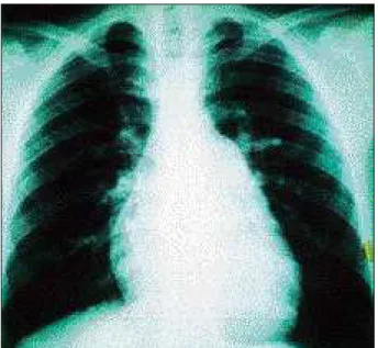
A chest X-ray in the anteroposterior projection sho-wed moderate enlargement of the cardiac silhouette due to the right atrium and bulging of the left pulmonary trunk segment (fig. 1).
The electrocardiography showed sinus rhythm, heart rate of 100bpm, QRS axis of +120°, morphology of right bundle-branch block in V1 with a QRS duration of 0.08s, and
a deep S wave in V5 and V6, suggesting right ventricular hypertrophy (figs. 2A and B).
The catheterization of the pulmonary artery through venous via revealed pulmonary artery pressure of 48mmHg (normal = 25mmHg) and pulmonary vascular resistance of 724 dines/s/cm5 (normal = 225 – 315 dines/s/cm5). The
pul-monary capillary pressure remained normal. The hemogram, assessment of the renal function, coagulation tests, and urinary sediment examination were all normal.
A few hours after hospitalization, the patient suddenly experienced shalon breathing, bradycardia, total atrioven-tricular block, and evolved with electromechanical disso-ciation. The patient did not respond to the resuscitation ma-neuvers and died.
Diagnostic hypotheses – 1) Heart failure due to rheu-matic valvular disease – mitral stenosis; 2) Primary pulmona-ry hypertension.
(Medical student Marcos Bianchini Cardoso)
Discussion
Clinical features – Pulmonary hypertension is defi-ned as a pressure in the pulmonary arterial system higher than 25mmHg at rest and higher than 30mmHg during exercise 1.
Primary pulmonary hypertension is a rare condition that is clinically defined after exclusion of all cardiac and pulmonary diseases that may cause elevation of the pres-sure in the pulmonary artery and elevation of vascular resis-tance. Its major symptoms are dyspnea, thoracic pain, and syncope, and, on examination, signs of different grades of cor pulmonale may be present. Among the causes of secon-dary pulmonary hypertension we emphasize pulmonary
Arq Bras Cardiol, volume 76 (nº 4), 333-6, 2001
Maria Aparecida Barne Teixeira, Sandrigo Mangini, Vera Demarchi Aiello
334 334
Clinicopathologic Session Arq Bras Cardiol
2001; 76: 233-6.
disorders (chronic obstructive pulmonary disease, conge-nital anomalies), heart diseases (congeconge-nital malformations, left heart failure), thromboembolic disorders, exogenous substances (cocaine, anorectics), portal hypertension, and HIV infection 1.
According to clinical data, we can exclude pulmonary hypertension secondary to mitral stenosis because of the
normal capillary pressure, allowing the suggestion of the diagnosis of primary pulmonary hypertension, because electrocardiographic data and the radiography did not dif-ferentiate them. The cardiac murmur of tricuspid insufficien-cy may be justified by a probable enlargement of the right ventricle.
(Dr. Maria Aparecida Barone Teixeira and medical student Sandrigo Mangini)
AA
BB
Fig. 2A and 2B –Electrocardiograms.
Fig. 4 – Section of the heart (4-chamber section) showing right ventricular dilation and hypertrophy. Ao- aorta.
Fig. 3 – External view of the anterior surface of the heart showing marked bulging of the right ventricular outflow tract.
Arq Bras Cardiol 2001; 76: 233-6.
Clinicopathologic Session
335 335
Fig. 5 –Microphotograph of the pulmonary parenchyma, where preacinar vessels with hypertrophy of the tunica media can be seen (arrows). Hematoxylin and eosin stain, original magnification 40 X.
Fig. 6 – Microphotograph of intraacinar pulmonary arterial branches sho-wing isolated hypertrophy of the tunica media. Müller stain, original magni-fication 400 X.
Autopsy
The heart weighed 600g and was enlarged due to dif-fuse thickening of the walls. The right ventricular outflow tract was very dilated (fig. 3). The ventricular cavities were slightly enlarged, and the atria were moderately dilated (fig. 4). No rheumatic valvular disease was found, and the tricus-pid valve did not show insufficiency when the reflux test was performed. The microscopic study of the myocardium revealed hypertrophy of the fibers.
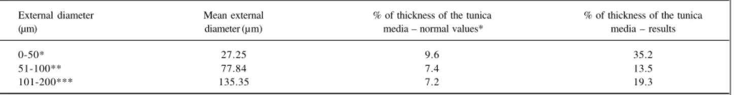
The lungs were slightly congested, and the major and peripheral branches of the pulmonary artery showed no thrombosis, embolism, or even lipoidosis (fig. 5). The liver had characteristics of passive congestion, which was confirmed on the microscopic examination. The histopathologic study of the lungs showed a normal parenchyma and arteries with no occlusive lesions of the tunica intima, on the hematoxylin and eosin stain (Fig. 6). The Müller stain for elastic fibers was performed, and the thicknesses of the medial layer of the arterioles with external diameters varying from 0 to 50µm, from 51 to 100µm, and from 101 to 200µm were measured and recorded as percentages. These measurements showed a 4-fold, 2-4-fold, and 3-fold increase, respectively, confirming the arteriopathy characterized by isolated medial hypertrophy. Measurements were performed with the aid of the Quantimet-500 Leica system of image analysis.
(Drs. Maria Aparecida Barone Teixeira and Vera Demarchi Aiello)
Comments
The anatomical and radiological findings are in accor-dance, and this was confirmed by the specimen that matched the cardiac silhouette on chest X-ray, mainly the prominence of the right ventricular outflow tract, ie, the pulmonary artery. Right ventricular hypertrophy, both grossly and microscopi-cally, is the reliable anatomical substrate of the electrocardio-graphic changes, ie, QRS axis shifted to the right, and mor-phology of right bundle-branch block. The electrocardiogram did not show hypertrophy of the left chambers, therefore, not matching the autopsy findings.
In regard to histopathology, no plexogenic pulmonary arteriopathy was observed in our case.
Even though in primary pulmonary hypertension no pathognomonic histologic lesion exists, most cases repor-ted show proliferative lesions of the tunica intima and dila-ted lesions, such as plexiform and angiomatoid lesions 2-4;
only few cases show isolated hypertrophy of the tunica me-dia, as our patient did.
The patient’s symptoms, dyspnea, cyanosis, and ede-ma, are the clinical equivalents of the development of arte-riopathy and consequent cor pulmonale. However, the lack of correlation between the severity of the histologic lesions (only hypertrophy of the tunica media, with no intimal oc-clusive lesions) and the patient’s symptoms is worthy of note 1,2. Even though a greater pulmonary resistance is
observed in patients with plexiform lesions, a lot of he-terogeneity exists between the hemodynamic measures and Table I – Mean percentage of medial thickness in regard to the external diameter of the peripheral pulmonary arteries.
External diameter Mean external % of thickness of the tunica % of thickness of the tunica
(µm) diameter (µm) media – normal values* media – results
0-50* 27.25 9.6 35.2
51-100** 77.84 7.4 13.5
101-200*** 135.35 7.2 19.3
336 336
Clinicopathologic Session Arq Bras Cardiol
2001; 76: 233-6.
1. Rubin LJ, et al. Primary pulmonary hypertension – ACCP Consensus Statement. Chest 1993; 104: 236-50.
2. Pietra GG, Edwards WD, Kay JM, et al. Histopathology of primary pul-monary hypertension – A qualitative and quantitative study of pulpul-monary blood vessels from 58 patients in the National Heart, Lung, and Blood Institute, Primary Pulmonary Hypertension Registry. Circulation 1989; 80: 1198-1206.
3. Heath D, Edwards JE. The pathology of hypertensive pulmonary vascular disease. A description of six grades of structural changes in the pulmonary
References
arteries with special reference to congenital cardiac septal defects. Circulation 1958; 18: 533-47.
4. Wagenvoort CA. Grading of pulmonary vascular lesions – a reappraisal. Histopathology 1981; 5: 595-8.
5. Haworth, SG, Hislop AA. Pulmonary vascular development: normal values of peripheral vascular structure. Am J Cardiol 1983; 52: 578-83.
6. Palevsky HI, Schloo BL, Pietra GG, et al. Primary pulmonary hypertension – vascular structure, morphometry, and responsiveness to vasodilator agents. Circulation 1989; 80: 1207-21.
the histologic pattern 2. In our case, we could not correlate
the grade of arteriopathy (isolated hypertrophy of the tunica media) with the pressure levels of the pulmonary artery. Pietra et al 2, in a study of 58 patients with primary
pul-monary hypertension, showed that those with plexiform arteriopathy had greater pressure levels in the pulmonary artery as compared with the ones with thrombotic arteriopa-thy or veno-occlusive disease; in their only case with isola-ted medial hypertrophy, no report exists about the pressure level in the pulmonary artery. Palevsky et al 6, studying 19
patients with primary pulmonary hypertension, 3 of whom with isolated medial hypertrophy, showed no significant difference between the pressure levels in the pulmonary artery and the grade of arteriopathy.
The sudden onset of clinical findings and death of our patient when compared with the exclusive
anatomicopatholo-gical findings of hypertrophy of the tunica media is a fact to be discussed, because the 3 patients of the study by Pale-vsky et al 6 with this type of arteriopathy did not evolve in the
same way. In the study by Pietra et al 2 no report exists about
the evolution of the patient with the same type of lesion. According to table I, a greater grade of medial hypertro-phy of the arterioles of smaller diameter (0-50µm) exists, and this could be related to the rapid evolution of the patient to death, because these arterioles account for the higher vascular resistance. However, we could not find any re-ference or report about this in the literature reviewed. It is worth noting that we could not find any other cause to ex-plain the decompensation of the hemodynamic status and consequent death of the patient.