6
Rev Dor. São Paulo, 2015 jan-mar;16(1):6-9
ABSTRACT
BACKGROUND AND OBJECTIVES: Physical therapy con-tributes to mitigate temporomandibular disorder symptoms because, in addition to stimulating proprioception and the production of joint synovial luid, it improves adhered muscle ibers elasticity. his study aimed at evaluating the stability of therapeutic results in a follow-up period post- physical therapy in temporomandibular disorder patients.
METHODS: Participated in the study 25 individuals of both genders, with temporomandibular disorder diagnosis. After a multimodal physical therapy program during 10 weeks, which included self-care guidance and home exercises, participants were re-evaluated by the Research Diagnostic Criteria for Tem-poromandibular Disorders and algometry. Results obtained im-mediately after treatment were compared to results of the evalu-ation carried out after two months of follow-up.
RESULTS: From 25 participants, with mean age of 31.6 years, 76% had no temporomandibular disorder diagnosis immedi-ately after treatment and from these, 68% have maintained this result in the two-month follow-up period. With regard to joint noises, 60% of participants have remained with no noises and pressure pain threshold values had no statistically signiicant dif-ferences between evaluations.
CONCLUSION: Multimodal physical therapy intervention, combined with self-care guidance and home exercises has pro-duced, in this study, positive and long-lasting efects on tem-poromandibular disorder symptoms, maintaining results for two months after treatment completion.
Keywords: Facial pain, Musculoskeletal manipulations, Physical ther-apy, Temporomandibular joint disorders, herapeutic approaches.
Stability of physical therapy effects on temporomandibular disorder*
Estabilidade dos efeitos da fisioterapia na disfunção temporomandibular
Muriel Priebe1, Ana Gabrieli Ferreira Antunes2, Eliane Castilhos Rodrigues Corrêa3
*Received from the Federal University of Santa Maria, Santa Maria, RS, Brazil.
1. Federal University of Santa Maria, Post-Graduation Program on Human Communication Disorders, Santa Maria, RS, Brazil.
2. Federal University of Santa Maria, Course of Physical herapy and Rehabilitation, Santa Maria, RS, Brazil.
3. Federal University of Santa Maria, Department of Physical herapy and Rehabilitation, Santa Maria, RS, Brazil.
Submitted in September 25, 2014.
Accepted for publication in February 10, 2015. Conlict of interests: none – Sponsoring sources: none.
Correspondence to: Muriel Priebe
Rua Tuiutí, 1155/301B – Centro 97015-190 Santa Maria, RS, Brasil. E-mail: [email protected]
© Sociedade Brasileira para o Estudo da Dor
RESUMO
JUSTIFICATIVA E OBJETIVOS: A isioterapia contribui para amenizar os sintomas da disfunção temporomandibu-lar, pois além de estimular a propriocepção e a produção do líquido sinovial na articulação, melhora a elasticidade das i-bras musculares aderidas. O objetivo deste estudo foi aval-iar a estabilidade dos resultados terapêuticos em um período de follow-up após a isioterapia, em pacientes com disfunção temporomandibular.
MÉTODOS: Vinte e cinco indivíduos, de ambos os gêneros, com diagnóstico de disfunção temporomandibular partici-param do estudo. Após um programa de isioterapia multi-modal, durante 10 semanas, que incluiu orientações de auto-cuidado e de exercícios domiciliares, foram reavaliados pelos Critérios Diagnósticos para Pesquisa em Desordens Temporo-mandibulares e por algometria. Os resultados obtidos logo após o tratamento foram comparados aos resultados da aval-iação realizada após 2 meses de follow-up.
RESULTADOS: Dos 25 participantes do estudo, com média de idade de 31,6 anos, 76% apresentaram ausência de diag-nóstico de disfunção temporomandibular logo após o trata-mento edestes, 68% mantiveram esse resultado no follow-up
de dois meses. Quanto aos ruídos articulares,60% dos partici-pantes permaneceram sem ruídos eos valores de limiar de dor à pressão não apresentaram diferença estatisticamente signii-cativa entre as avaliações.
CONCLUSÃO: A intervenção fisioterapêutica multimodal, combinada à orientação de autocuidado e exercícios domi-ciliares produziu, neste estudo, efeitos positivos e duradou-ros nos sintomas de disfunção temporomandibular manten-do os resultamanten-dos obtimanten-dos por manten-dois meses após o término manten-do tratamento.
Descritores: Condutas terapêuticas, Dor facial, Fisioterapia, Manipulações musculoesqueléticas, Transtornos da articula-ção temporomandibular.
INTRODUCTION
Temporomandibular disorder (TMD) is characterized by functional or pathological change affecting the temporo-mandibular joint (TMJ), which may impair masticatory muscles and stomatognathic system. The number of TMD cases is continually increasing, probably due to current psy-chological stress, based on current etiologic factors, physical and systemic conditions, as well as psychological factors are
ORIGINAL ARTICLE
7
Stability of physical therapy efects on temporomandibular disorder Rev Dor. São Paulo, 2015 jan-mar;16(1):6-9
responsible for TMD orientation and maintenance1,2.
TMD symptoms include persistent or recurrent pain in masticatory muscles or TMJ, jaw movement limitations or deviations, TMJ noises, joint discomfort and headache. In addition to impairing functionality, these factors consider-ably impact quality of life (QL) of these individuals3-5.
To minimize TMD symptoms, manual therapy aims, by means of manipulation techniques, mobilization and specific exercises, at stimulating proprioception, producing adhered fibers elasticity, stimulating synovial fluid and promoting pain relief. So, when associated to other physical therapy techniques, it is very useful for treatment outcomes6,7.
Guided home exercises and postural reeducation during daily life activities may help controlling TMD symptoms8.
If combined with therapeutic exercises and manual therapy, they may be effective to treat disc displacement patients and those refractory to conventional treatments9,10. It is also
worth stressing the effectiveness of the association of cervi-cal therapy and orofacial treatment in patients with cervico-genig headache associated to TMD signs and symptoms11.
Algometry is a widely used resource to study the effects of physical therapy on pain. It allows an objective measure-ment of pressure pain sensitivity, with high level of reliabil-ity, both to evaluate myofascial diseases and control group individuals12,13. In addition, algometry also contributes to
infer the importance of the evaluation of other regions of the body of TMD patients, in addition to the craniofacial region14.
More than evaluating results after physical therapy inter-vention, it is important to follow-up the maintenance of its therapeutic effects. So, this study aimed at evaluating thera-peutic effects stability of a multimodal physical therapy pro-gram, by comparing TMD signs and symptoms, in addition to evaluating pressure pain threshold observed immediately after treatment and after a 2-month follow-up period.
METHODS
Participants were referred by the Orofacial Motility Labora-tory where they were evaluated and treated with physical therapy for TMD. Participated in the study individuals of both genders, aged from 18 to 65 years and with diagnosis of TMD obtained by the Research Diagnostic Criteria for Temporomandibular Disorders (RDC/TMD). Patients were submitted to 10 weekly sessions lasting 45 minutes. All par-ticipants have signed the Free and Informed Consent Term. Data collected from participants’ evaluation cards were: results of RDC/TMD evaluation, presence of joint noises, pain in muscle and joint regions, as well as pressure pain threshold in 16 muscles bilaterally evaluated: anterior, me-dial and posterior temporal, superior, meme-dial and inferior masseter, sternocleidomastoid and superior trapezius. Pressure algometer – Force Dial Dynamometer® FDK/FDN
(Wagner Instruments) was used to evaluate pain thresh-old9,12. This tool may help diagnosis, in addition to checking
the efficacy of myofascial pain treatments13.
Physical therapy program included a combination of thera-peutic modalities, with focus on craniocervicomandibular system structures such as: therapeutic ultrasound, myofas-cial release, manual therapy, stretching and neuromuscular exercises, in addition to self-care and home exercises guid-ance15. This study has followed the application of modalities
of the same protocol.
This study has compared evaluation data immediately af-ter treatment to the evaluation of the same variables in the follow-up period, to observe the maintenance of treatment effects. With regard to RDC/TMD diagnoses, 96% of pa-tients have improved, not presenting any TMD diagnosis for the group without diagnosis, when evaluated immedi-ately after treatment, and just one out of 25 evaluated indi-viduals has maintained the same initial diagnosis.
Shapiro-Wilk test was used to check data normality. Because data were not parametric, Wilcoxon test was used to com-pare algometry results between both evaluations, consider-ing significant p<0.05 (95%).
This study was approved by the institution’s Ethics Com-mittee under protocol 0281.0.243.000-08.
RESULTS
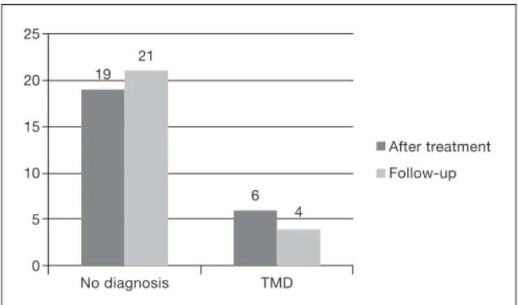
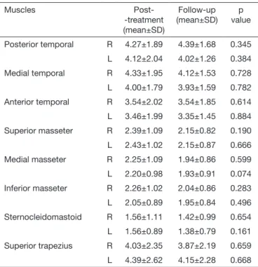
Participated in the study 25 individuals, being 20 females and 5 males, with mean age of 31.6±12.21 years and 19 (76%) with good post-treatment evolution, that is, they had no TMD immediately after treatment. From these, 17 (68%) have maintained such result in the 2-month follow-up peri-od, according to RDC/TMD evaluation. One patient (4%) has maintained the diagnosis of disc displacement with re-duction, 4 (16%) who had some group II diagnosis (disc displacement) have evolved to no diagnosis, and 8% of those who after treatment had no TMD started to present some group I disorders (muscle disorders) (Figure 1), thus total-ing 21 patients with no TMD diagnosis in this evaluation. There has been no significant difference in pressure pain threshold when comparing results immediately after treat-ment and two months after its completion (Table 1). Joint noises remained absent in 60% of patients. In 20% of
pa-Figure 1. Diagnosis of temporomandibular disorder after treatment
and during follow-up
No diagnosis TMD
19 21
6 4 25
20
15
10
5
0
8
Priebe M, Antunes AG and Corrêa EC Rev Dor. São Paulo, 2015 jan-mar;16(1):6-9
tients noises they have increased and in remaining patients they have decreased.
Table 1. Pressure pain threshold (kg/cm2) immediately after
treat-ment and follow-up
Muscles
Post--treatment (mean±SD)
Follow-up (mean±SD)
p value
Posterior temporal R 4.27±1.89 4.39±1.68 0.345 L 4.12±2.04 4.02±1.26 0.384 Medial temporal R 4.33±1.95 4.12±1.53 0.728 L 4.00±1.79 3.93±1.59 0.782 Anterior temporal R 3.54±2.02 3.54±1.85 0.614 L 3.46±1.99 3.35±1.45 0.884 Superior masseter R 2.39±1.09 2.15±0.82 0.190 L 2.43±1.02 2.15±0.87 0.666 Medial masseter R 2.25±1.09 1.94±0.86 0.599 L 2.20±0.98 1.93±0.91 0.074 Inferior masseter R 2.26±1.02 2.04±0.86 0.283 L 2.05±0.89 1.95±0.84 0.496 Sternocleidomastoid R 1.56±1.11 1.42±0.99 0.654 L 1.56±0.89 1.38±0.79 0.161 Superior trapezius R 4.03±2.35 3.87±2.19 0.659 L 4.39±2.62 4.15±2.28 0.668
R = righ; L = left.
With regard to pain at palpation, from 24 structures evalu-ated by RDC/TMD, 21 have maintained post-treatment results in the follow-up period, except to right inferior mas-seter, right lateral pterygoid and left temporal tendon.
DISCUSSION
In the evaluation of TMD diagnosis (RDC/TMD), 19 (76%) patients had no TMD diagnosis after treatment with a multimodal intervention protocol15 and, according
to results of this study, 17 (68%) patients had no TMD at 2-month follow-up evaluation. In 4 (16%) patients who still had the diagnosis after treatment, there has been disorder remission at 2-month follow-up evaluation. Through this intervention, authors have achieved a significant decrease in disorder severity, evaluated by the Temporomandibular index15, being that such therapeutic effects were maintained
after a 2-month follow-up period. Results in line with our study may be attributed, in addition to the multimodal ap-proach, to self-care and home exercises guidance also in-cluded in the intervention protocol, critical for the achieve-ment of short and long-term results.
Based on pressure pain threshold results in the follow-up period, there have been no statistically significant changes in any muscle evaluated as compared to values obtained im-mediately after treatment. A different study, although not presenting statistically significant differences, has shown immediate pressure pain threshold increase of masseter and
temporal muscles in patients with latent trigger points, im-mediately after treatment with manipulation or techniques for soft tissues, without observing the maintenance of such results13.
With regard to noises, 60% have remained without them, 20% have decreased them and remaining patients had joint noises after 2-month follow-up. Statistically significant re-sults show the positive effects of cervical manual therapy and of orofacial manual therapy associated to cervical man-ual therapy on TMD signs and cervical spine disorders11.
In agreement with our findings, authors have also observed that their results were maintained after a 6-month follow-up period.
Similar to our study, osteopathy and conventional treatment for TMD patients have shown that both were effective to relieve pain, increase maximum mouth opening amplitude and lateral head movement around its axis, being that such effects have remained after a 2-months follow-up period, considering the positive effects in the short and medium-term. Values of visual analog scale, mouth movement am-plitude and head rotation movements got worse for the os-teopathy group during the 2-months follow-up period, as compared to re-evaluation immediately after treatment16.
A different study with 70 volunteers, has compared one group receiving just self-care guidance with TMD symp-toms improvement in 57% of patients, to a group combin-ing physical therapy (home exercises) and self-care guidance, with 77% improvement. The group practicing physical ther-apy and regular self-care has obtained masticatory muscles relaxation, pain relief and improvement in depression symp-toms and sleep quality. Authors have indicated that self-care guidance, explanation of risk factors and training in home exercises provide physical and psychological gains, improv-ing symptoms and patients’ anxiety17.
Some authors have investigated the effects of multimodal interventions on TMD signs and symptoms, which have been maintained even after physical therapy treatment completion, especially when this included passive and active mandibular and cervical exercises, relaxation techniques, postural correction and directed exercises4,11,18. With this,
it is shown the importance of focusing on the craniocervi-calmandibular system for the treatment of TMD patients, involving the spine and cervical muscles, since this system is one functional unit. In addition, this approach introduced in our study, may be an important contributing factor to maintain therapeutic results.
Self-care exercises have proven benefits19 and it is considered
that, together with patients’ education, they are relevant factors to maintain treatment and therapeutic continuity20.
Also, they are not expensive and perpetuate physical therapy effects, the effect of which has durability, but its decrease is observed after two months15,16.
9
Stability of physical therapy efects on temporomandibular disorder Rev Dor. São Paulo, 2015 jan-mar;16(1):6-9
limited our discussion. It is suggested that, to confirm the stability of therapeutic results, longer follow-up periods, above 6 months, should be evaluated.
CONCLUSION
Most patients have maintained the same results with regard to TMD diagnosis and presence of joint noises after two months of treatment. Treatment effects on pain have also remained, since there has been no difference in pressure pain threshold values evaluated immediately after and two months after treatment completion. So, physical therapy intervention was effective and with long-lasting effects for these patients. This result may be attributed to improved muscle balance and decreased joint overload obtained with the treatment, including the whole craniocervicomandibu-lar system, as well as self-care and home exercises guidance, critical for the achievement and maintenance of therapeutic results.
REFERENCES
1. Tvrdy P. Methods of imaging in the diagnosis of temporomandibular joint disorders. Biomed Pap Med Fac Univ Palacky Olomouc Czech Repub. 2007;151(1):133-6. 2. Resende CM, Alves AC, Coelho LT, Alchieri JC, Roncalli AG, Barbosa GA. Quality of
life and general health in patients with temporomandibular disorders. Braz Oral Res. 2013;27(2):116-21.
3. Dworkin SF, Huggins K, Wilson L, Mancl L, Turner J, Massoth D, et al. A randomi-zed clinical trial using research diagnostic criteria for temporomandibular disorders: axis II to target clinic cases for a tailored self-care TMD program. J Orofac Pain. 2002;16(1):48-63.
4. La Touche R, Fernández-de-las-Peñas C, Fernández-Carnero J, Escalante K, Angulo--Días-Parreño S, Paris-Alemany A, et al. he efects of manual therapy and exercise directed at the cervical spine on pain sensitivity in patients with myofascial temporo-mandibular disorders. J Oral Rehabil. 2009;36(9):644-52.
5. Strini PJ, Souza GC, Bernardino Junior R, Fernandes Neto AJ. Alterações biomecâ-nicas em pacientes portadores de disfunção temporomandibular antes e após o uso de
dispositivos oclusais. Rev Odonto. 2009;17(33):42-7.
6. Grossi DB, Chaves TC. Physiotherapeutic treatment for temporomandibular disor-ders (TMD). Braz J Oral Sci. 2004;3(10):492-7.
7. Kalamir A, Pollard H, Vitello AL, Bonello R. Manual therapy for temporomandibular disorders: a review of literature. J Bodyw Mov her. 2007;11(1):84-90.
8. Matta MA, Honorato DC. Uma abordagem isioterapêutica nas desordens temporo-mandibulares: estudo restrospectivo. Rev Fisioter Univ São Paulo. 2003;10(2):77-83. 9. Maluf AS, Moreno BG, Alfredo PP, Marques A, Rodrigues G. Exercícios terapêuti-cos nas desordens temporomandibulares: uma revisão de literatura. Rev Fisioter Pesq. 2008;15(4):408-15.
10. Yoda T, Sakamoto I, Imai H, Honma Y, Shinjo Y, Takano A, et al. A randomized controlled trial of therapeutic exercice for clickink due to disc anterior displacement with reduction in temporomandibular joint. Cranio. 2003;21(1):10-6.
11. von Pierkartz H, Hall T. Orofacial manual therapy improves cervical movement im-pairment associated with headache and features of temporomandibular dysfunction: a randomized controlled trial. Man her. 2013;18(4):345-50.
12. Vedolin GM, Lobato VV, Conti PC, Lauris JR. he impact of stress and anxie-ty on the pressure pain threshold of myofascial pain patients. J Oral Rehabil. 2009;36(5):313-21.
13. Oliveira-Campelo NM, Rubens-Rebelatto J, Martí N-Vallejo FJ, Albuquerque-Sedí NF, Fernández-de-Las-Peñas C. he immediate efects of atlanto-occipital joint ma-nipulation and suboccipital muscle inhibition technique on active mouth opening and pressure pain sensitivity over latent myofascial trigger points in the masticatory muscles. J Orthop Sports Physher. 2010;40(5):310-7.
14. Silveira A, Armijo-Olivo S, Gadotti IC, Magee D. Masticatory and cervical muscle tenderness and pain sensitivity in a remote area in subjects with a temporomandibular disorder and neck disability. J Oral Facial Pain Headache. 2014;28(2):138-46. 15. Freire AB, De Nardi AT, Bouleur J, Chiodelli L, Pasinato F, Corrêa EC. Abordagem
isioterapêutica multimodal: efeitos sobre o diagnóstico e a gravidade da disfunção temporomandibular. Fisioter Mov. 2014;27(2):219-27.
16. Cuccia AM, Caradonna C, Annunziata V, Caradonna D. Osteopathic manual thera-py versus conventional conservative therathera-py in the treatment of temporomandibular disorders: a randomized controlled trial. J Bodyw Mov her. 2010;14(2):179-84. 17. Michellotti A, Steenks MH, Farella M, Parisini F, Cimino R, Martina R. he
addi-tional value of a home physical therapy regimen versus patient education only for the treatment of myofascial pain of the jaw muscles: short-term results of a randomized clinical trial. J Orofac Pain. 2004;18(2):114-25.
18. Nicolakis P, Erdogmus B, Kopf A, Nicolakis M, Piehslinger E, Fialka-Moser V. Efec-tiveness of exercise therapy in patients with myofascial pain dysfunction syndrome. J Oral Rehabil. 2002;29(4):362-8.
19. Michellott A, de Wijer A, Steenks M, Farella M. Home-exercise regimes for the management of non-speciic temporomandibular disorders. J Oral Rehabil. 2005;32(11):779-85.