(1) Universidade Federal de Pernambuco, UFPE, Recife, Pernambuco, Brasil. (2) Real Hospital Português, RHP, Recife,
Pernambuco, Brasil. (3) Departamento de Anatomia da
Universidade Federal de Pernambuco, UFPE, Recife, Pernambuco, Brasil. (4) Departamento de Medicina Clínica da
Universidade Federal de Pernambuco, UFPE, Recife, Pernambuco, Brasil. (5) Ambulatório de Neurologia do Hospital
das Clínicas da Universidade Federal de Pernambuco, HC/UFPE, Recife, Pernambuco, Brasil.
Source: Bolsa REUNI
Conlict of interest: non-existent
Contributions of the electromyography of needle
for the study of swallowing in humans
Contribuições da eletromiografia de agulha
para o estudo da deglutição em seres humanos
Luciana Rodrigues Belo(1) Sílvio Vasconcelos(2) Maria das Graças Wanderley de Sales Coriolano(3) Nadja Asano(4) Amdore Guesel Asano(5) Otávio Gomes Lins(1)
Received on: March 12, 2015 Accepted on: July 11, 2016
Mailing address:
Luciana Rodrigues Belo
Rua Abel de Sá Bezerra Cavalcanti, 161, apt 601, Casa Amarela
Recife – PE - Brasil CEP: 52051-270
E-mail: [email protected]
ABSTRACT
Intramuscular EMG is performed by the doctor, from the ixing needle electrodes or thin wire for the study of isolated muscles or deep muscles. This study aims to identify and describe the contributions of intra -muscular electromyography, for the evaluation of swallowing in humans. The search, carried out between April and March 2015, in the databases PubMed, BIREME, AND BANK OF THESES OF CAPES, resulted in 21 references, of which only seven met our inclusion criteria. Selected articles make important contribu-tions to the understanding of the electrophysiological behavior and electrophysiological during swallowing and it is believed that the lack of studies using this tool in humans should be the annoyance and risks caused by the introduction of the needle into the belly muscle and maybe the introduction of a thin wire (ine wire or wire cooper) is more interesting for the aid diagnosis of neuromuscular disorders and nerve that compromise swallowing, the ability to dramatically reduce the annoyance caused by the needle. Keywords: Electromyography; Deglutition; Muscles
RESUMO
A Eletromiograia intramuscular é realizada pelo médico, a partir da ixação de eletrodos de agulha ou de io ino para o estudo de músculos isolados ou músculos profundos. Esse estudo tem como objetivo identiicar e descrever as contribuições da eletromiograia intramuscular, para a avaliação da deglutição em seres humanos. A busca, realizada no período entre abril e março de 2015, nos bancos de dados da PUBMED, BIREME E BANCO DE TESES DA CAPES,resultaram em 21 referências, das quais, apenas sete se enquadraram nos critérios de inclusão. Os artigos selecionados trazem contribuições importantes para o entendimento do comportamento eletroisiológico e eletroisiopatológico durante a deglutição e acredita-se que a escassez de estudos utilizando essa ferramenta em seres humanos deva-se ao incô-modo e riscos causados pela introdução da agulha no ventre muscular e talvez a introdução de um io ino (ine wire ou cooper wire), seja mais interesante para o auxílio diagnóstico de denervações e trans-tornos neuromusculares que comprometam a deglutição, pela possibilidade de reduzir drasticamente o incômodo causado pela agulha.
Electromyography needle and swallowing | 1239
INTRODUCTION
The assessment of swallowing includes the use of
speciic clinical protocols and complementary tests,
which aim to analyze the integrity of the structures involved in this mechanism. In addition to endoscopy
and videoluoroscopy of swallowing, the literature
indicates that Surface Electromyography (sEMG) and Intramuscular Electromyography (SMEI) as important tools in electrophysiological evaluation of this function1-6.
Electromyography can be used for the diagnosis of
neuromuscular diseases or trauma, and the kinesio -logical study of muscles in certain motor activities. Portney and Roy (2004)7 differentiate clinical
electromy-ography of kinesiological electromyelectromy-ography. In clinical EMG, performed by physicians, nerve conduction velocity tests are performed. While in kinesiological EMG, it is possible to study muscle function while performing speciic tasks or therapeutic regimens8.
The kinesiological EMG can be performed from surface electrodes, needle electrodes, the ine wire type or hook wire electrodes. SEMG has been widely
studied and used by many researchers and health
professionals, and corresponds to a non-invasive and
without contraindications that aims to capture the
electrical activity of muscles or muscle groups, from ixing surface electrodes on the skin parallel to the muscle ibers2,3,9-11.
However, when studying the muscles of the face and neck by sEMG, there is a greater probability of crosstalk funding (activity of neighboring muscles), making the isolated assessment of certain muscles, in addition to the high variability within and between subjects, limiting analysis of electrophysiological indings12,13. The SMEI
has a great advantage over the EMGs, by minimizing
the capture activity from neighboring muscles and
signal interference, secondary to the displacement
between the electrode and the skin, in order, that the ixing is directly inside the muscle under study14.
This technique is performed by physicians from
the introduction of needle electrodes or ine Wire type
set in the muscle membrane14,15. Thus, the aim of this
review was to describe the contributions of SMEI, for
the evaluation of swallowing in humans.
METHODS
The research was conducted by three researchers. Two researchers (LB and MGWS) sought the data independently and blindly initially. The third researcher
(OGL), established as a proofreader. It has been
consulted in cases of doubt to establish agreement between ideas. Articles were included published in the
last 15 years (1999-2013), with human beings of both sexes, whose sample was made up of children, youth, adults, healthy elderly subjects (without neurological sequelae). Articles were excluded if they did not use
the data obtained from needle electromyography in
swallowing evaluation, review articles and studies with
animal models.
The search was carried out between April and March 2015. The descriptors were chosen according to the lists DeCS and MeSH. The list of DeCS descriptors were: Swallowing and electromyography. The MeSH
list descriptors were: déglutition, Swallowing and
electromyography.
Keywords were also used to extend the search
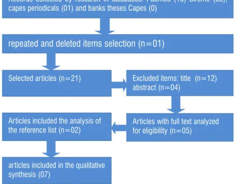
(Figure 1). References of selected articles were analyzed
for other studies, which could have been omitted from the electronic search. Banks portal Bireme data were used (Medline, Lilacs, IBECS, Scielo, Cochrane Library, and other banks that portal), the Pubmed, and bank
Capes theses. The search strategy applied followed Castro recommendations et al.16, Dickersin et al.17 and the Cochrane Collaboration.
#1.
(“fine wire” or “hook wire” or “laryngeal EMG” or “needle EMG”)#2
.
(“EMG” or “electromyography”)#3.
(“deglutition” or “swallowing” or “swallow”)Intersecction: #1 and #2 and #3
There were found 18 articles in PUBMED portal;
three articles in BIREME portal and work on PERIODIC
CAPES and no article in CAPES OF THESES BANK potentially relevant and were stored for analysis. Of the three selected items from PORTAL BIREME (one was repeated in the results of the survey in Pubmed
portal), getting 18 articles of PUBMED, two articles of BIREME portal, and another article of Periodicos. Capes for analysis. Among the 21 articles analyzed, 16 were excluded for not meeting the inclusion criteria. After analyzing the results of the ive articles that were selected, two were included. Finalizing the collection
with the addition of seven articles: Vitti & Basmajian (1977)18; Ertekin et al. (2000)19; Ertekin et al. (2001)20;
Baudon et al. (2002)21; Renault et al. (2011)22; Alkan et al. (2012)23; Inokuchi et al. (2014)24 (Figure 2, Figure 3).
REVIEW IN THE LITERATURE
Analyzing the articles and the instrumentation used
it was observed that only the items Ertekin (Ertekin et
al. (2000)19, and Ertekin et al. (2001)20 and Alkan et al. (2012)23, used a control group of subjects considered
normal, while Baudon et al. (2002)21 and Renault et al. (2011)22 used recommended normal data for previous studies.
Only items Ertekin (Ertekin et al, 2000)19 and Ertekin
et al, 2001)20 and Alkan et al. (2012)23 described the Records collected by research in databases: Pubmed (18) Bireme (03),
capes periodicals (01) and banks theses Capes (0)
repeated and deleted items selection (n=01)
Excluded items: title (n=12) abstract (n=04)
Selected articles (n=21)
Articles included the analysis of the reference list (n=02)
Articles with full text analyzed for eligibility (n=05)
articles included in the qualitative synthesis (07)
Figure 2. Search and selection of articles: Pubmed, Bireme and Capes theses database.
1B
Figure 3. Represents the number of articles found in each block, and the number of items that were analyzed after the crossroads of
As this review aims to describe the contributions of
EMG i for evaluation of swallowing, the line of reasoning
will focus on the results found in this function.
The data were analyzed according to the analog
method of classiication developed by Basmajian (1974), as follows: “O”, no activity; ‘’ ± “insigniicant
activity;” + “Slightly on,” ++ “, moderate activity,” +++ “very marked activity, and” ++++ “to marked activity.
Vitti & Basmagian (1977)18 studied the electrical activity in the following tests: saliva swallowing and swallowing a volume of water not described in the
article. As a result, there was found that during saliva
swallowing is the full participation of suprahyoid
muscles, conirming the indings of Cunningham and
Basmajian (1969)25, Lehr et al. (1971)26, and Hrycyshyn and Basmajian (1972)27. Marked activity occurred in the anterior belly of the digastric and the mylohyoid
muscle, followed by moderate activity of the geniohyoid
muscle.
The medial pterygoid was activated in 82% of the
sample, with wide participation. In other muscles, the
activity ranged from negligible to light. In the temporal
muscle, the activity was present in 23-36% while the
masseter in 55% of cases. The researchers believe that
the activity observed in the temporal muscle, masseter
and medial pterygoid is probably the result of contact of
the posterior teeth when swallowing occurs, as shown in cineluoroscopic studies28.
This study analyzed the swallowing of water in three
stages: 1-Sipping, 2- retaining water for 5 seconds and 3- swallowing. In the irst phase, there is no activity in the temporal muscle, masseter and medial pterygoid. Potential insigniicant was observed in the anterior belly of the digastric, mylohyoid and genius-hyoid, ranging
between 46 and 64% of cases. This activity is probably
a result of mild depression jaw, necessary to keep the
glass of water between his lips.
During the second stage, holding the water in the oral cavity, all muscles remained electrophysiological silence. In the last phase, swallowing, it found the total activity in the anterior belly of the digastric, mylohyoid and genius-hyoid. Insigniicant activities were observed in some cases, the anterior and posterior portions of
the temporal and masseter muscles while in the medial
temporal potential was inactive. On the other hand, the
pterygoid muscle showed moderate activity in 86% of cases. Vitti & Basmagian (1977)18 believe that this
activity may occur to keep the posterior tooth contact.
It was mentioned in the methods as given in the
command swallowing events. Only the results, the ilters that have been conigured on the EMG machine,
but did not report the sampling frequency, while Inokuchi et al. (2014)24 reported only the sampling frequency (Table 1).
The lack of agreement among researchers, as to the most appropriate technical speciication, hinders
the replication of their studies and can lead to different results. Only two (Vitti & Basmajian (1977)18 and
Inokuchi et al. (2014)24 used electrodes of the ine wire
type or hook wire, although the most suitable in kinesi -ology studies because they cause less interference and discomfort to patients (Table 1).
Electromyography is used to assess the scope of diseases or traumas as well as a tool for studying
kinesiological neuromuscular function. The kinesio
-logical EMG aims to examine the function and / or muscle performance, while performing speciic tasks purposeful, or therapeutic regimens. For this, the therapist examines patterns of muscle response onset and cessation of activity, muscle fatigue and
muscle response level in relation to the effort; type of muscle contraction and position. This type of test may be performed by surface electrodes for the study of
supericial muscles and needle or thin wire electrodes (ine wire) for detecting the electrical activity of speciic
muscles and / or deep 7.
Few items with humans who have studied the electromyographic activity from needle electrodes or
ine wire type during swallowing were found. In the search performed in the databases referred to above,
seven articles were selected to describe the contri-butions of intramuscular electromyography for the evaluation of swallowing in humans. Tables 1 and 2 are important methodological information to those provisions.
EMGi’s contributions to the study of swallowing in
humans
Vitti & Basmagian (1977)
18Vitti & Basmagian (1977)18 aimed to analyze compre-hensively intramuscular myoelectric activity of muscles:
temporal, masseter, medial pterygoid, anterior belly of the digastric, mylohyoid and geniohyoideous in “normal” individuals with bipolar type electrodes “ine wire” during jaw movements, chewing and swallowing. Evaluated 29 participants considered normal, with all
establishment of a limit of dysphagia. What proves to be possible to identify changes in electromyographic activity during swallowing of patients with ALS and thus promote the therapeutic planning and systematic monitoring of the treatment and/or management in these cases.
Ertekin et al. (2001)
20The study by Ertekin et al. (2001)20 aimed to show
the existence and frequency of subclinical electrophysi -ological abnormalities in oropharyngeal swallowing
in myotonic dystrophy, as well as to clarify the patho -physiological mechanisms in this disorder through the
following indings: displacement of the larynx, from a
piezoelectric sensor; electromyographic activity surface of suprahyoid muscles (EMG-MSH) and intramuscular electromyographic activity of the cricopharyngeal muscle (SMEI through needle electrodes) during swallowing of different water volumes (Table 1 and 2).
The electrophysiological behavior of the cricopha-ryngeal muscle was considered normal in eight patients
with myotonic dystrophy. Of these eight, two were diagnosed with dysphagia, and six were considered
non-dysphagia. The tonic activity of the
cricopha-ryngeal muscle rest was off by 400 to 500 msec, while performing the raising and lowering of the larynx. In the ive remaining patients, all with dysphagia, EMG
cricopharyngeal muscle was considered pathological and showed clinical signs and symptoms suggestive of CNS involvement.
In this study, the examination of the cricopharyngeal
muscle was considered normal in most patients with myotonic dystrophy with or without dysphagia. Although
for about 40%, all with dysphagia, the electrophysi
-ological examination of the cricopharyngeal muscle showed several abnormalities. During swallowing, early iring activity and silence recovery appeared in EMG
and tended to be high amplitude and long duration.
In three patients with congenital myotonia, EMG crico
-pharyngeal muscle was normal, both at rest and during swallowing. Ertekin et al. (2001) 20 concluded that the involvement of the CNS may contribute to the delay in
the swallowing relex shooting and abnormal EMG EES
can identify dysphagia in this disease.
Ertekin et al. (2001)20 bring as contributions, physiopathological features of swallowing in myotonic
dystrophy, with the analysis of electrophysiological
parameters obtained from the semg of the supra-hyoid
muscles, EMHi of the cricopharyngeal muscle and the establishment of a limit of dysphagia. As Ertekin et al. authors mention that the participants keep the glass
of water between his lips to sip; however, that time is
carried over? What volume of water administered? The
authors also did not mention the sample ilters and
frequency programmed into the device.
The study by Vitti and Basmagian (1977)18 back as a contribution demonstrating the EMG activation
of muscles: the anterior digastric Venter, milohyo
-lideous, geniohyoideous (suprahyoid muscles), medial pterygoid, masseter and temporal (muscles lifts the
jaw) in different times of swallowing in humans.
Ertekin et al. (2000)
19Ertekin et al. (2000)19 investigated the pathophysi-ology of dysphagia in amyotrophic lateral sclerosis (ALS) by clinical and electrophysiological
measure-ments such as: larynx displacement from a piezo -electric sensor; electromyographic activity surface of suprahyoid muscles (EMG-MSH) and intramuscular electromyographic activity of the cricopharyngeal muscle (SMEI through needle electrodes) during swallowing of different water volumes (Table 1 and 2).
As a result, they found that the activity of suprahyoid muscles in the dry swallowing (saliva) was signiicantly longer, while the larynx repositioning time remained
within the normal range in patients with ALS.
The cricopharyngeal muscle of patients with ALS showed severe abnormalities during the voluntary
swallowing, which, according to the authors, may be
due to the delay in the opening of the upper esoph-ageal sphincter and/or by premature closure of this
sphincter; the total duration of opening was short, with unexpected bursts of motor units and the lack between the muscles lifts of the larynx and the opening of the upper esophageal sphincter was signiicant.
Ertekin et al. (2000)19 concluded that there are two pathophysiological mechanisms in the dysphagia of
it: the irst concerns the delay or absence of triggering
the reaction in pharyngeal voluntary swallowing;
and preservation of activity in the relex automatic
swallowing; the second is related to the hypertonicity
and hyperrelexivity of the cricopharyngeal muscle.
The authors believe that this is due to the progressive
degeneration of the ibers corticobulbars.
Ertekin et al. (2000)19 brings as a contribution to a better understanding of the pathophysiologic charac-teristics of swallowing in Amyotrophic Lateral Sclerosis with the analysis of electrophysiological parameters
obtained from the semg of the supra-hyoid muscles,
(2000)19, allows us to believe that the EMG can identify changes in muscle activity during swallowing of these patients and thus promote the therapeutic planning and systematic monitoring of the treatment and/or management by a speech.
Baudon et al. (2002)
21The aim of the survey of Baudon et al. (2002)21 was to evaluate the motor dysfunction by manometry and
electromyography of intramuscular needle, the genio
-glossus muscle and tireohioideous, during sucking and swallowing of newborn infants, with a result of
Pierre Robin (SPR). These patients had upper airway obstruction and dysphagia disease (Table 1 and 2).
The EMG showed lack of sucking and swallowing in 24 of 28 patients. The riots were mild in six, moderate in six and severe in 12 patients. All patients demonstrated manometric disorders such as: relaxation is incomplete
or assichronous of upper esophageal sphincter (15).
Activity of the EES with wave multi-peaks (17), waves of amplitudes too high (14) and asynchronous relaxation
of the lower esophageal sphincter higher (19). The frequency of disturbances and mean blood pressures at rest of both: upper esophageal sphincter and less
were signiicantly higher than in patients with gastro
-esophageal relux disease.
These results were obtained from the comparison
of the data found in this study, with recommended
normal data in previous studies by Renault & Raimbault (1992)29. These researchers, in the study entitled
“Electromyographie faciale, linguale pharyngée et
chez l’enfant: une méthode d’étude des troubles de
succion-déglutition et de leur physiopathologie” found
the electromyographic activity from needle electrodes
of the genioglossus muscles and tireohyoideous, to
analyze the coordination between the suction and swallowing newborns considered normal.
They believed that the activity of the genioglossus is related to the suction process and the activity of the muscle tireohyodeous with swallowing. Baudon et al. (2002) concluded that the 21 manometry and EMG were able to identify malfunctions in the motor
organi-zation of the tongue, pharynx, and esophagus; even in
the absence of clinical disorders in swallowing.
The identiication from the SMEI, incoordination
between the suction and swallowing found in the search Baudon et al. (2002) 21 has important contributions to the objective record of evaluation and management of
patients with Pierre Robin Sequence. A lack of coordi
-nation between these two functions enhances the risk
of penetration and aspiration of salivary content and / or feed.
Renault (2011)
22Renault (2011)22 evaluated the relevance of EMG combined techniques in the evaluation and management of children with Pierre Robin sequence (SPR) from the needle electromyography of facial
muscles, tongue, pharynx and larynx (Table 1 and 2);
They followed 81 infants among them 57 with Pierre Robin sequence alone (SPRI) and 24 with Pierre Robin sequence associated (SPRA) (Table 2).
As a result, they found electrophysiological signs of
neural impairment in facial muscles in 17 of 24 patients with Pierre Robin sequence associated. The soft palate muscles showed traces of low amplitude in 41.4% of patients who required two surgical steps to the soft palate repair; 18.5% of those who needed only one surgical step. In electrophysiological studies during
bottle-feeding, patients with moderate or severe abnor -mality of oral coordination / pharyngeal required more
prolonged enteral feeding, patients with mild abnor -malities or regular coordination.
Neurogenic electromyographic signals were
detected in at least one of the facial muscles or oral, for 17 of 24 patients with Spra, and one of 57 patients
with SPRI (p <0.0001). Neurogenic signs were more
frequently detected in the face and the soft palate,
which in the language. Electromyographic signs of
denervation of the facial muscles have been identiied
in the muscles of the tongue and soft palate in four patients.
Abnormal coordination patterns between sucking and swallowing were classiied into three stages of severity, from, electromyography: (a) light: this suction, however, alternating between sucking and
swallowing irregular; (B) Moderate: Suction present with synchronous or random pharyngeal phase; or (c) severe: the language did not perform the rhythmic
sucking activity, and inactive or tonic pharyngeal phase. Renault et al. (2011), as well as Baudon et al. (2002)21 compared their results with proposed normal data in
previous studies, however, Renault et al. (2011)22, cites Renault (2001)30, as being the source of such data, and
in fact, this article describes the facial electromyog -raphy in newborns and young infants with congenital
facial weakness, which leads us to believe that failed to referencing these normal indings and it is likely, given the similarity in the studies and the authors, the normal
obtained in Renault & Raimbault29, as well as the study of Baudon et al. (2002)21
Renault et al. (2011) 22, bring relevant data to corroborate the results found by Baudon et al. (2002) 21 assisting the analysis and interpretation of electrophysi-ological data of the muscles involved in swallowing.
Alkan et al. (2012)
23The research of Alkan et al. (2012)23 presented to analyze the relationship between the severity level of
the gastroesophageal relux, patterns of contraction
and behavior of the cricopharyngeal muscle through the analysis of measures of electrophysiological
crico-pharyngeal muscle, from the intramuscular electromy
-ography with electrode of needle. For this purpose,
there were compared 24 patients with
gastroesoph-ageal relux disease, with 21 healthy volunteers. The severity of relux was diagnosed through endoscopy upper GI series,24 and the application of the protocol Demeester score (Table 1 and 2).
The gastroesophageal relux disease was mild in
15 patients and moderate to severe in nine patients. The record of the triggering of motor units was normal in both groups during the period
pre-swallow/post-swallowing. The research kinesiological revealed that
the number of patients who showed no shots of electro-myographic activity during the period of pre-swallowing
had a positive correlation with the severity of relux
and the quantity of liquid swallowed. Shots passed
were observed in patients with relux and in the control
group. The duration of the pre-swallowing and the shots passed (rebound burst) was similar in all groups.
The duration of swallowing, corresponding to the period of silence, electrophysiological was shorter in patients with gastroesophageal relux disease lightweight, when compared to the group of healthy subjects, and the group with moderate dysphagia and
severe. The swallowing in parts (piece meal deglutition) the volume of 10 ml was high in patients with
gastro-esophageal relux disease moderate to severe. Also,
found a positive correlation of the number of swallowing
with the severity of relux.
Alkan et al. (2012)23 concluded that
electromyog-raphy of needle of the upper esophageal sphincter, was normal in patients with gastroesophageal relux disease. The ratings kinesiological, showed an
increase in piecemeal deglutition and in the number of
swallowing, and correlated, thus, positively correlated with the severity of relux. And bring as contributions,
important results on the electrophysiological behavior
of the cricopharyngeal muscle during your rest and in
swallowing in patients with gastroesophageal relux
disease.
Inokuchi et al. (2014)
24In this study, the researchers analyzed the electro
-myographic signal, picked up by ine-wire electrodes, during swallowing, to determine the typical sequence
of activation of muscles involved in swallowing in
normal subjects (no dysphagia). From their results,
they determined the time of activation of the muscles
(of the geniohyoid, anterior belly of the digastric, ester -nohyoideous and masseter) and compared these times between the different consistencies (Tables 1 and 2).
Considering the “onset” of of the geniohyoid muscle as reference (irst moment of swallowing - 0s), these researchers found that the intake of thickened liquids, the masseter, of the geniohyoid and anterior digastric belly, they were activated always simultaneously; while
esternohyoideous muscle was activated later. With
solid foods, the contraction of the masseter precedes
the time of activation of of the geniohyoid muscle
and anterior digastric belly, while the activation of the
muscle was later esternohyoideous and lagging behind swallowing liquids.
The role of the masseter differs between solid and
liquid food so that the change in its delay is expected.
The timing contraction of of the geniohyoid muscles and anterior digastric belly was consistent with its important role in laryngeal elevation. The contraction of the muscle esternohyoideous was always after the other muscles studied both in swallowing liquids and solids.
This sequence conirms previous studies, which described the events related to swallowing. Initially,
there is the closing of the mouth and jaw stabili-zation by the action of the levator muscles of the jaw (masseter among them). Then the suprahyoid muscles (especially genius-hyoid and digastric anterior belly)
move the hyoid bone, upper and above. This action
facilitates the clearance of the mouth by the action of the tongue and opening of the upper esophageal
sphincter. Finally, infrahyoid muscles, represented by esternohyoideous, pull the hyoid bone inferiorly and
subsequently providing their return to resting position characterizing the end of swallowing 31,32.
of the central pattern generator to the pharyngeal swallowing (Table 2).
In this article, the authors did not mention what was offered in liquid consistency thickened, and also not mentioned the band pass ilter set in electromyog -raphy equipment. The absence of such information can hamper the realization of this protocol in other studies; besides the fact that different methodologies may lead to different results.
However, it can be considered that the study conirms the indings of different researchers and
brings as contributions activation sequence of muscles involved in swallowing. This information can come to
help the electrophysiological evaluations, with incoordi
-nation identiication muscle activation that would result in deicits in oral motor control and risk of penetration and/or aspiration of food content, requiring further
research to validate the results.
In the past, the needle electromyography was quite
used to further the understanding of the electrophysi-ological behavior of muscles involved in swallowing
and to be quite uncomfortable, most studies have been
conducted in animal models.
These studies are not recent; the irst survey was
conducted by Doty and Bosman in 195633. In this study, the electromyographic during swallowing was observed
in 22 muscles of the mouth area, pharynx and larynx of monkeys, dogs and cats through electromyography
needle (cooper wire type electrodes).
These scholars have identiied a complex of muscles that considered key to the mechanism of swallowing, they are: higher pharyngeal constrictor, palatopha
-ryngeal, palatoglossus, posterior intrinsic muscles of the tongue, styloglossus, stylohyoid, genius-hyoid and mylohyoid. Surprisingly, different from that found
in the study of Vitti and Basmajian (1977) 18, Doty and Bosman (1956) 33 found electrophysiological silence
of digastric and sternohyoideous muscles, besides
the previous sternothyreoid and intrinsic muscles of the tongue of cats and dogs during swallowing.
Thexton (2007)34 recreated the study Doty and Bosman (1956)33 evaluating swallowing eight young pigs from the radiographic simultaneous study the
electromyo-graphic examination, carried out with the hook type electrodes (ine wire) 16 muscles and get results that approached the description by Doty and Bosma, identifying a main complex of muscles involved in this mechanism. However, the mylohyoid muscle has not
been activated early on in relation to the other and of the geniohyoid muscle there was not part of this
complex. Some classically considered inactive muscles showed active in the pharyngeal phase of swallowing,
including the digastric.
There is disagreement on the results of these studies
which may be related to methodological differences, such as equipment speciications, different contents and volumes offered, various samples (different animal species with a variation in the thickness and length
of the studied muscles) and including the limitations
imposed by own method of analysis. However, it is
notorious and devoted the importance of mylohyoid
muscles, of the geniohyoid and anterior digastric belly swallowing humans, acting synergistically and jointly. The mylohyoid raises hyoid bone, the loor of
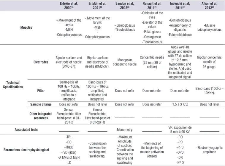
Table 1. Technical speciications of electromyography.
Ertekin et al, 200018
Ertekin et al, 200119
Baudon et al, 200220
Renault et al, 201121
Inokuchi et al, 201422
Alkan et al, 201223
Muscles
- Movement of the larynx -MSH -Cricopharynxeous
- Movement of the larynx -MSH - Cricopharynxeous
- Genioglossus -Tireohioideous
-Orbicular of the
eyes
-Elevator of the
velum
-Palatoglosso -Genioglosso -Tieohioideous
-Geniohioideous -Anterior belly of
digastric
-Esternohioideous
-Muscle cricopharynxeous
Technical
Speciications
Electrodes
Bipolar surface and electrode of needle
(DMC-37).
Bipolar surface and electrode of
needle (DMC-37).
Monopolar concentric needle
Concentric needle (25 mm 30 of
caliber)
Hook wire 40 gauge and needle
with 27 de caliber
of 12,5 mm, hypodermic and sterile. And used
the retiicated and
integrated signal.
Bipolar concentric needle of 26 gauge.
Filter
Band-pass of 100 Hz – 10kHz,
ampliicado, retiicado e
integrado
Band-pass of 100 Hz – 10kHz,
ampliied, retiicated and
integrated.
Does not refer Does not refer Does not refer Band-pass (100Hz – 10KHz).
Sample charge Does not refer Does not refer Does not refer Does not refer 1,5 à 3 Khz Does not refer
Other integrated resources
Sensor
Piezoelectric: ilter band-pass:
0.01-20 Hz
Sensor Piezoelectric:
Filter band-pass of 0.01-20 Hz
___________ ___________ ___________ ___________
Associated tests ___________ ___________ Manometry ___________ VF: Exposition de
5 min à 90 KV ___________
Parameters electrophysiological
-TRL -DD -TRDD - VD (jitter) -A EMG of MSH
-LD
-Coordination between the sucking and swallowing.
-Maximum
Amplitude of suction;
-Coordination between the sucking and swallowing
-Moments of
the beginning of muscle activation
(onset)
-DD -PD -PPD -DPD -DR -Nº D
-Electromyographic
amplitude
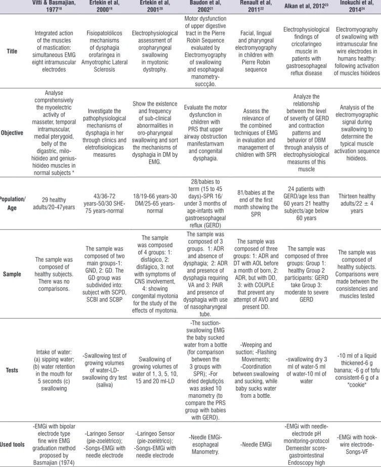
Table 2. Methodological characteristics of selected articles of BIREME and PUBMED.
Vitti & Basmajian, 197718
Ertekin et al, 200019
Ertekin et al, 200120
Baudon et al, 200221
Renault et al,
201122 Alkan et al, 2012
23 Inokuchi et al,
201424
Title
Integrated action of the muscles of mastication: simultaneous EMG eight intramuscular electrodes Fisiopatolólicos mechanisms of dysphagia orofaringea in Amyotrophic Lateral Sclerosis Electrophysiological assessment of oropharyngeal swallowing in myotonic dystrophy. Motor dysfunction of upper digestive tract in the Pierre Robin Sequence evaluated by Electromyography of swallowing and esophageal manometry-succção. Facial, lingual and pharyngeal electromyography
in children with
Pierre Robin sequence Electrophysiological indings of cricofaríngeo muscle in patients with gastroesophageal relux disease Electromyography
of swallowing with intramuscular ine wire electrodes in
humans healthy:
following activation
of muscles hióideos
Objective Analyse comprehensively the myoelectric activity of masseter, temporal intramuscular, medial pterygoid,
belly of the
digastric, milo-hióideo and
genius-hióideo muscles in
normal subjects "
Investigate the pathophysiological
mechanisms of dysphagia in her through clinics and
eletroisiologicas
measures
Show the existence
and frequency
of sub-clinical
abnormalities in
oro-pharyngeal swallowing and sort
the mechanisms of dysphagia in DM by
EMG.
Evaluate the motor dysfunction in
children with
PRS that upper
airway obstruction manifestamvam and congenital dysphagia. Assess the relevance of the combined techniques of EMG
in evaluation and management of
children with SPR
Analyze the relationship
between the level
of severity of GERD and contraction
patterns and behavior of DBM through analysis of electrophysiological measures of this
muscle
Analysis of the electromyographic signal during swallowing to determine the typical muscle activation sequence hióideos. Population/ Age 29 healthy adults/20-47years 43/36-72 years-50/30 SHE-75 years-normal 18/19-66 years-30 DM/25-65 years-normal 28/babies to term (15 to 45
days)-SPR 16/
under 3 months of
age-infants with
gastroesophageal
relux (GERD)
81/babies at the
end of the irst month showing the
SPR
24 patients with
GERD/age less than 60 years 21 healthy
subjects/age below
60 years
Thirteen healthy adults/22 ± 4
years
Sample
The sample was
composed of
healthy subjects. There was no
comparisons.
The sample was composed of two
main groups-1:
GND, 2: GD. The
GD group was
subdivided into:
subject with SCPD,
SCBI and SCBP
The sample
was composed
of 4 groups: 1: disfágico, 2: disfágico, 3: not
with symptoms of
CNS involvement,
4: showing
congenital myotonia for the study of the effects of myotonia.
The sample was
composed of 3 groups. 1: ADR and absence of dysphagia; 2: ADR
and presence of dysphagia requiring
VA and 3: PAIR and presence of
dysphagia with use
of nasopharyngeal tube.
The sample was
composed of three groups: 1: ADR and
DT with AOL before
a month of born, 2:
ADR, but with DD, 3: with COUPLE
that prevent any attempt of AVO and
present DD.
The sample was
composed of three groups: Group 1:
healthy Group 2 participants: GERD
take Group 3:
moderate to severe GERD
The sample was
composed of
healthy subjects. Comparisons were made between the
consistencies and muscles tested
Tests
Intake of water: (a) sipping water; (b) water retention
in the mouth for 5 seconds (c)
swallowing
-Swallowing test of growing volumes
of water-LD-swallowing dry test
(saliva)
Swallowing of growing volumes of water of 1, 3, 5, 10, 15 and 20 ml-LD
-The suction-swallowing EMG the baby sucked water from a bottle
(for comparison
between the 3 groups with
SPR); -For dried deglutiçõs
was asked 10
manometry (to compare the PRS
group with babies with GERD). -Weeping and suction; -Flashing Movements; -Coordination between swallowing
and sucking, while baby sucks water
from a bottle.
-swallowing dry 3 ml of water-5 ml of water-10 ml of
water
-10 ml of a liquid thickened-6 g banana; -6 g of tofu
consistent-6 g of a "cookie"
Used tools
-EMGi with bipolar
electrode type
ine wire EMG
graduation method proposed by Basmajian (1974) -Laringeo Sensor (pie-zoelétrico); -Songs-EMGi with needle electrode -Laringeo Sensor (pie-zoelétrico); -Songs-EMGi with needle electrode -Needle EMGi-esophageal Manometry. -Needle EMGi
-EMGi with
needle-electrode pH
monitoring-protocol Demeester
score-gastrointestinal Endoscopy high
-EMGi with hook-wire
5. Pelman A, Palmer P, McCulloch T, Vandaele D. Electromyography activity from human laryngeal, pharyngeal, and submental muscles during
swallowing. J Appl Phisiol. 1999;86:1663-9.
6. Logemann JA. Non-imaging techniques for the study of swallowing. Acta Otorhinolaryngol Belg. 1994;48:139-42.
7. Portney L, Roy SH. Eletromiograia e testes de
velocidade de condução nervosa. In: O`Sullivan
SB, Schmitz TJ. Fisioterapia: avaliação e tratamento. São Paulo: Manole, 2004. p. 213-56. 8. Preston DC, Shapiro BE. Electromyography
and Neuromuscular Disorders: Clinical Electromyography and Neuromuscular disorders.
In: Preston DC, Shapiro BE. Neuromuscular Junction Disorders, Elsevier- Butterworth – Heinemann, 2005. p. 407-24.
9. Vaiman M, Eviatar E, Segal S. Surface
electromyographic studies of swallowing in normal subjects: A review of 440 adults. Report 2. Quantitative data: Amplitude measures. Otolaryngol
Head Neck Surg. 2004;131(5):773-80.
10. Belo LR, Lins S, Cunha D, Lins O, Amorim C. Eletromiograia de superfície da musculatura
supra-hióidea durante a deglutição de idosos sem
doenças neurológicas e idosos com Parkinson.
Rev CEFAC. 2009;11(2):268-80.
CONCLUSION
The selected articles make important contributions to
the understanding of the electrophysiological behavior and electrophysiological during the mechanism of
swallowing, and it is believed that the lack of research
using intramuscular EMG for analysis of swallowing in
humans should be the trouble and risks caused by the
introduction of needle into the muscle belly and maybe
the introduction of a thin wire (ine wire or wire cooper)
is more interesting for the aid diagnosis of
neuromus-cular disorders and nerve that compromise swallowing,
the ability to dramatically reduce the annoyance caused by the needle.
REFERENCES
1. Crary MA, Baldwin BO. Surface electromyographic
characteristics of swallowing in dysphagia
secondary to brainstem stroke. Dysphagia.
1997;12:180-7.
2. Ertekin C, Palmer JB. Physiology and
electromyography of swallowing and its disorders. Suppl Clin Neurophysiol. 2000; 53:148-54.
3. Vaiman M. Standardization of surface
electromyography utilized to evaluate patients with dysphagia. Head Face Med. 2007;3: 26.
4. Gokyigit M, Pazarci N,Ercan I,Seker S,Turgut S, Ertekin C. Identiication of distinct swallowing
patterns for different bolus volumes. Clinical Neurophysiology. 2009;120(9):1750-4.
Conclusion
DS: activation occurs: the medial pterygoid and MSH,
ickle and take
masseter muscle contraction SAC: MSH activation
occurs so ickle
and vile. CHECK: there's the recrutamente of myoelectric activity.
FROM: recruit the contraction of
MSH and medial pterygoid muscle
There are 2 pathophysiological
mechanisms in dysphagia of SHE:
1-the iring of the relex, swallowing
volunteer is late or
absent; While in automatic relex, swallowing is preserved. 2-MCF:
hypertonic and can be found
hiperrelexivo. and
DPFC related
CNS involvement may contribute
to the delay in
the iring of the swallowing relex,
and abnormal EMG
indings from EES
cause dysphagia in this disease.
The EMG and
manometry were
able to identify shortcomings in the Organization of
motor language,
pharynx and
esofago even in the absence of clinical disorders in
swallowing.
The EMG on SPR revealed abnormalities in
swallowing, even in
cases considered
normal, making it
possible to evaluate both the gravity and the duration of the potential of dysphagia. And
the low amplitude
detected in MPM demonstrate the
need for two
surgical steps of repair of cleft
palate.
The EMGi ESS
needle was
normal in patients
with GERD.
Cinesiológicas assessments
showed increased
Peacemeal deglutition and number of
swallows and that there was a positively with the severity of the relux
The pattern of activation of hióideos muscles
found in swallowing
of different food consistencies
corroborates
with the default
generator concept central
to the swallowing
pharyngeal.
ALS: Amyotrophic Lateral Sclerosis; DM: muscular dystrophy; SPR: Sequence of Pierre Robin; EMG: Electromyography; GERD: Gastroesophageal Relux Disease;
ml: ml; CENTRAL NERVOUS SYSTEM: central nervous system; EES: Upper esophageal sphincter; LD: Limit of dysphagia; SEMG: Surface Electromyography; EMGi:
11. Coriolano MGW, Lins OG, Belo LR, Menezes DC, Moraes SRA, Asano AG et al. Monitorando a
deglutição através da eletromiograia de superfície.
Rev CEFAC. 2010;12(3):434-40.
12. Cram J, Kasman GS. The basics of
electromyography. In: Criswell E. Cram’s Introduction to surface electromyography. 2ª edição. Massachusets: Jones and Bartlett Publishers; 2011. p.35-61.
13. Hillel AD. The Study of Laryngeal Muscle Activity in Normal Human Subjects and in Patients With Laryngeal Dystonia Using Multiple Fine-Wire Electromyography. Laryngoscope. 2001;111:1-47. 14. Rudroff T. Kinesiological Fine Wire EMG: A practical
introduction to ine wire EMG applications. Arisona: Noraxon U.S.A; [citado em 2008]. Disponível: http:// www.noraxon.com/downloads/educational.php3
15. De Luca CJ. The Use of Surface Electromyography in Biomechanics. J Appl Biomech. 1997;13:135-63.
16. Castro AA, Clarck OAC, Atallah AN. Optimal search
strategy for clinical trials in the Latin American and Caribbean Health Science Literature Database (LILACS database): Update. Med J/Rev Paul Medm. 1999;117(3):138-9.
17. Dickersin K, Scherer R, Lefebvre C. Identifying relevant studies for systematic reviews – Systematic
Reviews. BMJ. 1994;309:1286-91.
18. Vitti M, Basmajian JV. Integrated actions of
masticatory muscles: Simultaneos EMG from Eight intramuscular electrodes. Anat Rec. 1977;187(2):173-89.
19. Ertekin C, Aydogdu I, Yüceyar N, Kiylioglu N, Tarlaci S, Uludag B. Pathophysiological mechanisms of
oropharyngeal dysphagia in amyotrophic lateral sclerosis. Brain. 2000;123:125-40.
20. Ertekin C, Yüceyar N, Aydogdu I, Karasoy H.
Electrophysiological evaluation of oropharyngeal swallowing in myotonic dystrophy. J Neurol Neurosurg Psychiatry. 2001;70:363-71.
21. Baudon JJ, Renault F, Goutet JM, Flores-Guevara R, Soupre V, Gold F et al. Motor dysfunction of the
upper digestive tract in Pierre Robin sequence as
assessed by sucking-swallowing electromyography
and esophageal manometry. J Pediatr. 2002;140:719-23.
22. Renault F, Baudon JJ, Galliani E, Flores-Guevara R, Marlin S, Garabedian EN, Vazquez MP. Facial, lingual, and pharyngeal electromyography in
infants with pierre robin sequence. Muscle & Nerve. 2011;43(6):866-71.
23. Alkan Z, Demir A, Yigit O, Adatepe T, Kesici B, Kocak
I et al. Cricopharyngeal Muscle Electromyography Findings in Patients with Gastroesophageal
Relux Disease. Otolaryngol Head Neck Surg.
2012;147(2):295-301.
24. Inokuchi H, Gonzáles- Fernandes M, Koichiro M, Brodsky M, Yoda M, Taniguchi H et al. Electromyography of swallowing with ine wire
intramuscular electrodes in healthy human: Activation Sequence of selected hyoid muscles. Dysphagia.2014;29:713-21.
25. Cunningham DP, Basmajian JV. Electromyography
of genioglossus and geniohyoid muscles during deglutition. Anat. Rec. 1969;165:401-10.
26. Lehr RP, Blanton PL, Biggs NL. An
Electromyographic study of the mylohyoid muscles. Anat Rec. 1971;169(4):651-60.
27. Hrycyshyn AW, Basmajian JV. Electromyography of
the oral stage of swallowing in man. Am. J. Anat. 1972;133:333-40.
28. Cleall JF. Deglutition: A Study of form and function. Am. J. Orthodont. 1965;51(8):566-94.
29. Renault F, Raimbault J. Electromyographie faciale,
linguale et pharyngée chez l’enfant: une méthode d’étude des troubles de succion-déglutition et de leur physiopathologie. Neurophysiol Clin. 1992;22:249-60.
30. Renault F. Facial electromyography in newborn
and young infants with congenital facial weakness.
Dev Med Child Neurol. 2001;43(6):421-7.
31. Ohsawa S, Yamamoto S, Kanda A. Lower lip-lifting
brace for bilateral facial nerve palsy: a case report. Arch Phys Med Rehabil. 2001;82(12):1737-9.
32. Tamura F, Fukui T, Kikutani T, Machida R, Yoshida M, Yoneyama T et al. Lip-closing function of elderly
people during ingestion: comparison with young adults. Int J Orofacial Myology. 2009;35:33-43.
33. Doty RW, Bosma JF. An electromyographic analysis of relex deglutition. J Neurophysiol. 1956;19:44-60. 34. Thexton AJ, Crompton AW, German RZ.
Electromyographic activity during the relex
pharyngeal swallow in the pig:Doty and Bosma (1956) revisited. J Appl Physiol. 2007;102:587-600.
35. Zemlim WR. Princípios de anatomia e isiologia em