1
Universidade Federal do Vale de São Francisco, Centro de Ciências da Saúde. R. da Aurora, s/n., Alves de Souza, 48608-170, Paulo Afonso, BA, Brasil. Correspondência para/Correspondence to: NG MORAIS. E-mail: <morais.ngm@gmail.com>.
2
Universidade Federal de Pernambuco, Centro de Ciências da Saúde, Programa de Pós-Graduação em Medicina Tropical. Recife, PE, Brasil.
3
University of California, Center for Disease Vector Research, Departament of Entomology. Riverside, CA, United States.
Long-term effects of neonatal malnutrition
on microbicide response, production of
cytokines, and survival of macrophages
infected by
Staphylococcus aureus
sensitive/resistant to methicillin
Efeitos tardios da desnutrição neonatal na
resposta microbicida, produção de
citocinas e viabilidade de macrófagos
na infecção por
Staphylococcus
aureus
sensível/resistente a meticilina
Natália Gomes de MORAIS1 Thacianna Barreto da COSTA2 Maiara Santos SEVERO3
Célia Maria Machado Barbosa de CASTRO2
A B S T R A C T
Objective
To assess microbicide function and macrophage viability after in vitro cellular infection by methicillin-sensitive/ resistant Staphylococcus aureus in nourished rats and rats subjected to neonatal malnutrition.
Methods
lipopolysaccharide; and two testing systems, macrophages plus methicillin-sensitive Staphylococcus aureus
and macrophages plus methicillin-resistant Staphylococcus aureus. The plates were incubated in a humid atmosphere at 37 degrees Celsius containing 5% CO2 for 24 hours. After this period tests the microbicidal response, cytokine production, and cell viability were analyzed. The statistical analysis consisted of analysis of variance (p<0.05).
Results
Malnutrition reduced weight gain, rate of phagocytosis, production of superoxide anion and nitric oxide, and macrophage viability. Production of nitrite and interleukin 18, and viability of macrophages infected with methicillin-resistant Staphylococcus aureus were lower.
Conclusion
The neonatal malnutrition model compromised phagocyte function and reduced microbicidal response and cell viability. Interaction between malnutrition and the methicillin-resistant strain decreased the production of inflammatory mediators by effector cells of the immune response, which may compromise the immune system’s defense ability.
Indexing terms: Macrophages. Malnutrition.Methicillin. Staphylococcus.
R E S U M O
Objetivo
Avaliar a função microbicida e a viabilidade de macrófagos, após infecção celular in vitro, com Staphylococcus aureus sensível/resistente a meticilina, em ratos nutridos ou submetidos a desnutrição neonatal.
Métodos
Ratos machos Wistar (n=40) foram divididos em dois grupos distintos: Nutrido (ratos amamentados por mães submetidas a dieta com 17% de caseína) e Desnutrido (ratos amamentados por mães submetidas a dieta com 8% de caseína). Os macrófagos foram recuperados após procedimento cirúrgico de traqueostomia, através da coleta do lavado broncoalveolar. Após o isolamento dos mononucleares, foram estabelecidos quatro sistemas: controle negativo, composto apenas pelos fagócitos; controle positivo, macrófagos mais lipopolissacarídeo; e dois sistemas teste, macrófagos mais Staphylococcus aureus sensível e resistente a meticilina. As placas foram incubadas por 24 horas, à temperatura de 37ºC, com atmosfera úmida e 5% de dióxido de carbono. Transcorrido esse período, foram realizados ensaios para análise da resposta microbicida, produção de citocinas e viabilidade celular. Na análise estatística, utilizou-se analysis of variance, admitindo-se p<0,05.
Resultados
A desnutrição acarretou redução do crescimento ponderal dos animais, da taxa de fagocitose, da produção de óxido nítrico, do ânion superóxido e da viabilidade de macrófagos. Houve menor produção de nitrito, de interleucina 18 e da viabilidade dos macrófagos infectados com Staphylococcus aureus meticilina-resistente.
Conclusão
O modelo de desnutrição neonatal adotado comprometeu a função dos fagócitos, com redução da resposta microbicida e da viabilidade celular. A interação de desnutrição com cepa resistente induziu baixa produção de mediadores inflamatórios por células efetoras da resposta imunológica, o que poderá resultar em compro-metimento da defesa.
Termos de Indexação: Macrófagos. Desnutrição. Meticilina. Staphylococcus.
I N T R O D U C T I O N
Environmental insults during vulnerable periods of an organism’s development can permanently affect the structure and function of organs and tissues. This vulnerability is associated
with the intense differentiation and maturation that organ systems undergo during these periods1.
According to Chandra2, neonatal
malnutrition affects the macrophages’ functional mechanism, causing enduring changes in the adult organism, even long after nutritional recovery. Malnourished individuals may have deficient phagocytic microbicidal function, such as low production of proinflammatory cytokines, free radicals (nitric oxide and superoxide anion), and macrophage viability, making the body more vulnerable to infections4,5.
Pathogenic invasion may deregulate the microbicidal responsiveness of immune6 cells. This
mechanism is triggered by various microorganisms to facilitate their growth and increase their survival time in the host7. In order to establish an infection, Staphylococcus aureus has developed several mechanisms designed to withstand the immune response.
Since the 1960’s, infection by Methicillin-Resistant Staphylococcus Aureus (MRSA) has been considered a public health problem worldwide, mainly because it is more deadly than Methicillin-Sensitive Staphylococcus Aureus (MSSA)8. MRSA
strains seem to have different virulence mechanisms, more intense than MSSA strains9.
Numerous clinical studies on morbidity and mortality rates have indicated that MRSA is more virulent than MSSA. However, laboratory studies that assessed the presence and magnitude of pathogenic mechanisms and virulence factors in MSSA and MRSA strains obtained conflicting results5,10.
Despite the various studies using malnutrition models, there is still a lot to be researched in terms of neonatal malnutrition and its late effects on the immune response. Also, there are hardly any studies evaluating the interaction between infection and malnutrition. The present study aimed to evaluate the impact of neonatal malnutrition on microbicidal function, cytokine production, and viability of alveolar macrophages infected in vitro by methicillin-sensitive and MRSA. In this context animal experiments may help to clarify morphological
changes in early life stages, their intimate relation with microbicidal response, and infectious disease emergence and evolution.
M E T H O D S
Animals and diet
Forty male Wistar rats (90-120 days) from the Universidade Federal do Pernambuco (UFPE) Department of Nutrition animal facility were used for the experiments. The experiments were conducted ethically as recommended by the
Colégio Brasileiro de Experimentação Animal
(COBEA, Brazilian College of Animal Experimentation) and the National Institute of Health Guide for Care and Use of Laboratory Animals and approved by the Animal Ethics Committee of the Center for Biological Sciences, UFPE under Protocol nº 23076.026684/2009-38. The animals were kept under controlled temperature (22°C, Standard Deviation-(SD)=1°C) in 12:12 hours light-dark cycles and had free access to water and chow.
One day after birth, litter size was standardized to six male pups per mother. On the same day, their first day of life, the litters were divided into two groups: a Nourished (N) group consisting of pups nursed by dams consuming a 17% casein diet (n=20); and a Malnourished (M) group consisting of pups nursed by dams consuming an 8% casein diet (n=20) for the first 21 days after birth. The 8% casein diet is widely used as an experimental model for the study of malnutrition because it induces protein malnutrition11 in animals.
During the neonatal period the animals were weighed daily by a digital electronic scale (Marte, model S-4000, with an accuracy of 0.1 g) to monitor their body weight during nutritional manipulation. From the 22nd day of life until the
separated from the dams, kept in cages in groups of three, and fed a standard chow (Vivarium, Labina Purina-Brazil) until adulthood (approximately 90 days).
Strains of Staphylococcus aureus
Strains of Methicillin-Sensitive Staphylococcus Aureus (The American Type Culture Collection - ATCC 33591) and methicillin-sensitive Staphylococcus aureus were used due to their distinctive resistance and importance in terms of public health. The bacteria were maintained in Tryptic Soy Broth (TSB) supplemented with 20% glycerol, at -20°C, until use. Twenty-four hours before each experiment, the strains were plated on blood agar (agar supplemented with 5% sheep blood) and incubated at 37°C. At the beginning of the test, some colonies were transferred to tubes containing Phosphate-Buffered Saline (PBS) to provide a turbidity of approximately 0.15 nm at a wavelength of 570 nm. This absorbance, according to Lu & McEwan12, corresponds to a
concentration of approximately 106 bacteria/mL
of PBS.
Bronchoalveolar lavage
Bronchoalveolar Lavage (BAL) was done as recommended by Castro et al.13. The animals
were anaesthetized with chloralose-urethane (0.5 and 12.5%, respectively) at 8 mL/kg ip. BAL was collected by injecting 0.9% saline through a plastic cannula into the trachea. Several aliquots of 3 mL were then injected and collected in 50 mL conical polypropylene tubes (Falcon, Sigma).
Culture of alveolar macrophages
Bronchoalveolar Lavage samples were centrifuged at 1500 rpm for 15 minutes. The precipitate that corresponds to the cells was resuspended in RPMI 1640 (Roswell Park
Memorial Institute medium - Gibco, Invitrogen Corporation) containing 3% fetal bovine serum (Gibco-Invitrogen Corporation) and antibiotics (100 U penicillin/mL and 100 µg streptomycin/mL).
The cells were transferred to 35 mm diameter (6-well Falcon) cell culture dishes, in which a 2 mL suspension was dispensed in a proportion of 106 cells/mL in RPMI 1640. After 1
hour in an incubator at 37°C and 5% CO2, the supernatant was discarded with non-adherent cells and 2 mL of half RPMI were added, leaving the plates for another 1 hour in the incubator in order to stabilize the cells.
Systems
Three systems were established in order to evaluate the rate of adherence, phagocytosis, and superoxide production: Control (C), with only alveolar macrophages; MSSA, Alveolar Macrophages (AM) plus 100 µL of bacterial inoculum on the methicillin-sensitive strain (ATCC 29213); and MRSA, AM plus 100 µL of bacterial inoculum of methicillin-resistant strain (ATCC 33591). To evaluate the kinetics of nitric oxide and alveolar macrophage viability, a positive control was added - PC containing AM plus 10 µL of Lipopolysaccharide (LPS) (Escherichia coli
serotype; 055: B5, Sigma). Subsequently, the plates were incubated at 37°C in a wet atmosphere containing 5% CO2.
Assessment of the adhesion rate
After incubation of cell cultures for 1 hour, aliquots were collected from the supernatant containing non-adherent cells and wells of the filtration plate were resuspended with RPMI. These aliquots containing non-adherent cells were added to trypan blue stain (1:10 dilution) and cells were counted using a hemocytometer. The Adhesion Rate (AR) was calculated using the formula described by De la Fuente et al.14: AR=100
Determination of the rate of phagocytosis
For this assay the bacterial inoculum was added to a suspension of 106 cells/mL in RPMI
1640 for both strains to a concentration of 106
CFU/mL in PBS, with a remainder volume of 1.5 mL in each tube. The contents of the tubes were homogenized and gently/evenly distributed on slides for optical microscopy. Slides were placed in an oven for 1 hour. After this period, they were washed to remove non-adherent cells and then stained and viewed by a trained, “blind” observer (for the system under analysis) using a light microscope. The result was expressed in percentage of phagocytic cells in a total count of 100 cells15.
Analysis of low superoxide anion (O2-) release
O2- was induced by adding phorbol
myristate acetate/PMA (Sigma) to Hank’s solution (HBSS, Gibco-Invitrogen Corporation®) at a
concentration of 2 µg/mL. Analysis of discontinuous systems was prepared with hourly assessments for 2 hours. Assay specificity was confirmed by the addition of Superoxide Dismutase (SOD) from bovine erythrocytes, containing 3000 U/mg protein in the final solution of 3 mg/mL in distilled water (Sigma)13.
Kinetics of nitric oxide production by alveolar macrophages
The production of NO was given by the concentration of nitrite in the culture supernatant10. Every two hours, 100 µL of the
supernatant were taken from the cultures, in a total incubation period of 24 hours. To quantify nitrates and nitrites, 50 mL of Griess reagent (1.5% sulfanilamide in 5% H3PO4, 0.1% in N-(1-naphthyl)ethylenediamine H20) were added to supernatants. After standing 15 minutes at room
temperature, an reader Enzyme Linked Assay (ELISA, ImmunonoSorbent - BIO-RAD, Model 680), with 550 nm filter, was used for the procedure. The nitrite concentration was calculated using average values of a NaNO2 standard curve, data expressed in µM.
Quantification of cytokines IL-1βββββ and IL-18 (interleukin-1βββββ and interleukin-18)
After 24 hours of cell culture incubation, 100 µL of supernatant were collected. From this, IL-1β and IL-18 cytokines were counted by ELISA immunoenzyme test, using the Quantikineâ m
(R&D Systems) kit.
Viability of alveolar macrophages
Cell viability was assessed by mitochondrial reduction of MTT (3-[4,5-dimethylthiazol-2-yl]-2,5-diphenyltetrazolium bromide) on formazan16.
After 24 hours of incubation, cell cultures were washed with PBS (1X) at room temperature They were incubated with 550 µL of PBS and 55 µL of MTT solution for two hours protected from light. After this period, 200 µL of PBS and 200 µL of DMSO were added and the cell monolayer was scraped. Quantification of solubilized formazam was performed in an ELISA reader (Bio-Rad, model 680) with 570 nm filter. Results were expressed in absorbance of formazan (1x10⁶ cells).
Statistical analysis
R E S U L T S
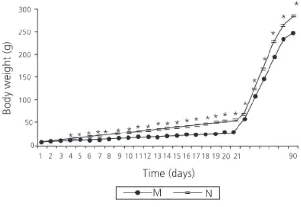
Body weight on malnutrition and nutritional recovery
The body weights (g) of the nourished and malnourished groups were similar until the 3rd day
of life. From the 4th to the 21st postnatal day, the
malnourished animals were lighter than the nourished animals (p<0.001). Between the 22nd
and 90th days of life the two groups were fed the
same chow, but the malnourished group remained lighter than the nourished group (p<0.001) (Figure 1).
Grip index
There were no differences between the nourished and malnourished groups (C-N=87.5±3,0; C-M=86±2,0; MSSA-N=90.3±2.6; MS S A - M = 9 1 ± 2 . 0 ; M R S A - N = 9 3.3±3.1; MRSA=93±3.0), p>0.05. Also, no differences were observed between the systems under analysis, p>0.05.
Rate of phagocytosis
The rate of phagocytosis was lower in the malnourished group (N=12.1±2.0; MSSA-M=4.1±3.2; N=10.4±3.1; MRSA-M=4.3±3.0), p<0.001. However, when analyzing the MSSA and MRSA systems, there was no difference in the rates of phagocytosis of alveolar macrophages (p>0.05).
Production of superoxide
The malnourished group produced less superoxide than the nourished group for all systems (p≤0.001) for both incubation times (p<0.05). However, the MSSA and MRSA systems did not differ (p>0.05) (Table 1).
Kinetic analysis of the nitric oxide production by alveolar macrophages
The quantification of NO was expressed in µM nitrite. The production of NO by macrophages was lower in malnourished animals in all systems (p<0.05). Differences were found between NC and PC systems in both groups after
Figure 1. Weight curve during the neonatal malnutrition period (21 days) and nutritional supplementation period (23-90 days) of the groups (N: Nourished- and M: Malnourished). Recife (PE), 2009.
Note: *p<0.05 in comparison between Nourished and Malnourished
groups.
Student’s t test. Values are expressed as Mean ± Standard Deviation (n=40).
Table 1. Absolute figures for superoxide production in the groups (N:Nourished; M:Malnourished), in the systems (C: Negative Control; MSSA: Methicillin-Sensitive Staphylococcus Aureus; MRSA: Methicillin-Resistant Staphylococcus Aureus) and incubation periods (1 and 2 hours). Recife (PE), 2009.
N M Time
Systems Groups
1.47 ± 0.08 1.07 ± 0.08*
M ± SD C
1.69 ± 0.07 1.10 ± 0.14*
M ± SD MSSA
1.70 ± 0.07 1.11 ± 0.08*
M ± SD MRSA 1 hour
2.79 ± 0.14# 2.12 ± 0.28#*
M ± SD C
2.88 ± 0.19# 2.34 ± 0.11#*
M ± SD MSSA
3.54 ± 0.17# 2.48 ± 0.14#*
M ± SD MRSA 2 hours
Note: *p<0.05 in comparison of Nourished and Malnourished groups; #p<0.05 in comparison of 1 hour and 2 hours.
8 hours of incubation, with high production of nitric oxide in the PC (p<0.001). The peak NO production for the PC system occurred after 22 hours, both for the nourished and the malnourished group (p<0.001). From 4 to 10 hours, the average NO production was lower than in the nourished group in the malnourished group in the MSSA and MRSA systems (p<0.05). The peak NO production occurred after a 4h incubation period in MSSA system, group N, after 6h for group D, and after 8h for MRSA in both groups. Up to 12 hours of incubation, both for the nourished and malnourished groups, there was a reduction of NO production for MSSA and MRSA systems (p<0.001), similar to those of CN (Figure 2).
IL-1βββββ Levels
The levels of IL-1β of nourished versus malnourished controls did not differ (p>0.05). However, when analyzing the testing systems, there was a lower concentration of IL-1β in the supernatant of the MRSA testing systems (MSSA-N=15.94±0.53 pg/mL; MSSA-M=11.81±3.01 pg/mL; MRSA-N=4.24±0.26 pg/mL; MRSA-M=6.41±0.3 pg/mL) p<0.05 (Figure 3).
IL-1βββββ Levels
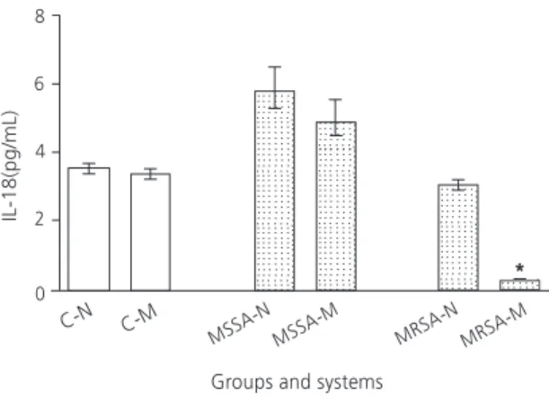
The levels of IL-18 of nourished versus malnourished controls did not differ (p>0.05). The production of IL-18 was higher in MSSA (MSSA-N=5.87±0.59 pg/mL; MRSA-N=3.11±0.23 pg/mL)
p<0.05. However, when analyzing the testing systems, there was a lower concentration of IL-18 in MRSA testing systems of the malnourished group (N=3.11±0.23 pg/mL; MRSA-M=0.27±0.01 pg/mL) p<0.05 (Figure 4).
Viability of alveolar macrophages
The malnourished group had lower macrophage viability than the nourished group in all systems under analysis (PC-N=69.2±0.8;
Figure 2. Nitric oxide production in the supernatant of alveolar macrophage cultures in groups (N: Nourished; M: Malnourished) and systems (PC: Positive Control, MSSA: Methicillin Sensible Staphylococcus Aureus; MRSA: Methicillin Resistant Staphylococcus Aureus). Recife (PE), 2009.
Note: *p<0.05 on the comparison of the Nourished and Malnourished groups.
Analysis of Variance and Tukey test. Values are expressed as Mean ± Standard Deviation (n=40).
Figure 3.Levels of IL-1â in the supernatant of alveolar macrophage cultures in groups (N: Nourished; M: Malnourished) and systems (C: Negative Control, MSSA: Methicillin Sensible
Staphylococcus Aureus; MRSA: Methicillin Resistant
Staphylococcus Aureus). Recife (PE), 2011.
Note: *p<0.05 in comparison of Nourished and Malnourished groups.
Analysis of Variance and Tukey test. Values are expressed as Mean ± Standard Deviation (n=40).
D I S C U S S I O N
Studies show that neonatal malnutrition models correlate with the deficiency of certain nutrients and gene expression, changing the genotype and phenotype17,18. The intensity and
duration of malnutrition will determine the extent of systemic consequences1.
In this study the experimental model of malnutrition consisted of an 8% casein diet, which is considered low protein. The low protein level on the diet offered to dams is characterized by the restricted amount of nutrients available to puppies. Thus, infants develop protein malnutrition, while puppies develop protein-calorie malnutrition. This fact is crucial for the genesis of the deleterious effects observed in the offspring19.
The animals nursed by these dams were stunted, evidenced by low weight at weaning that persisted to 90 days of age. From the fourth postnatal day, malnourished animals gained less weight than the nourished samples. This result is similar to that found by Melo et al.4, but they
used a regional basic diet low in all constituents. Costa et al.5 used an 8% casein diet to induce
malnutrition and also found that weight gain decreased after the fourth day of life.
Nutritional insults in the neonatal period seem to interfere with the programming of macrophage functional mechanisms, causing lasting changes detectable in adulthood, even after a long nutritional recovery3. Prestes-Carneiro et al.20 found that malnutrition from the first to
twelfth day of lactation compromised the microbicidal response, represented by lower rate of phagocytosis. Other researchers have reported a deficit in the production of nitric oxide4,21. Dong et al.22 reported lower production of free radicals
by alveolar macrophages after in vitro stimulation with LPS.
In the present study, the low-protein diet did not change alveolar macrophage adherence regardless of pathogenic stimulus, suggesting that this initial step of the macrophages’ immune response may not be impaired by neonatal malnutrition caused by an 8% casein diet. Chandra2 noticed that malnutrition changes
different stages of activated neutrophil and macrophage phagocytosis. Thus, malnutrition may change mechanisms that rely on macrophage activation not necessarily before activation.
According to this premise, the study demonstrated that malnutrition during lactation reduced macrophage phagocytosis in both systems. According to Prestes-Carneiro et al.20 in
the case of inflammatory stimuli, macrophages from malnourished animals do not respond with the same intensity as macrophages from nourished animals, which allows the development of inflammation and/or infection. Other studies have also reported low phagocytic capacity in animals subjected to malnutrition, whether neonatal or not4,22 .
Regarding oxidant activity, alveolar macrophages from neonatally malnourished rats produced less superoxide, both under normal conditions and under bacterial stimuli. Corroborating this finding, Kawakami et al.23
found that phagocytes’ antimicrobial systems are potentially affected by malnutrition.
Carneiro et al.20 also found that superoxide
production decreases during severe protein malnutrition.
In the present study, nitric oxide production was analyzed every two hours for a total of 24 hours of incubation. Both groups produced more than the PC, peaking at 22 hours, but malnutrition decreased production. Corroborating this result, Melo et al.4 found that alveolar and peritoneal
macrophages produced less nitric oxide after 24 hours of incubation with LPS in rats submitted to early malnutrition. Ferreira-Silva et al.24 also found
that nitrite concentration decreased in cell culture supernatant during nitric oxide production in the undernourished group after LPS stimulation. These data indicate that neonatal malnutrition induces changes in the macrophages, with significant repercussions during adulthood9.
Pumerantz et al.25 stated that the nitric
oxide produced by alveolar macrophages plays an important microbicide role against
Staphylococcus aureus. By comparing the nitric oxide release in the MSSA and MRSA systems of nourished and malnourished groups, the malnourished groups presented the lowest production. Low synthesis of this free radical may allow resistant bacteria to proliferate inside phagocytes because this important defense mechanism is compromised10.
According to Richardson et al.26, S. aureus
can evade multiple components of the innate immune response, including the microbicidal action of nitric oxide. These authors found that
S. aureus can adapt metabolically to nitrosative stress because it has an inducible NO-L-lactate dehydrogenase enzyme. The production of NO-L-lactate dehydrogenase enables S. aureus to keep homeostasis during nitrosative stress, and antibiotic resistance does not seem to interfere on this mechanism.
Based on analysis of IL-1β production, the nourished and malnourished groups differed only on the testing systems. High IL-1β production was detected in the MSSA system of the nourished group, but in the MRSA system, it was higher in
the malnourished groups. IL-1β is a potent endogenous pyrogen (a fever inducer), and a potent stimulator of leukocyte migration into tissues and cytokine and chemokine expression27.
IL-1β is an important mediator for defense against
Staphylococcus aureus. In S. aureus infection, the production of IL-1β acts in the recruitment of neutrophils and the subsequent degradation of the bacterial cell wall by lysozyme enzyme. However, S. aureus has an O-acetyltransferase enzyme that transforms the cell wall resistant to the action of lysozyme and thus escapes the microbicidal response28. These findings justify the
high MRSA-related mortality rates.
IL-18 production was higher in the positive than negative control. When analyzing the testing systems of the nourished and malnourished groups, the production in the malnourished groups was small and even smaller in the MRSA system. IL-18 induces the production of IFN-y (interferon-gamma) by cells of the immune system. This cytokine is important for the activation of macrophages, T lymphocytes, and other cells28. In MRSA infections of malnourished
animals, the pro-inflammatory profile (Th1) may be compromised, favoring the persistence of the bacteria in the host organism.
When comparing macrophage viability in the PC, MSSA, and MRSA systems, viability decreased intensely after infection with S. aureus.
This finding was more evident in the malnourished group infected by MRSA. This indicates that macrophage vulnerability is greater during MRSA infection, especially in immunocompromised individuals.
Protein-calorie deficiencies may induce irreversible cell damage that triggers the mechanism of programmed cell death20.
Ferreira-Silva et al.24 found a reduction in the viability of
alveolar macrophages after perinatal malnutrition. Corroborating these authors, Rivadeneira et al.29
alteration of nutritional and growth factors. These different routes induce activation of caspases, which generate the cleavage of structural proteins, impairing cytoskeleton integrity, resulting in cell death29.
Staphylococcus aureus is able to produce a variety of potent cytotoxins, allowing the bacteria to resist microbicidal response. Leucocidin is a toxin associated with new methicillin-resistant
Staphylococcus aureus strains that destroys leukocytes by forming pores in the cell membrane30.
Thus, we suggest that S. aureus infection induced phagocyte death by triggering cell lysis, and that neonatal undernutrition further promoted this effect. The study results may explain the high morbidity and mortality rates associated with MRSA infection in immunocompromised individuals.
C O N C L U S I O N
The study neonatal malnutrition model compromised some functional parameters of innate immunity, such as rate of phagocytosis and production of nitric oxide, superoxide anion, and IL-18. Phagocytosis and the production of these inflammatory mediators are critical for the effective destruction of invading microorganisms. Adherence rate and production of IL-1β were not affected, but neonatal nutrition does impact the programming of macrophage microbicidal mechanisms. Methicillin-sensitivity in Staphylococcus aureus strains seems to influence their ability to evade the microbicidal response, decreasing immune defense. Interaction between neonatal malnutrition and MRSA infection increased phagocyte susceptibility, which may allow severe and fatal infections. However, many gaps remain to be filled regarding the structure and performance of immune defense components during infections, such as those caused by
Staphylococcus aureus in adults who have endured environmental insults. Thus, it is important to conduct studies using more sensitive
and specific methods, such as biological molecular analyses. These may provide better data on this topic and contribute to the clarification of the morphological changes that occur in early life and the impact of such changes on the microbicidal response of phagocytes and on the emergence and evolution of infectious diseases.
C O L L A B O R A T O R S
NG MORAIS helped to design the study and experimental strategy; tabulate the data; discuss the results; and write the article. TB COSTA helped to prepare the experimental groups, maintain the animals, and collect the data. MS SEVERO helped to design the study and the experimental strategy. CMMB CASTRO helped to design the study, tabulate the data, discuss the results, and write the article.
R E F E R E N C E S
1. Pereira KNF, Vitoriano ILS, Melo MPP, Aragão RS, Toscano AE, Silva HJ, De Castro RM. Effects of malnutrition and/or neonatal inhibition of serotonin reuptake in neuromuscular development of the gastrointestinal tract: Review of literature. Neurobiology. 2009; 72(2):215-21.
2. Chandra RK. Nutrition and the immune system from birth to old age. Eur J Clin Nutr. 2002; 56(3):73-6. doi: 10.1038/sj.ejcn.1601492
3. Melo JF, Costa TC, Lima TDC, Chaves MEC, Vayssade LM, Nagel MD, et al. Long-term effects of a neonatal low-protein diet in rats on the number of macrophages in culture and the expression/ production of fusion proteins. Eur J Nutr. 2013; 52(5):1475-82. doi: 10.1007/s00394-012-0453-y 4. Melo JF, Macedo EMC, Silva RPP, Viana MT, Silva WTF, Castro CMMB. Efeito da desnutrição neonatal sobre o recrutamento celular e a atividade oxidante--antioxidante de macrófagos em ratos adultos endotoxêmicos. Rev Nutr. 2008, 21(6):683-94. doi: 10.1590/S1415-52732008000600007
5. Costa TB, Morais NG, Almeida TM, Severo MS, Castro CMMB. Early malnutrition and production of IFN-γ, IL-12 and IL-10 by macrophages/ lymphocytes: In vitro study of cell infection by methicillin-sensitive and methicillin-resistant
6. Tegnér J, Nilsson R, Bajic VB, Björkegren J, Ravasi T. Systems biology of innate immunity. Cell Immunol. 2007; 244(2):105-9. doi: 10.1016/j.cellimm.2007.0 1.010
7. Kubica M, Guzik K, Koziel J, Zarebski M, Richter W, Gajkowska B, et al. Potential new pathway for
Staphylococcus aureus dissemination: The silent survival of S. aureus Phagocytosed by human monocyte-derived macrophages. PLOS ONE. 2008; 3(1):1409-35. doi: 10.1371/journal.pone.0001409 8. Beam JW, Buckley B. Community-acquired methicillin-resistant Staphylococcus aureus: Prevalence and risk factors. J Athl. 2006; 41(3):337-40. 9. Mandell GL. Uptake, transport, delivery and intracellular activity of antimicrobial agents. Pharmacother. 2005; 25(12):130S-3S.
10. Morais NG, Costa TB, Almeida TM, Severo MS, Castro CMMB. Parâmetros imunológicos de macrófagos frente à infecção por Staphylococcus aureus meticilina sensível/resistente. J Bras Patol Med Lab. 2013; 49(2):84-90. doi: 10.1590/S1676-24 442013000200002
11. Passos MCF, Ramos CF, Moura EG. Short and long term effects of malnutrition in rats during lactation on the body weight of offspring. Nutr Res. 2000, 20(1):1603-12. doi: 10.1016/S0271-5317(00)00 246-3
12. Lu YF, McEwan NA. Staphylococcal and micrococcal adherence to canine and feline corneocytes: Quantification using a simple adhesion assay. Veterinary Dermatol. 2007; 18(1):29-35. doi: 10.1111/j.1365-3164.2007.00567
13. Castro CMMB, Manhães-de-Castro R, Medeiros AF, Santos AQ, Silva WTF, Lima Filho JLL. Effect of stress on the production of O2- in alveolar macrophages.
J Neuroimmunol. 2000; 108(1-2):68-72.
14. De la Fuente M, Del Rio M, Ferrandez MD, Hernanz A. Modulation of phagocytic function in murine peritoneal macrophages by bombesin, gastrin-releasing peptide and neuromedin C. Immunology. 1991; 73(2):205-11.
15. Malagueno E, Albuquerque C, Castro CMMB, Gadelha M, Inácio-Irmão J, Santana JV. Effect of biomphalaria straminea plasma of biomphalaria glabrata hemolymph cells. Mem Inst Oswaldo Cruz.
1998; 93(1):301-2. doi: 10.1590/S0074-027619 98000700059
16. Mosmann T. Rapid colorimetric assay for cellular growth and survival: Application to proliferation and cytotoxicity assays. J Immun Meth. 1983; 65(1/2):55-63.
17. Rodriguez L, Gonzalez C, Flores L, Jiménez-Zamudio L, Graniel J, Ortiz R. Assessment by flow cytometry
of cytokine production in malnourished children. Clin Diagn Lab Immunol. 2005; 12(4):502-7. doi: 10.1111/j.1365-2249.2007.03361
18. Waterland RA, Jirtle RL. Early nutrition, epigenetic changes at transposons and imprinted genes, and enhanced susceptibility to adult chronic diseases. Nutrition. 2004;20(1):63-8. doi: 10.1016/j.nut.20 03.09.011
19. Araujo FRG, De Castro CMMB, Rocha JA, Sampaio B, Diniz MFA, Evêncio LB, et al. Perialveolar bacterial microbiota and bacteraemia after dental alveolitis in adult rats that had been subjected to neonatal malnutrition. Br J Nutr. 2012; 107(1):996-1005. doi: 10.1017/S00 0711451100393X
20. Prestes-Carneiro LE, Laraya RD, Silva PRC, Moliterno RA, Felipe I, Mathias PC. Long-term effect of early protein malnutrition on growth curve hematological parameters and macrophage function of rats. J Nutr Scien Vitaminol. 2006; 52(6):414-20. doi: 10.3177/jnsv.52.414
21. Anstead GM, Chandrasekar B, Zhao W, Yang J, Perez LE, Melby PC. Malnutrition alters the innate immune response and increases early visceralization following Leishmania donovani infection. Infect Immun. 2001; 69:4709-18. doi: 10.1128/IAI.69.8.4 709-4718.2001
22. Dong W, Selgrade MJK, Gilmour MI, Lange RW, Park P, Luster MI, et al. Altered alveolar macrophage function in calorie-restricted rats. Am J Respir Cell Mol Biol. 1998; 19(1):462-9. doi: 10.1165/ajrcmb. 19.3.3114
23. Kawakami K, Kadota J, Lida K, Shirai R, Abe K, Kohno S. Reduced immune function and malnutrition in the elderly. Tohoku J Exp Med. 1999; 187(2):157-71.
24. Ferreira-Silva WT, Galvão BA, Ferraz Pereira KN, Castro CMMB, Manhaes-de-Castro R. Perinatal malnutrition programs sustained alterations in nitric oxide release by activated macrophages in response to fluoxetine in adult rats. Neuroimmunomodulation. 2009; 16(4):219-27. doi: 10.1159/000212382 25. Pumerantz A, Muppidi K, Agnihotri S, Guerrac C,
Venketaraman V, Wangb J, et al. Preparation of liposomal vancomycin and intracellular killing of Meticillin-Resistant Staphylococcus Aureus (MRSA). Internat J Antimicrob Ag. 2010; 37(2):140-4. doi: 10.1016/j.ijantimicag.2010.10.011
26. Richardson AR, Libby SJ, Fang FC. A nitric oxide-inducible lactate dehydrogenase Enables
27. Latz E. The inflammasomes: Mechanisms of activation and function.Curr Opin Immunol. 2010; 22(1):28-33. doi: 10.1016/j.coi.2009.12.004 28. Lalor SJ, Dugan LS, Sutton CE, Basdeo SA, Fletcher
JM, Mills KHG. IL-18 promote IL-17 production by gd and caspase-1-processed cytokines IL-1b and CD4 T cells that mediate autoimmunity. J Immunol. 2011; 6(1):55-63. doi: 10.4049/jimmunol.100 3597
29. Rivadeneira DE, Grobmyer SR, Naama HA, Mackrell PJ, Mestre JR, Stapleton PP, et al.
Malnutrition-induced macrophage apoptosis. Surgery. 2001; 129(5):617-25. doi: 10.1067/msy.2001.112963
30. Nizet V. Understanding how leading bacterial pathogens subvert innate immunity to reveal novel therapeutic targets. J Allergy Clin Immunol. 2007; 120(1):13 22. doi: 10.1016/j.jaci.2007.06.005


