Aspergillus usamii
b
-Mannanase to Improve Its
Thermostability and Cellulose-Binding Capacity by
In
Silico
Design
Cun-Duo Tang1., Jian-Fang Li2., Xi-Huan Wei2, Rou Min3, Shu-Juan Gao3, Jun-Qing Wang1, Xin Yin1,
Min-Chen Wu4*
1School of Biotechnology and Key Laboratory of Industrial Biotechnology, Ministry of Education, Jiangnan University, Wuxi, Jiangsu, China,2School of Food Science and Technology, Jiangnan University, Wuxi, Jiangsu, China,3School of Pharmaceutical Science, Jiangnan University, Wuxi, Jiangsu, China,4Wuxi Medical School, Jiangnan University, Wuxi, Jiangsu, China
Abstract
The AuMan5A, an acidophilic glycoside hydrolase (GH) family 5b-mannanase derived from Aspergillus usamii YL-01-78, consists of an only catalytic domain (CD). To perfect enzymatic properties of the AuMan5A, a family 1 carbohydrate-binding module (CBM) of theTrichoderma reeseicellobiohydrolase I (TrCBH I), having the lowest binding free energy with cellobiose, was selected byin silicodesign, and fused into its C-terminus forming a fusionb-mannanase, designated as AuMan5A-CBM. Then, its encoding gene,Auman5A-cbm, was constructed as it was designed theoretically, and expressed inPichia pastoris GS115. SDS-PAGE analysis displayed that both recombinant AuMan5A-CBM (reAuMan5A-CBM) and AuMan5A (reAuMan5A) were secreted into the cultured media with apparent molecular masses of 57.3 and 49.8 kDa, respectively. The temperature optimum of the reAuMan5A-CBM was 75uC, being 5uC higher than that of the reAuMan5A. They were stable at temperatures of 68 and 60uC, respectively. Compared with reAuMan5A, the reAuMan5A-CBM showed an obvious decrease inKmand a slight alteration inVmax. In addition, the fusion of a CBM of the TrCBH I into the AuMan5A contributed to its
cellulose-binding capacity.
Citation:Tang C-D, Li J-F, Wei X-H, Min R, Gao S-J, et al. (2013) Fusing a Carbohydrate-Binding Module into theAspergillus usamiib-Mannanase to Improve Its Thermostability and Cellulose-Binding Capacity byIn SilicoDesign. PLoS ONE 8(5): e64766. doi:10.1371/journal.pone.0064766
Editor:Jose M. Sanchez-Ruiz, Universidad de Granada, Spain
ReceivedJanuary 16, 2013;AcceptedApril 18, 2013;PublishedMay 31, 2013
Copyright:ß2013 Tang et al. This is an open-access article distributed under the terms of the Creative Commons Attribution License, which permits unrestricted use, distribution, and reproduction in any medium, provided the original author and source are credited.
Funding:This work was supported by National Nature Science Foundation of China (grant No. 31271811) (http://www.nsfc.gov.cn/); Doctoral Research Funds of Jiangnan University, China (grant No. JUDCF11011) (http://yjsb.jiangnan.edu.cn/); and Postgraduate Innovation Training Project of Jiangsu, China (grant No. CXZZ11_0480) (http://www.ec.js.edu.cn/). The funders had no role in study design, data collection and analysis, decision to publish, or preparation of the manuscript.
Competing Interests:The authors have declared that no competing interests exist.
* E-mail: [email protected]
.These authors contributed equally to this work.
Introduction
b-Mannanases (EC 3.2.1.78), abbreviated from b
-1,4-D-man-nan mannohydrolases, can hydrolyze the internal b
-1,4-D-mannosidic linkages of mannans. They could be applied in industrial processes, such as bleaching pulps, depolymerizing anti-nutritional factors in feedstuffs, the production of mannooligo-saccharides, and extracting oils from leguminous seeds [1]. To date, many researches have been performed on exploiting novelb -mannanases with good properties, improving their catalytic activities by mutating enzyme-producing strains and optimizing fermentation conditions, as well as producingb-mannanases on an industrial scale [2,3]. However, the applicability ofb-mannanases, exemplified by preparing feedstuffs and bleaching pulps, was limited by their low stability at the high temperature and/or extreme pH. To meet the increasing needs for b-mannanases, more interests are being focused on modifying their molecular structures by means of genetic engineering [4].
Based on the sequence alignment, 25,580 CBMs in the CAZY database (July, 2012) were classified into 64 families. The recognized function of CBMs was to bring enzymes into close vicinity to their substrates by binding carbohydrates [5]. Some studies also demonstrated that the fusion of CBMs into enzymes improved the catalytic activity and/or thermostability. For instances, delignification efficiency of the Pycnoporus cinnabarinus
laccase for softwood kraft pulp bleaching was improved by fusing a family 1 CBM of theA. nigercellobiohydrolase B into the laccase [6]. After two CBMs of theThermobifida fuscaandCellulomonas fimi
cellulases were fused into theT. fusca cutinase, respectively, both fusion cutinases exhibited a dramatic increase of up to 3-fold in the amount of fatty acids released from cotton fiber [7].
Almost allb-mannanases reported have been classified into GH families 5, 26 and 113 (http://www.cazy.org/fam/acc_GH.html) [8]. The family 5b-mannanases either contain an only CD [9] or, besides a CD, carry an additional CBM located at the C-terminus such asT. reeseib-mannanase [10] or at the N-terminus such as
an AuMan5A-encoding gene (Auman5A) was cloned and analyzed. The multiple sequence alignment among family 5b-mannanases displayed that no CBM was found in the AuMan5A [12]. To perfect the AuMan5A’s properties, our present work designed an AuMan5A-CBM by fusing a CBM of the TrCBH I into the
AuMan5A. Then, the Auman5A-cbm was constructed as it was
designed theoretically, and expressed in P. pastoris GS115. Moreover, enzymatic properties of the reAuMan5A-CBM and reAuMan5A were analyzed and compared.
Materials and Methods
Strains, Vectors and Media
A. usamiiYL-01-78 andT. reeseiLW-22, isolated from the soil in China, were used as donors of theAuman5Aand CBH I-encoding gene (Trcbh), respectively. E. coli JM109 and pUCm-T (Sangon, Shanghai, China) were used for gene cloning and DNA sequencing. Two recombinant T-vectors, pUCm-T-Auman5Aand pUCm-T-Trcbh, were constructed according to the sequences of the Auman5A (GenBank accession: HQ839639) and Trcbh
(GL985084).E. coliDH5a and pPIC9K (Invitrogen, San Diego, CA) were used for the construction of recombinant expression vectors.E. coliJM109 and DH5awere cultured in a LB medium.
P. pastorisGS115 was cultured in a yeast extract peptone dextrose (YPD) medium, and its transformant cultured and induced in following media that were prepared as described in the manual of Multi-CopyPichia Expression Kit (Invitrogen): minimal dextrose (MD), geneticin G418-containing YPD, buffered glycerol-complex (BMGY) and buffered methanol-complex (BMMY).
Bioinformatics Analysis of theb-mannanase and CBM Sequences
The multiple protein sequence alignment of the CBMs was performed using the ClustalW2 program (http://www.ebi.ac.uk/ Tools/msa/clustalw2/). The putative N-glycosylation site of theb -mannanase was located using the NetNGlyc program (http:// www.cbs.dtu.dk/services/NetNGlyc/). The Protparam program (http://au.expasy.org/tools/protparam.html) was used for pre-dicting theb-mannanase physicochemical properties. The phylo-genetic tree of the CBMs was constructed using both the ClustalW2 program and MEGA 4.0 software. The three-dimensional (3-D) structures of the CBMs were predicted by homology modeling using the MODELLER 9.9 program (http:// salilab.org/modeller/).
In silico Design of the Fusionb-mannanase
The candidate CBMs were chosen from the phylogenetic tree, and their 3-D structures were modeled based on a CBM crystal one of the TrCBH I (PDB code: 1CBH). While the 3-D structural information of cellobiose, used as the ligand, was handled using the GlycoBioChem PRODRG (http://davapc1.bioch.dundee.ac. uk/prodrg/submit.html). Then, the interaction between the candidate CBM and cellobiose was predicted by molecular docking (MD) simulation. The binding free energy of the CBM with cellobiose, contrary to their affinity [13], was calculated using the AutoDock 4.2 program (http://autodock.scripps.edu), which combines a rapid binding free energy evaluation through precalculated grids of affinity potentials with a variety of search algorithms to find the most suitable binding position for a ligand on a given macromolecule [14]. Finally, a CBM having the lowest binding free energy was selected, and fused into the AuMan5A forming an AuMan5A-CBM.
Construction of the Fusionb-mannanase Gene
TheAuman5A-cbmwas constructed by fusing the 39-end region (lcbm) of theTrcbh, which encodes both a Ser/Thr/Pro-rich linker and a CBM, into 39-end of theAuman5Aby overlapping PCR. The
Auman5Awas amplified from the pUCm-T-Auman5Awith primers
F1 (59-GAATTCTCCTTCGCCAGCACCTC-39with anEcoR I
site, underlined) and R1 (59
-GAGGGTTGCCGGCACTAT-CAATAGCAGC-39). Thelcbm was amplified from the
pUCm-T-Trcbh with primers F2 (59
-TGATAGTGCCGG-CAACCCTCCCGGCG-39) and R2 (59
-GCGGCCGCTTA-CAGGCACTGAGAGTAG-39, with a Not I site, underlined).
Using theAuman5Aand lcbmas primers and templates, the first-round overlapping PCR for the Auman5A-cbmwas performed as follows: a denaturation at 94uC for 4 min; 10 cycles of at 94uC for 30 s, 52uC for 30 s, 72uC for 75 s; an elongation at 72uC for 10 min. Then, F1 and R2 were added to the above PCR system to run the second-round PCR under the same conditions, except 30 cycles and an annealing temperature of 56uC. The amplified target band was cloned into pUCm-T, and transformed intoE. coli
JM109. The recombinant T-vector, pUCm-T-Auman5A-cbm, was confirmed by DNA sequencing.
Transformation of the Recombinant Expression Vectors
TheAuman5A-cbmandAuman5Awere excised from the pUCm-T-Auman5A-cbmand pUCm-T-Auman5Aby digestion withEcoR I and NotI, and inserted into pPIC9K, followed by transforming them into E. coli DH5a, respectively. Recombinant expression vectors, pPIC9K-Auman5A-cbmand pPIC9K-Auman5A, were con-firmed by sequencing. The resulting recombinant vectors were separately linearized withSac I, and transformed into P. pastoris
GS115 using a Gene Pulser Apparatus (Bio-Rad, Hercules, CA).
Screening and Expression of theP. pastoris Transformants
AllP. pastoristransformants were primarily screened based on their ability to grow on a MD plate, then inoculated successively on G418-containing YPD plates at increasing concentrations of 1.0, 2.0 and 4.0 mg/mL to screen multiple copies of the Auman5A-cbmandAuman5A, respectively.P. pastorisGS115 transformed with pPIC9K vector without any insert was used as the negative control. Expression of the gene Auman5A-cbm orAuman5A in P. pastoris GS115 was performed according to the instruction of Multi-Copy Pichia Expression Kit (Invitrogen) with some modi-fications [15].
Enzyme Activity and Protein Assays
b-Mannanase activity was determined by measuring the
amount of reducing sugars from locust bean gum (Sigma, St. Louis, MA), using the 3,5-dinitrosalicylic acid (DNS) method as reported previously [15]. One unit (U) ofb-mannanase activity was defined as the amount of enzyme liberating 1.0mmol of reducing sugar equivalent per min under the standard assay
conditions (at pH 3.6 and 60uC for 10 min). The protein
concentration was measured with the BCA-200 Protein Assay Kit (Pierce, Rockford, IL). Sodium dodecyl sulfate-polyacrylamide gel electrophoresis (SDS-PAGE) was carried out on a 12.5% gel using the reported method [16], and the isolated proteins were visualized by staining with Coomassie Brilliant Blue R-250 (Sigma).
Purification of the Recombinantb-mannanases
was brought to 75% saturation by adding solid ammonium sulfate. The resulting precipitate was collected, dissolved in 6.0 mL of 20 mM Na2HPO4-NaH2PO4 buffer (pH 7.0), and dialyzed against the same buffer overnight. The dialyzed solution was concentrated to 1.5 mL by ultrafiltration using a 30-kDa cutoff membrane (Millipore, Billerica, MA), and then loaded onto a Sephadex G-75 column (Amersham Pharmacia Biotech, Sweden; 1.6680 cm), followed by elution with the same buffer at a flow rate of 0.3 mL/min. Aliquots of 1.5 mL eluent only containing the reAuMan5A-CBM or reAuMan5A were pooled and concentrated for further studies.
Temperature Optimum and Stability Assays
The temperature optima of the reAuMan5A-CBM and reAuMan5A were determined under the standard assay condi-tions, except temperatures ranging from 50 to 80uC. To estimate the thermostability, the reAuMan5A-CBM or reAuMan5A was incubated at pH 3.6 and various temperatures (45–75uC) for 1.0 h, then the residual enzyme activity was measured under the standard assay conditions. The thermostability here was defined as a temperature, at or below which the residual reAuMan5A-CBM or reAuMan5A activity retained more than 85% of its original activity.
Enzymatic Kinetic Parameter Assays
Reaction rate (U/mg) of the reAuMan5A-CBM or reAuMan5A was measured under the standard assay conditions, except locust bean gum concentrations ranging from 1.0 to 10 mg/mL. The reaction rate versus the substrate concentration was plotted to confirm whether the catalyzing mode of the reAuMan5A-CBM or reAuMan5A conforms to the Michaelis-Menten equation. The enzymatic kinetic parameters, Km andVmax values, were graph-ically determined from the Lineweaver-Burk plotting.
Cellulose-binding Capacity Assay
The cellulose-binding test was performed according to the method [17] with some modifications. The cultured supernatant of the transformant was dialyzed against the 20 mM Na2HPO4 -NaH2PO4buffer (pH 7.0), and loaded onto a glass column (0.86 10 cm), where 1.0 g crystalline cellulose Avicel PH-101 (Sigma) was filled, at a flow rate of 0.2 mL/min, followed by washing with the same buffer. Total volume of the effluent was scaled. The binding capacity of the reAuMan5A-CBM or reAuMan5A was estimated by measuring the activity of unboundb-mannanase in the effluent.
Results
In silico Design of the Fusionb-mannanase
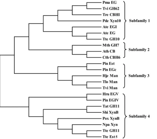
From the CAZY database, we randomly selected 23 family 1 CBM sequences. Then, the CBM phylogenetic tree was constructed (Fig. 1). Based on the topology of the tree, those CBMs were further grouped into 4 subfamilies. One to three CBMs from each subfamily were chosen as the candidate CBMs. The interaction between 3-D structures of the candidate CBM and cellobiose was predicted by MD simulation, and then the binding free energy was calculated using the AutoDock 4.2 program (Table 1). As a result, a CBM of the TrCBH I having the lowest binding free energy of 22.27 kcal/mol was selected, and fused into the C-terminus of AuMan5A. The molecule-docked confor-mation of the CBM with cellobiose was illustrated (Fig. 2).
Construction of the Fusionb-mannanase Gene
About 1050- and 210-bp bands of theAuman5Aandlcbmwere
amplified from the pUCm-T-Auman5A and pUCm-T-Trcbh,
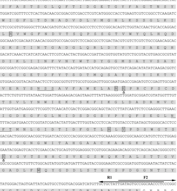
respectively. Then, they were subjected to the overlapping PCR to construct the fusion gene. As a result, an about 1250-bp band of theAuman5A-cbmwas amplified and inserted into pUCm-T. DNA sequencing result verified that the clonedAuman5A-cbmis 1241 bp in length (containing EcoR I and Not I sites), encoding an AuMan5A-CBM of 408 amino acid residues with a theoretical molecular mass of 43,798 Da and a pI of 4.22. The AuMan5A-CBM consists of a 345-aa CD from the AuMan5A, and both a 27-aa linker and a 36-27-aa CBM from the TrCBH I (Fig. 3).
Screening and Expression of theP. pastoris Transformants
A total of 30P. pastoristransformants withAuman5Arespectively resistant to 1.0, 2.0 and 4.0 mg/mL of geneticin G418, numbered as P. pastoris GSAuM1-1 to GSAuM1-10, GSAuM2-1 to GSAuM2-10 and GSAuM4-1 to GSAuM4-10, and 30 transfor-mants withAuman5A-cbm, numbered asP. pastorisGSAuMC1-1 to GSAuMC1-10, GSAuMC2-1 to GSAuMC2-10 and GSAuMC4-1 to GSAuMC4-10, were picked out for flask expression tests. P. pastorisGS115 transformed with pPIC9K, numbered asP. pastoris
GSC, was used as the negative control. After induction by adding 1.0% (v/v) methanol at 24 h intervals for 96 h, the cultured supernatants were used forb-mannanase activity assay. As a result, two transformants with the highest reAuMan5A-CBM and reAuMan5A activities of 40.6 and 44.1 U/mL, numbered asP. pastorisGSAuMC4-5 and GSAuM4-8, were selected, respectively. Nob-mannanase activity ofP. pastorisGSC was detected under the same expression conditions.
Purification of the Recombinantb-mannanases
The amount of the reAuMan5A-CBM or reAuMan5A in the cultured supernatant of the GSAuMC4-5 or GSAuM4-8 account-ed for more than 85% of that of the total protein. So they were purified to homogeneity only by a simple combination of ammonium sulfate precipitation, ultrafiltration and Sephadex G-75 gel filtration (Fig. 4). Specific activities of the purified reAuMan5A-CBM and reAuMan5A, towards locust bean gum under the standard assay conditions, were 312.3 and 341.3 U/mg, respectively.
Enzymatic Properties of the Recombinant Enzymes
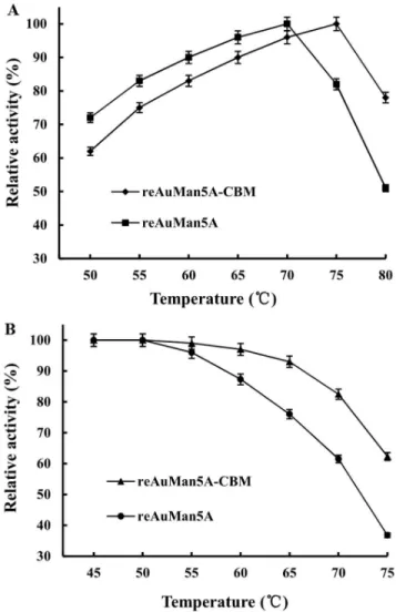
The temperature optima of the reAuMan5A-CBM and reAuMan5A were 75 and 70uC (measured at pH 3.6), respectively
(Fig. 5A). They were highly stable at temperatures of 68 and 60uC, respectively, but over which their residual activities declined
Figure 1. The phylogenetic tree showing the evolutionary relativity and homology among family 1 CBMs.The CBM-containing hydrolases are as follows: Pma EG,P. marneffeiendoglucanase (XP_002152969); Tvi GH62,T. virensGH family 62 enzyme (EHK19840); Tre CBH I,T. reeseicellobiohydrolase I (EGR44817); Pde Xyn10,P. decumbensfamily 10 xylanase (ADX86896); Ate EG I,A. terreusendoglucanase I (XP_001217291); Ate EG,A. terreusendoglucanase (AAW68436); Tte GH10,T. terrestrisGH family 10 enzyme (XP_003653793); Mth GH7,M. thermophilaGH family 7 enzyme (XP_003663441); Ath CB,A. thermophilumcellobiosidase (CAM98445); Cth CBH6,C. thermophilumfamily 6 cellobiohydrolase (AAY88915); Pin Est,P. indicaacetylxylan esterase (CCA73570); Pin EGc,P. indicaendoglucanase c (CCA67649); Hje Man,H. jecorinamannanase (AAA34208); Tlo Man,T. longibrachiatummannanase (ADN93457); Tvi Man,T. viridemannanase (AFP95336); Hru EG V,H. rufaendoglucanase V (AAQ21385); Pin EG IV,P. indica
endoglucanase IV (CCA70703); Tat GH11,T. atrovirideGH family 11 enzyme (EHK46770); Shi XynB,S. hirsutumxylanase B (EIM91441); Pox XynB,P. oxalicumxylanase B (ADV31286); Npa Xyn,N. patriciarumxylanase (ABW04217); Tte GH11,T. terrestrisGH family 11 enzyme (XP_003649436); Tte Est5,
T. terrestrisfamily 5 carbohydrate esterase (XP_003653797). doi:10.1371/journal.pone.0064766.g001
Table 1.The binding free energy of the candidate CBM combining with cellobiose.
CBM-containing hydrolase GenBank accession no.
Length of CBM (aa)
Aromatic residues in the groove surface
Apolar area/energy of CBM(A˚2)
Binding free energy (kcal/ mol)
Pma EG XP_002152969 34 Trp3, Tyr29, Tyr30 1775.44
21.56 Tre CBH I EGR44817 36 Tyr5, Tyr13, Tyr31, Tyr32 1644.23 22.27
Ate EG AAW68436 33 Trp10, Tyr20, Trp28, Tyr29 1780.38 21.21
Pin Est CCA73570 33 Tyr2, Tyr10, Tyr23, Trp27, Tyr28
1753.77 21.64
Tvi Man AFP95336 34 Tyr2, Tyr8, Tyr23, Tyr26,
Trp27, Tyr28 1776.76 21.98
Hru EG V AAQ21385 33 Tyr2, Trp10, Trp28, Tyr29 1727.44 21.65
Pox XynB ADV31286 20 Trp15, Tyr16 1275.21 21.78
Tte GH11 XP_003649436 33 Trp3, Trp11, Trp28, Tyr29 1725.45
21.16
rapidly and only retained 62.3 and 36.8% of the original ones at 75uC, respectively (Fig. 5B).
The Km and Vmax values of the reAuMan5A-CBM were
determined to be 0.66 mg/mL and 389.1 U/mg, respectively, and those of the reAuMan5A were 1.36 mg/mL and 415.8 U/mg, respectively. The reAuMan5A-CBM displayed an obvious de-crease inKmbut a slight alteration inVmax, as compared with the reAuMan5A.
Cellulose-binding Capacity
A total of 20 mL of dialyzed cultured supernatant, in which the reAuMan5A-CBM or reAuMan5A activity was adjusted to 20 U/ mL, was loaded onto the cellulose column. The test results showed that cellulose-binding capacity of the reAuMan5A-CBM with Avicel PH-101 was up to 92.3%, whereas that of the reAuMan5A was not detected.
Discussion
Mannans, the major hemicelluloses in plant cell walls and the specific substrates forb-mannanases, are always cross-linked with various kinds of carbohydrates, such as celluloses, xylans and arabinans [19]. CBMs often existed in some hydrolases decom-posing plant cell walls, such as b-mannanases, xylanases and arabino furanosidases. Being natively in enzymes or by fusing CBMs into the N- or C-termini of other enzymes, the CBMs could obviously enhance the catalytic efficiency of enzymes by increasing their local concentrations around the polysaccharide substrates, and/or improve the thermostability [17]. Because the AuMan5A consists of an only CD, it is reasonable to perfect its enzymatic properties, such as thermostability and cellulose-binding capacity, by fusing a CBM into the AuMan5A. However, there are a total of 25,580 CBMs classified into 64 families in the CAZY database, so the choice of a suitable CBM from so numerous CBMs is time-consuming and laborious. It was reported that the family 1 CBMs are mainly cellulose-binding modules, while celluloses are often covalently and non-covalently attached to mannans [5]. There-fore, our present work was focused on the family 1 CBMs.
The MD simulation can rapidly predict the most suitable binding position for a ligand on a given macromolecule and
calculate the binding free energy, which was used as a powerful tool in indirectly reflecting the affinity between ligand and receptor, such as the design of human phospholipase A2 or HIV-1 protease inhibitors [20,21]. In this work, we selected 23 family 1 CBMs to construct the phylogenetic tree, from which 8 candidate CBMs were chosen. And their 3-D structures were modeled based on a CBM crystal one of the TrCBH I. Next, the interaction between 3-D structures of the candidate CBM and cellobiose was predicted, and the binding free energy was calculated. And finally, a CBM having the lowest binding free energy was selected, and then theAuman5A-cbmwas constructed and expressed inP. pastorisGS115. As can be seen from Fig. 2, several aromatic amino acids are exposed to the groove surface of a CBM of the TrCBH I. In recent years, the 3-D structures of representative members from 22 CBM families have been resolved. Data from these 3-D structures speculated that these aromatic amino acids may play a key role in binding cellobiose or carbohydrates [22], which needs to be proved by our further studies.
P. pastoristransformant that can resist a higher concentration of G418 might have multiple copies of integration of a heterologous gene intoP. pastoris genome, which could potentially result in a high expression level of a heterologous protein as explained in the manual of Multi-Copy Pichia Expression Kit (Invitrogen). However, the expression level was not directly proportional to the concentration of G418 [9,23]. Due to those reasons, in this work, a total of 30 transformants withAuman5A-cbm resistant to different concentrations of G418 and 30 transformants with
Auman5Awere picked out for flask expression tests. This screening procedure has been applied to conduct the over-expression of other recombinant proteins or enzymes inP. pastoris[24,25].
TheP. pastorisexpression system has many advantages, one of which is that the purity of expressed recombinant proteins or enzymes is high. It was reported that the purity of recombinantA. sulphureusb-mannanase expressed inP. pastorisX-33 was 97% [9]. In this work, the purity of reAuMan5A-CBM or reAuMan5A was more than 85%. So they were purified to homogeneity only by a simple combination of ammonium sulfate precipitation, ultrafil-tration and Sephadex G-75 gel filultrafil-tration (Fig. 4). The purified reAuMan5A-CBM and reAuMan5A were N-glycosylated
pro-Figure 2. The molecule-docked conformation between 3-D structures of a CBM of the TrCBH I and cellobiose.(A) a cartoon model and (B) a surface model.
Figure 3. Nucleotide sequence of theAuman5A-cbmand its deduced amino acid sequence of the AuMan5A-CBM.The amino acid residues of the AuMan5A are marked in grayed background. A linker of theT. reeseiCBH I is underlined and its CBM is boxed. Two triangles below the boxed letters indicate the catalytic residues (E168 and E276) and five active site residues (R52, N167, H241, Y243, and W306) are located in grayed boxes. Two putative N-glycosylation sites of the AuMan5A-CBM are underlined in grayed background. The bold arrows above the letters represent the PCR primers.
teins, which were confirmed by N-deglycosylation and carbohy-drate content assays.
The temperature optimum and stability of the reAuMan5A-CBM were 75 and 68uC, which were 5 and 8uC higher than those of the reAuMan5A, respectively, indicating that the fusion of a CBM of the TrCBH I conferred the elevated tolerance to high temperature on the AuMan5A. TheKmvalue (0.66 mg/mL) of the reAuMan5A-CBM was much lower than that (1.36 mg/mL) of the reAuMan5A, implying that the former has a higher affinity towards locust bean gum. The cellulose-binding capacity of the reAuMan5A-CBM with Avicel PH-101 was up to 92.3%, suggesting that it can be gathered around the natural substrate (that is, mannan cross-linked with cellulose, xylan and arabinan) by exclusively binding cellulose [26].
Conclusions
In this work, the fusionb-mannanase gene,Auman5A-cbm, was constructed as it was designed theoretically, and functionally expressed inP. pastorisGS115. The fusion of a family 1 CBM of the TrCBH I into the C-terminus of AuMan5A obviously improved its thermostability and cellulose-binding capacity, which are beneficial for industrial applications. The superior properties of the reAuMan5A-CBM make it a good candidate in industrial
processes. To our knowledge, this work first established a novel strategy for molecular modification of enzymes byin silicodesign. Acknowledgments
We are grateful to Prof. Xianzhang Wu (School of Biotechnology, Jiangnan University) for providing technical assistance.
Author Contributions
Conceived and designed the experiments: MCW JFL CDT. Performed the experiments: CDT XHW. Analyzed the data: RM SJG JQW. Contributed reagents/materials/analysis tools: XY. Wrote the paper: CDT MCW.
References
1. van Zyl WH, Rose SH, Trollope K, Go¨rgens JF (2010) Fungalb-mannanases: Mannan hydrolysis, heterologous production and biotechnological applications. Proc Biochem 45: 1203–1213.
2. Heck JX, de Barros Soares LH, Ayub MAZ (2005) Optimization of xylanase and mannanase production byBacillus circulansstrain BL53 on solid-state cultivation. Enzyme Microb Technol 37: 417–423.
Figure 4. SDS-PAGE analysis of the purified reAuMan5A-CBM and reAuMan5A. Lane M, standard protein marker; lane 1, the AuMan5A-CBM with an apparent molecular mass of 57.3 kDa; lane 2, the reAuMan5A with an apparent molecular mass of 49.8 kDa. doi:10.1371/journal.pone.0064766.g004
Figure 5. Effects of temperature on (A) activity and (B) stability of the reAuMan5A-CBM and reAuMan5A.Temperature optima of the reAuMan5A-CBM and reAuMan5A were determined, respectively, under the standard assay conditions, except temperatures ranging from 50 to 80uC. Their thermostabilities were evaluated, respectively, by incubating them at pH 3.6 and various temperatures from 45 to 75uC for 1.0 h, and the residual enzyme activities were measured under the standard assay conditions.
3. Wu MC, Tang CD, Li JF, Zhang HM, Guo J (2011) Bimutation breeding of
Aspergillus nigerstrain for enhancing b-mannanase production by solid-state fermentation. Carbohydr Res 346: 2149–2155.
4. Roth R, Moodley V, van Zyl P (2009) Heterologous expression and optimized production of anAspergillus aculeatusendo-1,4-b-mannanase inYarrowia lipolytica. Mol Biotechnol 43: 112–120.
5. Guille´n D, Sa´nchez S, Rodrı´guez-Sanoja R (2010) Carbohydrate-binding domains: multiplicity of biological roles. Appl Microbiol Biotechnol 85: 1241– 1249.
6. Ravalason H, Herpoe¨l-Gimbert I, Record E, Bertaud F, Grisel S, et al. (2009) Fusion of a family 1 carbohydrate binding module ofAspergillus nigerto the
Pycnoporus cinnabarinuslaccase for efficient softwood kraft pulp biobleaching. J Biotechnol 142: 220–226.
7. Zhang Y, Chen S, Xu M, Cavaco-Paulo A, Wu J, et al. (2010). Characterization ofThermobifida fuscacutinase-carbohydrate-binding module fusion proteins and their potential application in bioscouring. Appl Environ Microbiol 76: 6870– 6876.
8. Zhang YL, Ju JS, Peng H, Gao F, Zhou C, et al. (2008) Biochemical and structural characterization of the intracellular mannanase AaManA of
Alicyclobacillus acidocaldariusreveals a novel glycoside hydrolase family belonging to clan GH-A. J Biol Chem 283: 31551–31558.
9. Chen XL, Cao YH, Ding YH, Lu WQ, Li DF (2007) Cloning, functional expression and characterization ofAspergillus sulphureusb-mannanase inPichia pastoris. J Biotechnol 128: 452–461.
10. Stalbrand H, Saloheimo A, Vehmaanpera J, Henrissat B, Penttila M (1995) Cloning and expression in Saccharomyces cerevisiae of a Trichoderma reesei b -mannanase gene containing a cellulose binding domain. Appl Environ Microbiol 61: 1090–1097.
11. Benech RO, Li XM, Patton D, Powlowski J, Storms R, et al. (2007) Recombinant expression, characterization, and pulp prebleaching property of aPhanerochaete chrysosporiumendo-b-1,4-mannanase. Enzyme Microb Technol 41: 740–747.
12. Tang CD, Guo J, Wu MC, Zhao SG, Shi HL, et al. (2011) Cloning and bioinformatics analysis of a novel acidophilicb-mannanase gene,Auman5A, from
Aspergillus usamiiYL-01–78. World J Microbiol Biotechnol 27: 2921–2929. 13. Timmers LF, Ducati RG, Sanchez-Quitian ZA, Basso LA, Santos DS, et al.
(2012) Combining molecular dynamics and docking simulations of the cytidine deaminase fromMycobacterium tuberculosisH37Rv. J Mol Model 18: 467–479.
14. Phosrithong N, Ungwitayatorn J (2010) Molecular docking study on anticancer activity of plant-derived natural products. Med Chem Res 19: 817–835. 15. Li JF, Zhao SG, Tang CD, Wang JQ, Wu MC (2012) Cloning and functional
expression of an acidophilicb-mannanase gene (Anman5A) fromAspergillus niger
LW-1 inPichia pastoris. J Agric Food Chem 60: 765–773.
16. Laemmli UK (1970) Cleavage of structural proteins during the assembly of the head of bacteriophage T4. Nature 227: 680–685.
17. Thongekkaew J, Ikeda H, Iefuji H (2012) Increases thermal stability and cellulose-binding capacity ofCryptococcus sp. S-2 lipase by fusion of cellulose binding domain derived fromTrichoderma reesei. Biochem Biophys Res Commun 420: 183–187.
18. Dubois M, Gilles KA, Hamilton JK, Rebers PA, Smith F (1956) Colorimetric method for determination of sugars and related substances. Anal Chem 28: 350– 356.
19. Moreira LRS, Filho EXF (2008) An overview of mannan structure and mannan-degrading enzyme systems. Appl Microbiol Biotechnol 79: 165–178. 20. Ortiz AR, Pisabarro MT, Gago F, Wade RC (1995) Prediction of drug binding
affinities by comparative binding energy analysis. J Med Chem 38: 2681–2691. 21. Perez C, Pastor M, Ortiz AR, Gago F (1998) Comparative binding energy analysis of HIV-1 protease inhibitors: incorporation of solvent effects and validation as a powerful tool in receptor-based drug design. J Med Chem 41: 836–852.
22. Shoseyov O, Shani Z, Levy I (2006) Carbohydrate binding modules: biochemical properties and novel applications. Microbiol Mol Biol Rev 70: 283–295.
23. Li JF, Tang CD, Shi HL, Wu MC (2011) Cloning and optimized expression of a neutral endoglucanase gene (ncel5A) from Volvariella volvacea WX32 inPichia pastoris. J Biosci Bioeng 111: 537–540.
24. Tan ZB, Li JF, Wu MC, Tang CD, Zhang HM, et al. (2011) High-level heterologous expression of an alkaline lipase gene fromPenicillium cyclopiumPG37 inPichia pastoris. World J Microbiol Biotechnol 27: 2767–2774.
25. Shi HL, Yin X, Wu MC, Tang CD, Zhang HM, et al. (2012) Cloning and bioinformatics analysis of an endoglucanase gene (Aucel12A) from Aspergillus usamiiand its functional expression inPichia pastoris. J Ind Microbiol Biotechnol 39: 347–357.