359
Gomes LM et al. Primary bone lymphoma simulating sarcomatous degeneration
Radiol Bras. 2012 Nov/Dez;45(6):359–361
Primary bone lymphoma simultaneous to osteochondroma
simulating sarcomatous degeneration: case report
*
Linfoma ósseo primário simultâneo a osteocondroma simulando degeneração sarcomatosa: relato de caso
Laura de Moraes Gomes1, Felipe Augusto Rozales Lopes2, Décio Valente Renck3
In the literature, there is no evidence of relationship between primary bone lymphoma and osteochondroma or of coexistence of both of them in a single bone. The present report describes an uncommon case of primary bone lymphoma occurring simultaneously with osteochondroma in the proximal third of the tibia. In the present case, magnetic resonance imaging signs simulated the presence of sarcomatous degeneration.
Keywords: Primary bone lymphoma; Osteochondroma; Sarcomatous degeneration; Bone tumors.
Não há evidências relatadas na literatura de associação entre linfoma ósseo primário e osteocondroma ou da coexis-tência deles em uma mesma região óssea. Este relato de caso descreve um caso raro de linfoma ósseo primário ocor-rendo juntamente com um osteocondroma no terço proximal de tíbia. Os sinais de imagem na ressonância magnética neste caso simulam uma degeneração sarcomatosa do osteocondroma.
Unitermos: Linfoma ósseo primário; Osteocondroma; Degeneração sarcomatosa; Tumores ósseos.
Abstract
Resumo
* Study developed in the Center of Imaging Diagnosis at Hos-pital Santa Casa de Misericórdia de Pelotas, Pelotas, RS, Brazil. 1. Master, MD, Radiologist, Hospital Santa Casa de Miseri-córdia de Pelotas, Pelotas, RS, Brazil.
2. Graduate Student (6th year) of Medicine, Universidade Católica de Pelotas (UCPel), Pelotas, RS, Brazil.
3. Master, MD, Radiologist, Chief of the Center of Imaging Diagnosis, Hospital Santa Casa de Misericórdia de Pelotas, Pe-lotas, RS, Brazil.
Mailing Address: Felipe Augusto Rozales Lopes. Rua Emílio Jorge dos Reis, 512, Três Vendas. Pelotas, RS, Brazil, 96020-440. E-mail: [email protected]
Received February 24, 2012. Accepted after revision July 5, 2012.
Gomes LM, Lopes FAR, Renck DV. Primary bone lymphoma simultaneous to osteochondroma simulating sarcomatous degeneration: case report. Radiol Bras. 2012 Nov/Dez;45(6):359–361.
0100-3984 © Colégio Brasileiro de Radiologia e Diagnóstico por Imagem
CASE REPORT
tion secondary to osteochondroma (Figures 2, 3 and 4).
Additionally to the infiltrative lesion identified in the tibia, a well-defined focal lesion was found in the bone marrow of the scribing a case of primary bone lymphoma
simultaneous to osteochondroma which, at imaging studies, simulated sarcomatous degeneration in the proximal third of the tibia.
CASE REPORT
A 65-year-old, male patient with no his-tory of trauma presented intense and dis-abling pain in the proximal third of his tibia for nine months. At physical examination a small palpable mass was found with lo-cal sensation of heat.
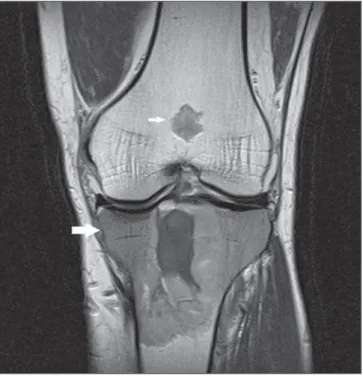
Radiography demonstrated the presence of an osteochondroma in the posterosupe-rior portion of the tibia with radiolucent areas and centrally located radiodense ar-eas suggesting the presence of calcifica-tions within the osteochondroma (Figure 1). Magnetic resonance imaging (MRI) re-vealed the presence of an infiltrative bone lesion with heterogeneous contrast en-hancement and marked bone marrow infil-tration extending to the adjacent soft tis-sues, with no noticeable cortical bone de-struction. MRI also demonstrated intra-ar-ticular compromise by the lesion and bone marrow involvement by the osteochon-droma. Such imaging findings were ini-tially interpreted as sarcomatous degenera-INTRODUCTION
Primary bone lymphoma and sarcoma-tous degeneration secondary to osteochon-droma are rare tumors and no relation be-tween such entities is found in the litera-ture.
Primary bone lymphoma represents 1% of extranodal non-Hodgkin’s lymphomas, occurring predominantly in the sixth and seventh decades of life(1).On the other
hand, osteochondroma represents 20% of benign bone tumors and usually is found in the second and third decades of life. Ma-lignant transformation if found in 1% of cases of solitary osteochondromas(2).
Since no study reporting the concomi-tant presence of these two tumors in a single bone region has been found in the lit-erature, the present report is aimed at
360
Gomes LM et al. Primary bone lymphoma simulating sarcomatous degeneration
Radiol Bras. 2012 Nov/Dez;45(6):359–361 DISCUSSION
According to Nava et al.(3), MRI has been utilized for staging lymphomas be-cause of its high sensitivity for detecting malignant tumors, constituting an excellent alternative to the method utilized in the present study, CT.
Primary bone lymphoma is character-ized by intermittent local pain that may persist for months(4). It is commonly lo-cated in long bones, preferentially in the femur and pelvic bones. Primary bone lym-phomas originated in the center of the bone may disrupt the cortical bone as the med-ullary cavity is already occupied by the le-sion and there is a risk for involvement of soft tissues(5).
As regards the radiological appearance of bone lymphomas, there is a permeative or moth-eaten pattern, causing periosteal reaction. The most common pattern is the lytic-destructive one, with radiolucent foci on an ill delimited area. A blastic/sclerotic pattern may be observed, although rarely as compared with bone metastases from lym-phoma. At MRI, T1-weighted sequences are the most appropriate to demonstrate medullary changes, showing areas of Figure 3. Coronal MRI T1-weighted image showing an infiltrative lesion in the proximal epiphysis and diaphysis of the tibia (larger arrow). A focal, well-de-fined bone lesion is observed in the femoral metaphysis (smaller arrow) with lobular configuration and presence of calcifications, suggestive of chondroma. Figure 2. Sagittal MRI T1-weighted image with fat saturation demonstrating
neoplastic involvement of osteochondroma (black arrow), intra-articular com-promise by the tumor (smaller white arrow), as well as lesion extension to-wards soft tissues (larger white arrow).
femoral metaphysis, with hyposignal on T1-weighted sequences, lobular configura-tion and presence of calcificaconfigura-tions sugges-tive of chondroma.
Biopsy identified a diffuse peripheral B-cell non-Hodgkin’s lymphoma, with posi-tive CD20, CD3, CD5 markers; and stag-ing computed tomography (CT) did not demonstrate the presence of enlarged
lymph nodes, which confirmed primary bone lymphoma.
It is important to note that among the de-scribed imaging findings, the cortical bone was relatively preserved, even with the adjacent soft tissues infiltration, corrobo-rating the histopathological diagnosis of lymphoma.
361
Gomes LM et al. Primary bone lymphoma simulating sarcomatous degeneration
Radiol Bras. 2012 Nov/Dez;45(6):359–361 hyposignal within the bone marrow. Both on T2-weighted sequences and fluid-sen-sitive MRI sequences with fat suppression demonstrate areas of hypersignal. In cases of radiological permeative pattern, soft tis-sues involvement is observed. Both CT and MRI reveal cortical bone erosion(6).
Like bone lymphomas, osteochondro-mas are most commonly found in long bones, particularly the humerus and the femur, and are preferentially located in metaphyseal regions. Osteochondromas present easily recognisable radiological signs at conventional radiography, demon-strating cortical and spongy bone continu-ity with adjacent bone tissues. Both MRI and CT confirm such characteristic.
The radiological aspect of sarcomatous degeneration includes characteristics of osteochondroma growth with irregular sur-face, focal regions of radiolucency within the lesion, erosion or destruction of adja-cent bone tissues. MRI characterizes low-grade chondrosarcomas with hypersignal on T2-weighted sequences and hyposignal
on tumor septations with post-contrast en-hancement(7).
The definitive diagnosis of bone lym-phoma is achieved by means of histologi-cal study of the lesion. For immunohis-tochemical diagnosis, LCA, CD20 and CD3 markers should be investigated(8). The
differential diagnosis includes osteosar-coma, Ewing’s tumor and neoplastic me-tastases(5).
Finally, the occasional finding of an os-teochondroma simultaneous to a bone lym-phoma in a single bone region has raised doubts regarding the imaging diagnosis, initially leading to the belief that the find-ings corresponded to sarcomatous degen-eration secondary to osteochondroma. Ad-ditionally, the diagnostic difficulty was determined by the disease rarity, and hence the relevance of differential diagnoses should be emphasized.
REFERENCES
1. Krishnan A, Shirkhoda A, Tehranzadeh J, et al. Primary bone lymphoma: radiographic-MR imag-ing correlation. Radiographics. 2003;23:1371–83.
2. Kurimori CO, Bordalo-Rodrigues M, Cerri GG. Degeneração maligna para condrossarcoma de um osteocondroma pediculado da tíbia. Radiol Bras. 2010;43(1):xi–xii.
3. Nava D, Oliveira HC, Luisi FA, et al. Aplicação da ressonância magnética de corpo inteiro para o es-tadiamento e acompanhamento de pacientes com linfoma de Hodgkin na faixa etária infanto-juve-nil: comparação entre diferentes sequências. Ra-diol Bras. 2011;44:29–34.
4. Pires de Camargo O, Machado TMS, Croci AT, et al. Primary bone lymphoma in 24 patients treated between 1955 and 1999. Clin Orthop Relat Res. 2002;(397):271–80.
5. Edeiken J. Diagnóstico radiológico de las enferme-dades de los huesos. 3ª ed. Buenos Aires: Edito-rial Medica Panamericana; 1984; p. 66–73, 303– 14.
6. Gomes FSE, Lewin F, Mariotti GC, et al. Osteo-condromas: avaliação por imagem das complica-ções. Rev Imagem. 2007;29:53–9.
7. Hicks DG, Gokan T, O’Keefe RJ, et al. Primary lymphoma of bone. Correlation of magnetic reso-nance imaging features with cytokine production by tumor cells. Cancer. 1995;75:973–80. 8. Neupatimagem-Unicamp. Linfoma não-Hodgkin