Anastomotic leaks after Roux-en-Y gastric bypass
surgery by Higa’s technique for treatment of morbid
obesity: radiological findings*
Fístulas de anastomose superior pós-gastroplastia redutora pela técnica de Higa para tratamento da obesidade mórbida: aspectos por imagem
Ester Moraes Labrunie1, Edson Marchiori2, Jean-Michel Tubiana3
OBJECTIVE: The present study was aimed at describing main radiological findings in patients who developed leaks as a complication of Roux-en-Y gastric bypass surgery by the Higa’s technique. MATERIALS AND METHODS: Twenty-four patients with post-gastric bypass anastomotic leaks were evaluated by means of computed tomography or gastrointestinal series. RESULTS: Leaks of superior anastomoses generally occurred within 30 postoperative days. Nineteen patients were radiologically diagnosed with anastomotic leaks, and ten of them presented contrast material extravasation, which is considered as a direct sign of the presence of anastomotic leaks. Seven of the remainder nine patients demonstrated contrast extravasation on subsequent studies, and indirect signs were also observed in six cases. Indirect signs were also observed in the patients with contrast material extravasation, the most frequent finding being pneumoperitoneum. Later follow-up examinations demonstrated contrast extravasation in one, and indirect signs in four of the five patients who had not been radiologically diagnosed. CONCLUSION: The most frequent radiological finding was contrast material extravasation (a direct sign of leak). Indirect signs were: unusual air-fluid levels, intracavitary fluid collections, disproportionate postoperative pneumoperitoneum, presence of fluid in the peritoneal cavity, anastomotic edema and small-bowel distention.
Keywords: Morbid obesity; Bariatric surgery; Anastomotic leak; Complication; Radiology; Computed tomogra-phy.
OBJETIVO: Descrever os principais aspectos radiológicos encontrados nas fístulas pós-operatórias de anas-tomose superior em pacientes submetidos a derivação gastrintestinal em Y de Roux pela técnica de Higa. MATERIAIS E MÉTODOS: Foram estudados 24 pacientes com fístula de anastomose no pós-operatório de gastroplastia redutora, avaliados por tomografias computadorizadas e/ou seriografias esofagogastrojejunais. RESULTADOS: As fístulas de anastomose superior ocorreram até o 30º dia de pós-operatório. Dezenove pacientes realizaram exame radiológico no momento do diagnóstico, sendo observado extravasamento de contraste, considerado sinal direto de fístula de anastomose, em dez pacientes. Dos nove restantes, em sete foi evidenciado extravasamento em exames subseqüentes, sendo ainda identificados sinais indiretos de fís-tula em seis destes. Sinais indiretos foram observados também em pacientes com extravasamento de con-traste nos exames iniciais, sendo o pneumoperitônio o aspecto mais freqüente. Dos cinco pacientes sem exame radiológico no momento do diagnóstico, exames subseqüentes evidenciaram extravasamento de contraste em um e sinais indiretos em quatro pacientes. CONCLUSÃO: O achado radiológico mais comum foi o extravasamento de contraste (sinal direto de fístula). Os sinais indiretos foram: nível líquido bizarro, coleção intracavitária, pneumoperitônio desproporcional ao tempo pós-operatório, líquido na cavidade peri-toneal, edema da anastomose inferior e distensão de delgado.
Unitermos: Obesidade mórbida; Cirurgia bariátrica; Fístula de anastomose; Complicação; Radiologia; Tomo-grafia computadorizada.
Abstract
Resumo
* Study developed in the Department of Radiology at Univer-sidade Federal do Rio de Janeiro (UFRJ) and Casa de Saúde São José, Rio de Janeiro, RJ, Brazil.
1. PhD, Associate Professor, Universidade Federal do Rio de Janeiro (UFRJ), MD, Radiologist at Casa de Saúde São José, Rio de Janeiro, RJ, Brazil.
2. Full Professor, Department of Radiology, Universidade Fe-deral Fluminense (UFF), Niterói, RJ, Adjunct Coordinator for the Course of Post-Graduation in Radiology, Universidade Federal do Rio de Janeiro (UFRJ), Rio de Janeiro, RJ, Brazil.
INTRODUCTION
Obesity is a chronic disease character-ized by excessive body fat, frequently as-sociated with a series of related diseases, with increased risk of physical disability and early mortality. An endemic increase of
Labrunie EM, Marchiori E, Tubiana JM. Anastomotic leaks after Roux-en-Y gastric bypass surgery by Higa’s technique for treatment of morbid obesity: radiological findings. Radiol Bras. 2008;41(2):75–79.
3. Service de Radiologie, Assistance-Publique-Hôpitaux de Pa-ris, Faculté de Médicine Pierre et Marie Curie, Hôpital Saint-Antoine, Paris, France.
Mailing address: Dr. Edson Marchiori. Rua Thomaz Cameron, 438, Valparaíso. Petrópolis, RJ, Brazil, 25685-120. E-mail: [email protected]
this disease has been observed world-wide(1,2). Morbid obesity is defined as a body mass index of > 40 kg/m², or yet ≥ 35 kg/m² in patients with comorbidity(1,3).
Non-surgical treatments outcomes have been disappointing, and bariatric surgery has been indicated as the only reliable method allowing significant weight loss, extended weight maintenance and manage-ment and reversion of some obesity-related comorbidities(1,4,5).
Postoperative radiological evaluation, aiming at an early diagnosis of possible complications in patients submitted to bariatric surgery, is frequently part of radi-ologists in their daily practice. The diagno-sis may be difficult because of technical limitations imposed by these patients’ bio-type and clinical condition. A detailed and careful analysis represents a challenge both to the surgical team and the radiologist. An incorrect evaluation or late diagnosis of complications may delay the treatment or even put the patients’ life at risk.
Postoperative anastomotic leak with peritonitis is the most severe early compli-cation of the Roux-en-Y gastric bypass surgery, occurring in 1% to 6% of cases(6,7). In these cases the hospitalization period is extended, and the morbidity and mortality rates are significantly increased(7–9), par-ticularly in cases of high output leaks(6). Patients with this complication are submit-ted to a higher number of diagnostic pro-cedures and, frequently require surgical reintervention(8).
The present study was aimed at evalu-ating the imaging findings observed in 24 patients who developed anastomotic leaks as a complication resulting from gastro-plasty by the Higa’s technique for treatment of morbid obesity.
MATERIALS AND METHODS
Twenty-four patients submitted to Roux-em-Y gastric bypass surgery by the Higa’s technique, with postoperative anas-tomotic leaks were studied in the period between November 2001 and April 2006. The surgeries were performed by eight dif-ferent surgical teams in eight hospitals/in-stitutions, and the study was approved by the Committee for Ethics in Research of Casa de Saúde São José, Rio de Janeiro, RJ.
The sample of the present study in-cluded 20 female (83.4%) and four male (16.6%) patients with ages ranging be-tween 23 and 56 years (mean, 38 years). Twenty-one patients underwent video-laparoscopy surgery (87.5%), and three, laparotomy (12.5%). A total of 74 iodi-nated-contrast enhanced radiological stud-ies — 55 computed tomographstud-ies (CT) and 19 upper gastrointestinal series (UGIS) — were evaluated.
The confirmation of the diagnoses of anastomotic leaks was based on one or more of the following criteria: image finding of oral contrast extravasation, opacification of surgical drain by oral methylene blue and/ or surgical confirmation. Both direct and indirect signs of leaks were investigated. Contrast material extravasation was consid-ered as a direct sign of leak. Indirect signs considered as suggestive of leak were: dis-proportionate postoperative pneumoperito-neum or progressive increase in the gas volume; intracavitary fluid collections, with or without fluid or contrast material level inside; significant amount of fluid in the subphrenic and/or perihepatic spaces.
Five patients had not been radiologi-cally diagnosed. Four of them underwent surgical reintervention, and one was sub-mitted to conservative clinical treatment. All of them were subsequently submitted to radiological examination, with detection of contrast material extravasation in one, and indirect signs of leak in the other four patients.
The leaks were classified as early (oc-curring up to the 7th postoperative day), and intermediate (occurring between the 8th and the 30th postoperative days).
RESULTS
Leakage was the first postoperative complication in 22 patients. The initial complications in the remainder two pa-tients were bowel obstruction (one case), and anastomotic edema (one case). As re-gards the moment of the leak onset, the cases were divided into: early (18 cases) and intermediate (six cases). In the present casuistic, leaks onset after the 30th postop-erative day was not observed.
Contrast material extravasation was detected at the initial examination — either
UGIS (Figure 1) or CT (Figure 2A). Sub-sequent studies demonstrated contrast ma-terial extravasation in eight of the other 14 patients. In four patients, only indirect sings of leaks were found (Figures 2B and 2C). Two patients were clinically/surgically di-agnosed.
Fifty-five CT studies were performed in the 24 patients with anastomotic leaks (one to seven studies per patient). Seventeen CT studies were performed at the moment of the complication diagnosis, eight of them demonstrating contrast material extravasa-tion, and three were normal. The other six patients demonstrated only indirect signs of leak. Alterations considered as indirect signs and observed at the initial examina-tion of these 24 patients were: pneumoperi-toneum (tem patients), anastomotic edema (five patients), intracavitary fluid collec-tions (four patients) and free intraperitoneal fluid (three patients). Follow-up CT stud-ies could demonstrate contrast material extravasation which had not been detected at the first examination in seven patients.
Thirteen patients with anastomotic leaks were submitted to upper gastrointes-tinal series. Eight studies were performed at the time of the diagnosis, with contrast material extravasation being detect in only three. Follow-up examinations allowed the detection of the leak which had not been visualized at the initial examination in two patients.
The amount and sites of contrast mate-rial extravasation were variable, these be-ing quite evident in some studies and more subtle in others. Many times, small extrav-asations through the surgical drain adjacent to the stapling line were difficult to be vi-sualized; sometimes, contrast material was permeated with gas bubbles along the drain passage.
Fifteen of the 24 patients were submit-ted to surgery after being diagnosed with anastomotic leaks either because of the extent of contrast material extravasation, because of the complications diagnosed at this first moment, or because of the neces-sity to review the anastomosis and place surgical drains. In nine patients, the treat-ment was conservative and/or by interven-tional radiology.
Figure 1. Aspects of leaks on gastrointestinal series: A: Major leakage of contrast material into the left subphrenic space (arrow) B: Minor anastomotic leak towards a surgical drain (arrow). C: Non-conclusive, unusual aspect of contrast material at UGIS (arrow) – extraluminal collection or ectactic loop? The CT image (not shown) has not demonstrated extraluminal collection of contrast material surrounding the excluded stomach.
B
A C
A B C
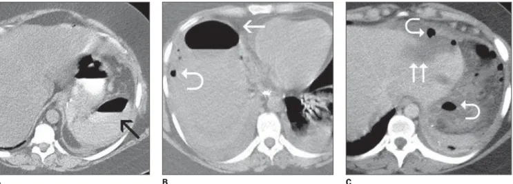
Figure 2. Aspects of leaks on CT. A: Perisplenic collection with extraluminal oral contrast material and fluid level (arrow). Percutaneously drained abscess (not shown). B: Right subphrenic collection, with fluid-gas level (arrow). Note small gas bubbles (pneumoperitoneum)(curved arrow). C: Free fluid in the subphrenic space (two arrows) and air/gas bubbles (pneumoperitoneum (curved arrows).
stays, with one case progressing to death because of septic and pulmonary compli-cations. Eleven patients evolved with sig-nificant intracavitary fluid collections, eight of them being treated by interven-tional radiology (abscesses). Other compli-cations observed were: evisceration (two cases), intracavitary hematoma after surgi-cal reintervention (two cases), lasting leaks observed at clinical follow-up for more than two months (two cases), multiple enterocutaneous fistulas (in patients with obstruction by debris and volvulus anterior to the fistula). A patient with anastomotic dehiscence was submitted to resection of
the small gastric chamber and evolved with stenosis of the esophagojejunal anastomo-sis, requiring endoscopic treatment.
DISCUSSION
Several surgical techniques are avail-able for the management of morbid obesity, each of them with possible complications, where radiological studies play an ex-tremely significant role in the diagnosis and follow-up(10,11).
Anastomotic leaks constitute one of the more severe and feared postoperative com-plications of this type of surgery, as far as
its high morbidity and mortality rates are concerned(8,12). Patients with this compli-cation require longer hospital stay, remain-ing in intensive care units because of sep-tic shock, multiple organs failure or intra-cavitary abscesses(8).
drain is a simple test frequently utilized in these cases; however, the absence of com-munication between the leak and the sur-gical drain may give false-negative re-sults(13). Patients without a surgical drain cannot be submitted to this test. Frequently, a delay in the treatment of anastomotic leaks results in progression to sepsis, or-gans failure and death(14). In the present casuistic, one of the patients diagnosed with anastomotic leak by methylene blue test, had the leak demonstrated only at the follow-up UGIS, where the filiform pas-sage of the contrast medium into the sur-gical drain was identified. Contrast mate-rial extravasation was not identified at the CT study performed in the same day, since a spontaneously dense surgical drain was visualized.
CT plays a complementary role to the upper gastrointestinal series (UGIS) in the diagnosis of anastomotic leaks(3). CT seems to be more sensitive for this evaluation, allowing the association between direct and indirect signs, as well as the investigation of secondary complications such as fluid collections(6). On the other hand, iodinated contrast-enhanced UGIS can demonstrate small contrast material extravasations into surgical drains which cannot be visualized at CT, as observe in one of the patients in the present casuistic.
Undetected small leakages may result in an extemporaneous withdrawal of the sur-gical drain and initiation of the oral diet, with harmful consequences for the patient. Small leaks oriented towards the surgical drain and with no sign of a resistant sepsis can be clinically treated by means of anti-biotics and parenteral nutrition, and usually resolve spontaneously(15). However, they also may result in intracavitary fluid collec-tions and development of a chronic fistu-lous tract(6), most frequently found in ma-jor leaks(7). Gonzalez et al.(8) have reported that the presence of a surgical drain allows the diagnosis of leaks in 50% of cases and, most of time, a clinic, conservative treat-ment of this complication in hemodynami-cally stable patients.
Several signs suggestive of anastomotic leak are described in the literature. The presence of oral contrast extravasation is a direct sign of leak. Other aspects consid-ered as indirect signs and already described
in the literature(8,16) are collections (espe-cially those adjacent to the gastric reser-voir) and free fluid in the abdominal cav-ity. In the present casuistic, such findings have also been observed. Pneumoperito-neum may be observed, although in a small volume, in the early postoperative period by the presence of a surgical drain. A higher-than-expected volume of pneumo-peritoneum, or even its increase at subse-quent examinations has also been consid-ered as an indirect sign indicating a leak. This was the indirect sign most frequently found in the present casuistic.
Blachar et al.(3) have reported that the majority of leak-related collections occur in the perianastomotic region and in the upper left quadrant of the abdomen, par-ticularly in the perisplenic region. In the present study, 13 abscesses were observed in ten patients with the following localiza-tions: left perisplenic/subphrenic (six cases), left anterior subphrenic (two cases), right subphrenic (two cases), posterior to the excluded stomach (one case), Douglas’ pouch (one case), and pararectal fossa (one case). Oral contrast extravasation into the collection was observed in two cases. Eight patients with abscesses were successfully submitted to CT-guided percutaneous puncture and drainage.
Oligosymptomatic patients may not present any evidence of leakage at an early postoperative UGIS. Later complementary studies may be required to demonstrate the presence of a leak, as well as to evaluate its extent, orientation, or not, to surgical drains, presence of collections and evalu-ation of the possibility of treatment by means of percutaneous drainage(8,12).
Ten of the 24 patients studied in the present casuistic demonstrated the presence of contrast material extravasation at the first examinations. Six of the remainder 14 patients presented solely indirect signs of leakage at the moment of the diagnosis. Subsequent CT studies and/or UGIS dem-onstrated the presence of extraluminal con-trast material in these six patients. No ab-normality was found on the initial CT/ UGIS of other three patients, whose diag-nosis was based on their clinical signs. Follow-up examination of one of these three patients demonstrated contrast mate-rial extravasation; the diagnosis was
clini-cal/surgical in two patients. Five patients included in the present study were not ra-diologically diagnosed, and active contrast extravasation was found in one of them during a follow-up examination. In four patients the diagnosis was based on the presence of indirect signs.
According to Hamilton et al.(12), the peak of tension on tissues and anastomo-sis occurs between the 5th and 7th postop-erative day, and therefore this is the period where a leakage onset probability is high-est. These data are coincidental with the present casuistic where 17 of the 24 pa-tients with anastomotic leaks presented the complication up to the 7th postoperative day, and seven between the 8th and 12th postoperative days. No leak developed af-ter this period. It is an assumption of these authors(12) that this can be a reason for the presence of leaks not being identified on UGIS performed within the first 48 post-operative hours. Such an argument cor-roborates the idea that asymptomatic pa-tients should be submitted to early routine postoperative studies.
Luján et al.(15) have highlighted that a loop distention above the intestinal ob-structions either in the alimentary or biliary branch, submit the anastomosis to a higher tension with a risk for development of leaks and/or dehiscence progressing to death in some patients. In this casuistic, leakage has been observed in two patients with intesti-nal obstruction and in five with anasto-motic edema.
pneu-moperitoneum as well as intracavitary col-lections should be taken into consideration.
REFERENCES
1. Blachar A, Federle MP. Gastrointestinal compli-cations of laparoscopic Roux-en-Y gastric by-pass surgery in patients who are morbidly obese: findings on radiography and CT. AJR Am J Roentgenol. 2002;179:1437–42.
2. Moura Jr LG, Guimarães SB, Castro-Filho HF, et al. Capella’s gastroplasty: metabolites and acute phase proteins changes in midline and bilateral arciform approaches. Arq Gastroenterol. 2004;41: 215–9.
3. Blachar A, Federle MP, Pealer KM, et al. Gastrointestinal complications of laparoscopic Roux-en-Y gastric bypass surgery: clinical and imaging findings. Radiology. 2002;223:625–32. 4. Pareja JC, Pilla VF, Callejas-Neto F, et al. Gas-tric bypass Roux-en-Y gastrojejunostomy conver-sion to distal gastrojejunoileostomy for weight loss failure – experience in 41 patients. Arq Gastroenterol. 2005;42:196–200.
5. Pories WJ, Swanson MS, MacDonald KG, et al. Who would have thought it? An operation proves to be the most effective therapy for adult-onset diabetes mellitus. Ann Surg. 1995;222:339–52. 6. Blachar A, Federle MP, Pealer KM, et al. Radio-graphic manifestations of normal postoperative anatomy and gastrointestinal complications of bariatric surgery, with emphasis on CT imaging findings. Semin Ultrasound CT MRI. 2004;25: 239–51.
7. Carucci LR, Turner MA. Radiologic evaluation following Roux-en-Y gastric bypass surgery for morbid obesity. Eur J Radiol. 2005;53:353–65.
8. Gonzalez R, Nelson LG, Gallagher SF, et al. Anastomotic leaks after laparoscopic gastric bypass. Obes Surg. 2004;14:1299–307.
9. See C, Carter PL, Elliott D, et al. An institutional experience with laparoscopic gastric bypass complications seen in the first year compared with open gastric bypass complications during the same period. Am J Surg. 2002;183:533–8. 10. Labrunie EM, Marchiori E. Obstrução intestinal
pós-gastroplastia redutora pela técnica de Higa
para tratamento da obesidade mórbida: aspectos de imagem. Radiol Bras. 2007;40:161–5.
11. Francisco MC, Barella SM, Abud TG, et al. Aná-lise radiológica das alterações gastrintestinais após cirurgia de Fobi-Capella. Radiol Bras. 2007; 40:235–8.
12. Hamilton EC, Sims TL, Hamilton TT, et al. Clinical predictors of leak after laparoscopic Roux-en-Y gastric bypass for morbid obesity. Surg Endosc. 2003;17:679–84.
13. Onopchenko A. Radiological diagnosis of internal hernia after Roux-en-Y gastric bypass. Obes Surg. 2005;15:606–11.
14. Singh R, Fisher BL. Sensitivity and specificity of postoperative upper GI series following gastric bypass. Obes Surg. 2003;13:73–5.
15. Luján JA, Frutos MD, Hernández Q, et al. Laparoscopic versus open gastric bypass in the treatment of morbid obesity: a randomized prospective study. Ann Surg. 2004;239:433–7. 16. Yu J, Turner MA, Cho SR, et al. Normal anatomy