Introduction
Systemic hypertension is a multifactorial clinical condition
and an independent risk factor for mortality in patients with
cardiovascular diseases
1. Increase in blood pressure is linked to
neural mechanisms, e.g., autonomic dysfunction, sympathetic
hyperactivity and disarray in arterial baroreceptors and
chemo-receptors
2,3,4. The arterial baroreceptor in hypertensive subjects
adapts to high levels of blood pressure, via receptors
depolar-ization reduction, diminishing thus its functional capacity
5,6.
The role of physical exercise in blood pressure reduction has
been well documented in animal models
7and humans
8,9, and
this phenomenon occurs due to adjustments in the neurohumoral
mechanisms. Post-exercise hypotension [PEH] is a prolonged
decrease in arterial blood pressure after a single bout of exercise
7.
The autonomic mechanisms attributed to PEH are
well-documented, such as reduced peripheral sympathetic activity
10,11,
modiications in the cardiac autonomic activity
12,13and
adjust-ments in the barorelex sensitivity
14,15.
On the other hand, the
central neural mechanisms have only recently been discovered
and investigated. This review emphasizes on evidence of synaptic
mechanisms in the central barorelex pathway that contribute
to development of PEH. Therefore, we will summarize studies
that deine important areas of central nervous system (CNS)
involved in physical activities and blood pressure regulation.
Central barorelex arc, blood pressure, and sympathetic
activity
Regulation of the cardiovascular system by the barorelex in
-volves multiple components of the barorelex arc, such as sensors
(baroceptors), afferents pathway (depressor nerve aortic), central
circuit (nucleus tractus solitarii (NTS) and others brain areas), and
efferent pathway (heart, vessels). The afferent ibers baroreceptor,
which carries blood pressure information, makes an excitatory
synaptic contact with second-order neurons in the NTS. The NTS
integrates and receives information from arterials baroceptors and
through connections with caudal ventral lateral medulla (CVLM),
rostral ventral lateral medulla (RVLM), and dorsal nucleus of
the vagus nerve promotes the control of hemodynamics to adjust
blood pressure
16. Within the NTS, glutamate, a primary excit
-atory neurotransmitter, acts on ionotropic glutamate receptors
to mediate fast synaptic transmission
17. The afferent ibers from
skeletal muscle also project the NTS through a poly-synapse
pathway.
These ascending ibers, which carry information from
the muscles, make an excitatory synapse releasing the substance
P closer to the GABAergic interneurons in the NTS
18.
The NTS
output neurons convey signals from the baroreceptors and muscles
afferent to neurons in the CVLM via excitatory glutamatergic
synapses. The neuronal output of the CVLM provides inhibitory
(GABAergic) inputs to the cardiovascular sympathetic neurons
in the RVLM, projecting to the sympathetic pre-ganglionic
neurons in the intermediolateral cell column in the spinal cord.
Therefore, increase in blood pressure activate the baroreceptors,
which increases NTS neuronal activity, increasing GABAergic
neuronal activity in the CVLM, which decreases neurons activity
of the RVLM and reduces the sympathetic nerve activity that
returns blood pressure to the control level.
Blood pressure is determined by product of vascular
pe-ripheral resistance with cardiac output, and efferent pathways
of sympathetic vasomotor outlow control both determinants
factors. This sympathetic outlow presents tonic activity and
has source in intermediolateral cell column in preganglionic
neurons located in the spinal cord. Moreover, this sympathetic
tone activity controls the cardiovascular function through
va-soconstrictors and cardioaccelerator adjustments
19.
Direct projections of the intermediolateral column
origi-nates predominantly from at least ive areas of the brain: a)
rostral ventrolateral medulla (RVLM); b) rostral ventromedial
medulla; c) caudal raphe nuclei; d) A5 cell group in the pons;
and e) paraventricular nucleus of the hypothalamus (PVN). The
Mini review
Neural mechanisms
and post-exercise
hypotension: The importance of experimental
studies
Maria do Socorro Brasileiro-Santos
Amilton da Cruz Santos
Universidade Federal da Paraíba, João Pessoa, PB, Brasil
Abstract —
A single bout of exercise can decrease blood pressure level in hypertensive individuals and this phenomenon
is known as post-exercise hypotension (PEH). PEH is clinically important and reduces blood pressure after physical
exercise in hypertensive subjects. This reduction has been attributed to autonomic mechanisms, e.g., reduced peripheral
sympathetic activity, adjustments in cardiac autonomic balance and barorelex sensitivity. Besides, evidence has
suggested that the central barorelex pathway has an important role in the occurrence of PEH. Therefore, the aim of this
study was to review the effects of physical exercise on areas of the central nervous system involved in the regulation
of blood pressure.
RVLM has a great relevance in sympathetic regulation to the
cardiovascular system and PVN may provide a tonic excitatory
drive to the RVLM neurons. PVN neurons send direct projections
to sympathetic preganglionic neurons of the intermediolateral
column
20, therefore, PNV neurons can affect the sympathetic
tonus through its direct and indirect connections. Consequently,
both RVLM and PVN could adjust sympathetic vasomotor tone
and regulates blood pressure
21,22,23.
Post-exercise hypotension
Post-exercise hypotension (PEH) has been observed in
normotensive and hypertensive humans
8,9, likewise, in
ani-mal models of hypertension
7, being greater in hypertensive
than normotensive subjects
24,25,26. In humans, PEH has been
documented following various types of dynamic exercise
(walking, running, cycling, and swimming), as well as in
resistance exercise
7,25,26,27,28.
The duration of PEH occurs from ten minutes and persists
until 24 hours after a bout of exercise
29,30. Different methods
have been used to assess it, such as the auscultatory method
31,32,
intra-arterial measurement method
33,34, and ambulatory blood
pressure monitoring (24h assessment)
29,30,31,35. In the recent
meta-analysis, Cassonatto et al.
8showed that a single bout of
resistance exercise elicited small-to-moderate reductions in
systolic blood pressure at 60 and 90 minutes after exercise, and
in 24-hour ambulatory blood pressure compared to control
ses-sion. They concluded that a single bout of resistance exercise
could have a blood pressure-lowering effect that lasts up to 24
hours. Marques-Silvestre et al.
9found in their systematic review,
relevant reductions of systolic/diastolic blood pressure after a
session of dynamic aerobic exercise and it was maintained for
several hours. Therefore, these post-exercise blood pressure
reductions could have an impact on cardiovascular health, since
a 5 mm Hg reduction is clinically signiicant and is associated
with risk reduction for stroke and heart disease of 15%–20%
36.
Also, it is interesting to note that the time and magnitude of
PEH depends on the subject’s characteristics
37, physical
activ-ity type
34,38, muscle mass involved
39, duration, volume and/or
intensity of physical activity performed
25,40,41,42.
The use of physical exercise to reduce blood pressure in
hypertensive subjects is already well emphasized in clinical
trials and systematic reviews
8,43,44,45. In addition, several clinical
trials identiied the effectiveness of physical training to reduce
blood pressure levels
46,47,48, as well as in meta-analysis
7,49,50,51.
Meta-analysis of Halbert et al.
51published for two decades,
showed reductions of – 4.7mmHg and – 3.1mmHg in systolic
and diastolic blood pressure, respectively. Fagard
49identiied
reductions of – 3.3 mmHg in systolic blood pressure and – 3.5
mmHg in diastolic blood pressure. Similarly, Whelton et al.
50observed a signiicant reduction in systolic and diastolic blood
pressure of – 3.84 mmHg and – 2.58 mmHg, respectively.
It is known that neural mechanisms are associated with PEH,
such as reduction of sympathetic nervous activity, increase of
vagal modulation, and improved barorelex sensitivity
10,11,26,46.
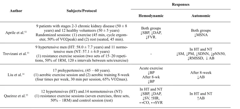
In table 1 are presented post-exercise autonomic responses
as-sessed by different methods. Overall, there were reductions of
muscle sympathetic nerve activity after aerobic exercise
10,11,52,
reduction of cardiac autonomic balance
14,53, and increase of
barorelex sensitivity and heart rate variability
54. Contrarily,
other studies reported increased cardiac and vasomotor
sym-pathetic modulation, decreased parasymsym-pathetic modulation
and/or attenuation of barorelex sensitivity
12,13,15,55however,
those studies used resistance exercise
13,15,55or maximal
aero-bic exercise
12.
Table 1. The effect of physical exercise on autonomic and hemodynamic parameters.
Author Subjects/Protocol
Responses
Hemodynamic Autonomic
Aprile et al.52
9 patients with stages 2-3 chronic kidney disease (50 ± 8 years) and 12 healthy volunteers (50 ± 5 years) Randomized sessions: (1) exercise (45 min, cycle ergom
-eter, 50% of VO2peak) and (2) rest (seated, 45 min).
Both groups
↓SBP, ↓DAP, ↓FVR.
Both groups
↓MSNA
Trevizani et al.53
9 hypertensive men (HT: 58.0 ± 7.7 years) and 11 normo
-tensive men (NT: 57.1 ± 6.0 years)
(1) resistance exercise session (two sets of 15–20 repeti
-tions, 50% of 1RM, 120 s intervals between sets/exercise)
-In HT and NT ↓SM, ↓PM, ↓SDNN, ↓pNN50,
↓RMSSD, ↓AB
Liu et al.14
17 prehypertensive, (45 – 60 years).
(1) aerobic exercise session and (2) aerobic training 8-week (four times per week, 30 min per session, 65% VO2max).
Acute exercise
↓BP
After 8-wk
↓BP
After 8-week
↓AB
Queiroz et al.55 (1) resistance exercise sessions (seven exercises, three sets, 12 hypertensives (HT) and.14 normotensives (NT) 50% – 1RM) and control session (rest)
In HT and NT ↓SBP, ↓DAP, ↓SV, ↑HR, ↔CO, ↔SVR
Neural mechanisms and PEH
The hemodynamic changes induced by physical exercise depends
on cardiovascular autonomic activity and CNS. Regarding the
areas of CNS, studies have shown that the RVLM
56,57,
NTS
58,59,
and PVN
58,60,61are involved with this cardiovascular control.
Evidence has shown that there are important mechanisms
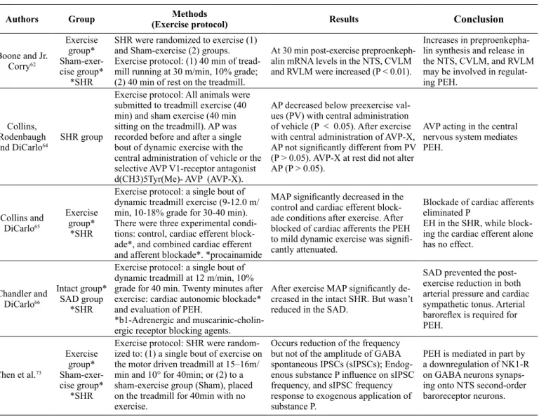
working in the nervous system to adjust the PEH. Table 2 shows
studies that investigated the contribution of this nervous system
to the occurrence of PEH. Boone and Jr. Corry
62demonstrated
that the expression of the gene preproenkephalin (PPK) increases
in the CNS after treadmill exercise in SHRs, suggesting that
increase in PPK synthesis and release in the NTS, CVLM, and
RVLM may be involved in regulating PEH. Previous studies on
both humans and SHRs indicate that endorphin systems (opioid
receptor antagonist, naloxone) attenuate PEH, and they trigger the
transient depression of blood pressure immediately after running
period in the SHR
63.
In addition, injections of vasopressin V1
receptor antagonist into the lateral cerebral ventricle impedes
the manifestation of PEH
64. This result supports the
participa-tion of central mechanisms in the development of PEH, even
though the exact role of each receptor system and the speciic
site of interaction are still indeterminate.
On the other hand, there is evidence suggesting crucial role
of the central barorelex pathway in PEH. Disturbance of inputs
from the cardiopulmonary and arterial barorelex to the CNS
prior to exercise attenuates the development of PEH. Blocking
the cardiac afferents and efferent ibers with intrapericardial
procainamide prevents PEH
65. Correspondingly, Chandler and
DiCarlo
66observed that sinoaortic denervation, which eliminates
arterial barorelex afferents, participates in development of PEH
in SHRs. These authors postulated that probably an enhanced
inhibitory inluence of cardiopulmonary afferents might alter the
arterial barorelex by modulating the response of barosensitive
neurons in the NTS to arterial baroreceptor input. These altera
-tions, through resetting of the arterial barorelex with a reduction
in gain, would account for the hypotension, sympathoinhibition,
and absence of relex tachycardia that occurs after a single bout
of dynamic exercise in hypertensive rats. Therefore, this data
has demonstrated the importance of a functioning barorelex for
occurrence of PEH. Previous investigations have shown
reduc-tion of sympathetic nerve activity after exercise
67, as might be
Author Subjects/Protocol
Responses
Hemodynamic Autonomic
Niemela et al.15
12 healthy male subjects. (31 ± 3 years) Randomized sessions: (1) aerobic exercise session on a
bicycle, (2) light resistance exercise session, (3) heavy resistance exercise session, and (4) control intervention
with no exercise.
-After 30 and 60 min (aerobic and light resistance exercise)
↓BRSLF
After heavy exercise
↓PM, ↑SM-SBP
Resk et al.13
17 normotensives Experimental sessions: (1) control (C-40 min of rest), (2) low – (E40% – 1RM), and (3) high-inten
-sity (E80% – 1 RM) resistance exercises.
After E40%
↓SBP, ↓DBP, ↔SVR, ↓SV, ↑HR, ↓CO
After E80%
↔DBP, ↓SV, ↑SVR, ↑HR, ↓CO
After E40% and E80%
↑SM, ↓PM,
Bisquolo et al.10 (1) aerobic exercise session on a bicycle (50% VO2peak) 21 healthy young men Randomized sessions: and (2) control session.
Between sessions
↔BP, ↔HR
Exercise session
↑FBF
Exercise session
↓MSNA
Raczak et al.54 (1) aerobic exercise session on a treadmill for 30 min at 18 healthy males (20 – 24 years) 65% – maximal HR.
↓SBP ↑BS, ↑SDNN, ↔HRV
Halliwill, Taylor and Eckberg11
9 healthy subjects (22 – 27 years)
(1) aerobic exercise session on a bicycle, 60 min at 60% VO2peak or (2) control session (60 min seated rest).
↓SBP, ↓MBP,
↓TPR, ↑CO. ↓MSNA
Piepoli et al.12
10 normal subjects Randomized sessions: (1) aerobic exer
-cise (maximal upright bicycle) and (2) control session (no exercise day, 30 min of upright rest)
After 60min
↓DBP, ↑CO, ↔SV, ↓SVR
After 60min
↑SM, ↓PM,
After 10min
↓BS
expected during the lower blood pressure and the
barorelex-mediated regulation of the sympathetic nerve activity must be
reset to a lower operating point during PEH.
In the same way, the fundamental role of the RVLM neurons
in controlling sympathetic vasomotor tone and blood pressure,
the regulatory role of GABA in controlling baseline activity
of those neurons and reduced barorelex function during PEH.
Thus, Kajekar et al.
68brilliantly suggested evaluating the role of
RVLM cardiovascular sympathetic neuronal activity with PEH,
as well as the relationship between RVLM sympathetic output
with RVLM GABAA-receptor mechanisms, and with barorelex
sympathetic nerve activity. They concluded that upregulation of
GABA signaling at sympathetic cardiovascular RVLM neurons
lead to a decreased neuronal output that may contribute to the
decrease in sympathetic outlow and hence PEH. Several evi
-dences suggest that muscle afferent ibers release the substance
P to activate NK1-R on GABA neurons in the NTS to modify
barorelex function during exercise
21,69,70. The data raised the
possibility that the unique interaction between the substance
P and GABAergic signal transmission systems may contribute
to PEH. Based on this information, Chen et al.
71in an elegant
study conirmed that microinjection of a substance P–NK1-R
antagonist in the NTS immediately before exercise attenuates
the development of PEH in spontaneously hypertensive rats
(SHRs). Since that activation of the NK1-R has been shown
to result in the receptor internalization
72, exercise-induced
substance P NK1-R internalization on GABA neurons may pro
-vide the unique interaction between the two neurotransmission
systems to trigger PEH. Later, Chen et al.
73proposed testing the
hypothesis of how a single bout of dynamic exercise decreases
the GABA inhibitory synaptic inputs in the NTS baroreceptor
second-order neurons via substance P NK1-R internalization on
GABA neurons in SHRs, and evidence that a decrease in blood
pressure induced by a single bout of exercise in hypertension
is mediated in part by downregulation of NK1-R on GABA
neurons synapsing on NTS second-order baroreceptor neurons.
They concluded that a single bout of dynamic exercise decreases
the GABA inhibitory synaptic inputs in the NTS baroreceptor
second-order neurons via substance P NK1-R internalization
on GABA neurons, and suggest that exercise-induced NK1-R
downregulation could provide a potential target for lowering
blood pressure in hypertensive subjects.
Table 2. Experimental studies and neural mechanisms associated with PEH.
Authors Group Methods
(Exercise protocol) Results
Conclusion
Boone and Jr.
Corry62
Exercise group* Sham-exer-cise group*
*SHR
SHR were randomized to exercise (1) and Sham-exercise (2) groups. Exercise protocol: (1) 40 min of tread
-mill running at 30 m/min, 10% grade; (2) 40 min of rest on the treadmill.
At 30 min post-exercise
preproenkeph-alin mRNA levels in the NTS, CVLM and RVLM were increased (P < 0.01).
Increases in preproenkepha-lin synthesis and release in
the NTS, CVLM, and RVLM
may be involved in regulat-ing PEH.
Collins,
Rodenbaugh
and DiCarlo64
SHR group
Exercise protocol: All animals were submitted to treadmill exercise (40 min) and sham exercise (40 min sitting on the treadmill). AP was
recorded before and after a single bout of dynamic exercise with the central administration of vehicle or the
selective AVP V1-receptor antagonist d(CH3)5Tyr(Me)- AVP (AVP-X).
AP decreased below preexercise
val-ues (PV) with central administration of vehicle (P < 0.05). After exercise with central administration of AVP-X, AP not signiicantly different from PV (P > 0.05). AVP-X at rest did not alter AP (P > 0.05).
AVP acting in the central
nervous system mediates PEH.
Collins and DiCarlo65
Exercise group*
*SHR
Exercise protocol: a single bout of dynamic treadmill exercise (9-12.0 m/ min, 10-18% grade for 30-40 min).
There were three experimental
condi-tions: control, cardiac efferent block -ade*, and combined cardiac efferent and afferent blockade*. *procainamide
MAP signiicantly decreased in the
control and cardiac efferent block-ade conditions after exercise. After blocked of cardiac afferents the PEH
to mild dynamic exercise was signii -cantly attenuated.
Blockade of cardiac afferents eliminated P
EH in the SHR, while block-ing the cardiac efferent alone has no effect.
Chandler and DiCarlo66
Intact group* SAD group
*SHR
Exercise protocol: a single bout of
dynamic treadmill at 12 m/min, 10% grade for 40 min. Twenty minutes after
exercise: cardiac autonomic blockade*
and evaluation of PEH.
*b1-Adrenergic and muscarinic-cholin-ergic receptor blocking agents.
After exercise MAP signiicantly de -creased in the intact SHR. But wasn’t reduced in the SAD.
SAD prevented the post-exercise reduction in both arterial pressure and cardiac sympathetic tonus. Arterial
barorelex is required for
PEH.
Chen et al.73
Exercise group* Sham-exer-cise group*
*SHR
Exercise protocol: SHR were random
-ized to: (1) a single bout of exercise on
the motor driven treadmill at 15–16m/
min and 10° for 40min; or (2) to a sham-exercise group (Sham), placed
on the treadmill for 40min with no exercise.
Occurs reduction of the frequency
but not of the amplitude of GABA
spontaneous IPSCs (sIPSCs); Endog
-enous substance P inluence on sIPSC frequency, and sIPSC frequency
response to exogenous application of substance P.
PEH is mediated in part by
a downregulation of NK1-R
on GABA neurons
synaps-ing onto NTS second-order
Considerations
The paraventricular nucleus of the hypothalamus (PVN) and the
rostral ventrolateral medulla (RVLM) promotes tonic effect on
the control of sympathetic vasomotor tone that triggers blood
pressure responses. Thus, there are evidences on barorelex
system and central mechanisms related to PEH. The main
cen-tral mechanisms are (1) interaction between substance P and
the GABAergic system in the NTS that contributes to PEH,
and (2) baroreceptor neurons disinhibited in the NTS increases
RVLM inhibition, via activation of the GABAergic neurons
in the CVLM, reduces PEH. We concluded that the nervous
system has an important contribution to reduce postexercise
blood pressure. In addition, the barorelex system is important
to adjust PEH, as well as nucleus tractus solitarii and RVLM
involvement is fundamental to its occurrence.
References
1. Mancia G, Fagard R, Narkiewicz K, Redon J, Zanchetti A, Bohm
M, et al. 2013 ESH/ESC Practice Guidelines for the Management of Arterial Hypertension. Blood Press. 2014;23(1):3-16.
2. Iturriaga R, Andrade DC, Del Rio R. Crucial Role of the Carotid
Body Chemoreceptors on the Development of High Arterial Blood Pressure During Chronic Intermittent Hypoxia. Adv Exp Med Biol. 2015;860:255-60.
3. Smith PA, Graham LN, Mackintosh AF, Stoker JB, Mary DA.
Relationship Between Central Sympathetic Activity and Stages of Human Hypertension. Am J Hypertens. 2004;17(3):217-22.
4. Chapleau MW, Li Z, Meyrelles SS, Ma X, Abboud FM.
Mechanisms Determining Sensitivity of Baroreceptor Afferents in Health and Disease. Ann N Y Acad Sci. 2001;940:1-19.
5. Consolim-Colombo FM, Fiorino P. Sistema Nervoso Simpático
e Hipertensão Arterial Sistêmica: Aspectos Clínicos. Rev Bras Hiperten. 2005;12(4):251-5.
6. Krieger EM. Arterial Baroreceptor Resetting in Hypertension (the
JW McCubbin memorial lecture). Clin Exp Pharmacol Physiol Suppl. 1989;15:3-17.
7. Kenney MJ, Seals DR. Postexercise hypotension. Key fea
-tures, Mechanisms, and Clinical Signiicance. Hypertension. 1993;22(5):653-64.
8. Casonatto J, Goessler KF, Cornelissen VA, Cardoso JR, Polito
MD. The blood pressure-lowering effect of a single bout of resis
-tance exercise: A systematic review and meta-analysis of random
-ized controlled trials. Eur J Prev Cardiol. 2016;23(16):1700-14.
9. Marques-Silvestre ACO, Brasileiro-Santos MS, Oliveira AS,
Maciel da Silva FT, Santos AC. Magnitude da Hipotensão Pós-Exercício Aeróbio Agudo: Uma Revisão Sistemática dos Estudos Randomizados. Motricidade. 2014;10(3):99-111.
10. Bisquolo VA, Cardoso CG Jr., Ortega KC, Gusmao JL, Tinucci T,
Negrao CE, et al. Previous Exercise Attenuates Muscle Sympathetic
Activity and Increases Blood Flow During Acute Euglycemic
Hyperinsulinemia. J Appl Physiol (1985). 2005;98(3):866-71.
11. Halliwill JR, Taylor JA, Eckberg DL. Impaired Sympathetic
Vascular Regulation in Humans After Acute Dynamic Exercise. J Physiol. 1996;495(Pt 1):279-88.
12. Piepoli M, Coats AJ, Adamopoulos S, Bernardi L, Feng
YH, Conway J, et al. Persistent Peripheral Vasodilation and Sympathetic Activity in Hypotension After Maximal Exercise. J Appl Physiol (1985). 1993;75(4):1807-14.
13. Rezk CC, Marrache RC, Tinucci T, Mion D Jr., Forjaz CL. Post-Resistance Exercise Hypotension, Hemodynamics, and Heart Rate
Variability: Inluence of Exercise Intensity. Eur J Appl Physiol. 2006;98(1):105-12.
Authors Group Methods
(Exercise protocol) Results
Conclusion
Chen et al.71 SHR group
Exercise protocol: treadmill at 15 m/
min, 10°, for 40 min. AP was taken
during and after (2 h) exercise. The vehicle and substance P (NK-1)
receptor antagonist injections were made randomly.
The antagonist, in a dose that blocked
substance P signiicantly attenuated the PEH (P < 0.05). Vehicle microin -jection had no effect.
PEH is mediated, at least in
part, by a substance P (NK-1) receptor mechanism in the NTS.
Kajekar et al.68
Exercise group* Sham-exer-cise group*
*SHR
Exercise protocol: treadmill at 15 m/
min, 10° for 40 min. Randomly
as-signed to a (1) single bout of exercise;
or to a sham-exercise group placed on the treadmill for 40 min with no
exercise. MAP was measured every 10
min during 40 min of exercise or sham exercise and over the next 10 h after exercise or sham exercise
PEH lasted 10 h in SHR.
Extracel-lular Resting RVLM neuronal activity
was lower and was increased largely by GABAA-receptor antagonism in
PEH versus Sham PEH (P < 0.05). Barorelex control of RVLM neuronal
activity operated with a reduced gain
(P < 0.05).
Upregulation of aspects of
GABA signaling at
sympa-thetic cardiovascular RVLM
neurons lead to a decreased neuronal output that may contribute to the decrease
in sympathetic outlow and
hence PEH.
Kulics,
Collins and DiCarlo67
Exercise group*
*SHR
Exercise protocol: treadmill exercise (9–12 m/min, 10% grade for 40 min). Protocol 1: CO and TPR were
determined before, during, and after exercise.
Protocol 2: LSNA was recorded before
and after exercise.
Associated with the PEH there was a reduction in TPR and an elevation in
CO (P < 0.05, all comparisons). The
reductions in arterial pressure and TPR were associated with a decrease
in LSNA (P < 0.05)
PEH is mediated by
reduc-tions in TPR and SNA
14. Liu S, Goodman J, Nolan R, Lacombe S, Thomas S. Blood
Pressure Responses to Acute and Chronic Exercise Are Related in Prehypertension. Med Sci Sports Exerc. 2012;44(9):1644-52.
15. Niemela TH, Kiviniemi AM, Hautala AJ, Salmi JA, Linnamo
V, Tulppo MP. Recovery Pattern of Barorelex Sensitivity After Exercise. Med Sci Sports Exerc. 2008;40(5):864-70.
16. Machado BH. Neurotransmission of the cardiovascular relexes
in the nucleus tractus solitarii of awake rats. Ann N Y Acad Sci. 2001;940:179-96.
17. Baude A, Strube C, Tell F, Kessler JP. Glutamatergic neurotrans
-mission in the nucleus tractus solitarii: structural and functional characteristics. J Chem Neuroanat. 2009;38:145-53.
18. Potts JT. Inhibitory neurotransmission in the nucleus tractus
solitarii: implications for barorelex resetting during exercise. Exp Physiol. 2006;91:59-72.
19. Guyenet PG. The sympathetic control of blood pressure. Nat Rev
Neurosci. 2006; 7:335-46.
20. Sawchenko PE, Swanson LW. Immunohistochemical identiica -tion of neurons in the paraventricular nucleus of the hypothalamus
that project to the medulla or to the spinal cord in the rat. J Comp Neurol. 1982;205:260-72.
21. Allen AM. Inhibition of the hypothalamic paraventricular nucleus in spontaneously hypertensive rats dramatically reduces
sympa-thetic vasomotor tone. Hypertension 2002; 39:275-80.
22. Badoer E. Hypothalamic paraventricular nucleus and
cardiovas-cular regulation. Clin Exp Pharmacol Physiol. 2001;28:95-9.
23. Strack AM, Sawyer WB, Hughes JH, Platt KB, Loewy AD. A
general pattern of CNS innervation of the sympathetic outlow
demonstrated by transneuronal pseudorabies viral infections.
Brain Res. 1989;491:156-62.
24. Santaella DF, Araújo EA, Ortega KC, Tinucci T, Mion Jr.D,
Negrão CE, et al. Aftereffects of exercise and relaxation on blood pressure. Clin J Sport Med (Print). 2006;16:341-7.
25. Pescatello LS, Guidry MA, Blanchard BE, Kerr A, Taylor AL,
Johnson AN, et al. Exercise intensity alters postexercise hypoten
-sion. J Hypertens. 2004;22:1881-8.
26. Halliwill JR. Mechanisms and clinical implications of post-exercise
hypotension in humans. Exerc Sport Sci Rev. 2001;29(2):65-70.
27. Brito A de F, de Oliveira CV, Brasileiro-Santos M do S, Santos
A da C. Resistance exercise with different volumes: blood pres
-sure response and forearm blood low in the hypertensive elderly. Clin Interv Aging. 2014;9:2151-828. Polito MD, Farinatti PT.
The effects of muscle mass and number of sets during resistance
exercise on postexercise hypotension. J Strength Cond Res. 2009;23:2351-7.
28. Ciolac EG, Guimaraes GV, D’Avila VM, Bortolotto LA, Doria
EL, Bocchi EA. Acute effects of continuous and interval aerobic
exercise on 24-h ambulatory blood pressure in long-term treated
hypertensive patients. Int J Cardiol. 2009;133(3):381-7.
29. Ciolac EG, Guimaraes GV, D’Avila VM, Bortolotto LA, Doria
EL, Bocchi EA. Acute aerobic exercise reduces 24-h ambulatory
blood pressure levels in longterm-treated hypertensive patients.
Clinics (São Paulo). 2008;63(6):753-8.
30. Brandao Rondon MU, Alves MJ, Braga AM, Teixeira OT,
Barretto AC, Krieger EM, et al. Postexercise blood pressure reduction in elderly hypertensive patients. J Am Coll Cardiol. 2002;39(4):676-82.
31. Cunha GA, Rios ACS, Moreno JR, Braga PL, Campbell CSG,
Simões HG, Denadai MLDR. Post-exercise hypotension in hy -pertensive individuals submitted to aerobic exercises of alternated
intensities and constant intensity-exercise. Rev Bras Med Esporte. 2006;12(6):313-7.
32. MacDonald JR, Hogben CD, Tarnopolsky MA, MacDougall JD.
Post exercise hypotension is sustained during subsequent bouts
of mild exercise and simulated activities of daily living. J Hum
Hypertens. 2001;15(8):567-71.
33. MacDonald JR, Rosenfeld JM, Tarnopolsky MA, Hogben CD,
Ballantyne CS, MacDougall JD. Post exercise hypotension is not
mediated by the serotonergic system in borderline hypertensive
individuals. J Hum Hypertens. 2002;16(1):33-9.
34. Queiroz AC, Gagliardi JF, Forjaz CL, Rezk CC. Clinic and ambulatory blood pressure responses after resistance exercise. J
Strength Cond Res. 2009;23(2):571-8.
35. Lawes CM, Bennett DA, Feigin VL, Rodgers A. Blood pres
-sure and stroke: an overview of published reviews. Stroke. 2004;35(4):1024-33.
36. Forjaz CLM, Tinucci T, Ortega KC, Santaella DF, Mion Jr.D, Negrão
CE. Factors Affecting Post-Exercise Hypotension in Normotensive and Hypertensive Humans. Blood Press Monit. 2000;5:255-62.
37. Christofaro DG, Casonatto J, Fernandes RA, Cucato GG,
Gonçalves CGS, Oliveira AR, et al. Efeito da Duração do Exercício Aeróbio sobre as Respostas Hipotensivas Agudas Pós-Exercício. Rev SOCERJ. 2008;21:404-8.
38. MacDonald JR, MacDougall JD, Hogben CD. The effects of exercising muscle mass on post exercise hypotension. J Hum
Hypertens. 2000;14(5):317-20.
39. Carvalho RS, Pires CM, Junqueira GC, Freitas D, Marchi-Alves
LM. Hypotensive response magnitude and duration in hyper
-tensives: continuous and interval exercise. Arq Bras Cardiol. 2015;104(3):234-41.
40. Mach C, Foster C, Brice G, Mikat RP, Porcari JP. Effect of exer
-cise duration on postexer-cise hypotension. J Cardiopulm Rehabil. 2005;25:366-9.
41. Forjaz CL, Cardoso CG Jr, Rezk CC, Santaella DF, Tinucci T.
Postexercise Hypotension and Hemodynamics: The Role of Exercise Intensity. J Sports Med Phys Fitness. 2004;44(1):54-62.
42. Cardoso Jr. CG, Gomides RS, Queiroz AC, Pinto LG, Silveira
Lobo F, Tinucci T, et al. Acute and Chronic Effects of Aerobic and Resistance Exercise on Ambulatory Blood Pressure. Clinics. 2010;65(3):317-25.
43. Mota MR, Pardono E, Lima LC, Arsa G, Bottaro M, Campbell CS, et al. Effects of Treadmill Running and Resistance Exercises on
Lowering Blood Pressure During the Daily Work of Hypertensive Subjects. J Strength Cond Res. 2009;23(8):2331-8.
44. Moraes MR, Bacurau RF, Ramalho JD, Reis FC, Casarini DE,
Chagas JR, et al. Increase in Kinins on Post-Exercise Hypotension in Normotensive and Hypertensive Volunteers. Biol Chem. 2007;388(5):533-40.
45. Laterza MC, Matos LD, Trombetta IC, Braga AM, Roveda
F, Alves MJ, et al. Exercise Training Restores Baroreflex Sensitivity in Never-Treated Hypertensive Patients. Hypertension. 2007;49(6):1298-306.
46. Pinto A, Di Raimondo D, Tuttolomondo A, Fernandez P,
Pressure Monitoring to Evaluate Effects on Blood Pressure of Physical Activity in Hypertensive Patients. Clin J Sport Med. 2006;16(3):238-43.
47. Syme AN, Blanchard BE, Guidry MA, Taylor AW, Vanheest
JL, Hasson S, et al. Peak Systolic Blood Pressure on a Graded Maximal Exercise Test and the Blood Pressure Response to an Acute Bout of Submaximal Exercise. Am J Cardiol. 2006;98(7):938-43.
48. Fagard RH. Exercise is Good for Your Blood Pressure: Effects of
Endurance Training and Resistance Training. Clin Exp Pharmacol Physiol. 2006;33(9):853-6.
49. Whelton SP, Chin A, Xin X, He J. Effect of aerobic exercise on
blood pressure: a metaanalysis of randomized, controlled trials. Ann Intern Med Ann Intern Med. 2002; 136(7):493-503.
50. Halbert JA, Silagy CA, Finucane P, Withers RT, Hamdorf PA,
Andrews GR. The Effectiveness of Exercise Training in Lowering Blood Pressure: A Meta-Analysis of Randomised Controlled Trials of 4 Weeks or Longer. J Hum Hypertens. 1997;11(10):641-9.
51. Aprile DC, Oneda B, Gusmão JL, Costa LA, Forjaz CL, Mion D
Jr, et al. Post-Exercise Hypotension Is Mediated by a Decrease in Sympathetic Nerve Activity in Stages 2-3 CKD. Am J Nephrol. 2016;43(3):206-12.
52. Trevizani GA, Peçanha T, Nasario-Junior O, Vianna JM, Silva LP,
Nadal J. Cardiac autonomic responses after resistance exercise in treated hypertensive subjects. Front Physiol. 2015;6:1-7.
53. Raczak G, Pinna GD, La Rovere MT, Maestri R,
Danilowicz-Szymanowicz L, Ratkowski W, et al. Cardiovagal response to acute mild exercise in young healthy subjects. Circ J. 2005;69(8):976-80.
54. Queiroz ACC, Sousa JCS, Cavalli AAP, Silva Jr ND, Costa
LAR,Tobaldini E, et al. Post-resistance exercise hemodynamic and autonomic responses: Comparison between normotensive and hypertensive men. Scand J Med Sci Sports. 2015;25:486-94.
55. Mueller PJ. Exercise training attenuates increases in lumbar sympathetic nerve activity produced by stimulation of the rostral
ventrolateral medulla. J Appl Physiol. 2007;102:803-13.
56. Martins-Pinge MC, Becker LK, Garcia MR, Zoccal DB, Neto
RV, Basso LS, et al. Attenuated pressor responses to amino acids
in the rostral ventrolateral medulla after swimming training in
conscious rats. Auton Neurosci. 2005;122:21-8.
57. Michelini LC, Stern JE. Exercise-induced neuronal plasticity in
central autonomic networks: role in cardiovascular control. Exp Physiol. 2009; 94:947-60.
58. De Souza CG, Michelini LC, Fior-Chadi DR. Receptor changes in the nucleus tractus solitarii of the rat after exercise training.
Med Sci Sports Exerc. 2001;33:1471-6.
59. Mastelari RB, de Souza HC, Lenhard A, de Aguiar Correa FM,
Martins-Pinge MC. Nitric oxide inhibition in paraventricular
nucleus on cardiovascular and autonomic modulation after
exer-cise training in unanesthetized rats. Brain Res. 2011;1375:68-76.
60. de Abreu SB, Lenhard A, Mehanna A, de Souza HC, Correa FM,
Hasser EM, et al. Role of paraventricular nucleus in exercise
training-induced autonomic modulation in conscious rats. Auton
Neurosci. 2009;148:28-35.
61. Boone JB Jr. Corry JM. Proenkephalin gene expression in the
brainstem regulates post-exercise hypotension. Brain Res Mol Brain Res. 1996;42:31-8.
62. Shyu BC, Thorén P. Circulatory events following spontaneous muscle exercise in normotensive and hypertensive rats. Acta
Physiol Scand. 1986;128:515-24.
63. Collins HL, Rodenbaugh DW, DiCarlo SE. Central blockade of
vasopressin V(1) receptors attenuates postexercise hypotension. Am J Physiol Regul Integr Comp Physiol. 2001;281:R375-80.
64. Collins HL, DiCarlo SE. Attenuation of postexertional hypotension
by cardiac afferent blockade. Am J Physiol. 1993;265:H1179-83.
65. Chandler MP, DiCarlo SE. Sinoaortic denervation prevents postexercise reductions in arterial pressure and cardiac
sympa-thetic tonus. Am J Physiol. 1997;273:H2738-45.
66. Kulics JM, Collins HL, DiCarlo SE. Postexercise hypotension is mediated by reductions in sympathetic nerve activity. Am J
Physiol Heart Circ Physio. 1999;276(1):H27-32.
67. Kajekar R, Chen CY, Mutoh T, Bonham AC. GABA(A) recep -tor activation at medullary sympathetic neurons contributes to
postexercise hypotension. Am J Physiol Heart Circ Physiol. 2002;282:H1615-24.
68. Boscan P, Paton JF. Excitatory convergence of periaqueductal
gray and somatic afferents in the solitary tract nucleus: role for neurokinin 1 receptors. Am J Physiol Regul Integr Comp Physiol. 2005;288:R262-69.
69. Potts JT, Fuchs IE, Li J, Leshnower B, Mitchell JH. Skeletal
muscle afferent ibres release substance P in the nucleus tractus solitarii of anaesthetized cats. J Physiol. 1999; 514:829-41.
70. Chen CY, Munch PA, Quail AW, Bonham AC. Postexercise hypotension in conscious SHR is attenuated by blockade of
substance P receptors in NTS. Am J Physiol Heart Circ Physiol. 2002; 283:H1856-62.
71. Marvizón JC, Wang X, Matsuka Y, Neubert JK, Spigelman I. Relationship between capsaicin-evoked substance P release and neurokinin 1 receptor internalization in the rat spinal cord.
Neuroscience. 2003;118:535-45.
72. Chen CY, Bechtold AG, Tabor J, Bonham AC. Exercise reduces GABA synaptic input onto nucleus tractus solitarii baroreceptor
second-order neurons via NK1 receptor internalization in spontane
-ously hypertensive rats. J Neurosci. 2009;29:2754-61.
Acknowledgments
We gratefully acknowledge the Brazilian National Research Council (CNPq) to fellowship the MSBS (SwB 2015/2016 – No. 202310/2014-0) and ACS (SwB 2015/2016 – No. 202309/2014-2), Facult of Kinesiology and Physical Education at the University of Toronto, Canada, and Department of Physical Education at the Federal University of Paraiba, Brazil.
Corresponding author
Maria do Socorro Brasileiro-Santos. Universidade Federal da Paraíba. Departamento de Educação Física.
Cidade Universitária. CEP: 58051-900. João Pessoa, PB, Brasil Email: [email protected]
Manuscript received on September 15, 2016 Manuscript accepted on November 01, 2016