385 Daud DF et al. Cardiac tamponade at chest CT in an infant
Radiol Bras. 2013 Nov/Dez;46(6):385–386
Cardiac tamponade in an infant during contrast infusion
through central venous catheter for chest computed
tomography
*
Tamponamento cardíaco durante infusão de contraste em acesso venoso central para realização de tomografia computadorizada de tórax em lactente
Danilo Felix Daud1, Marcos Menezes Freitas de Campos1, Luciano Augusto de Pádua Fleury Neto2
Complications from central venous catheterization include infectious conditions, pneumothorax, hemothorax and venous thrombosis. Pericardial effusion with cardiac tamponade hardly occurs, and in infants is generally caused by umbilical catheterization. The authors describe the case of cardiac tamponade occurred in an infant during chest computed tomography with contrast infusion through a central venous catheter inserted into the right internal jugular vein. Keywords: Cardiac tamponade; Radiology; Thoracic surgery.
Complicações decorrentes do cateterismo venoso central são infecciosas, pneumotórax, hemotórax e trombose venosa. O derrame pericárdico com tamponamento cardíaco é mais difícil de acontecer e, quando ocorre em lactentes, é geral-mente ocasionado por cateterismo umbilical. Descrevemos um caso de tamponamento cardíaco ocorrido durante to-mografia computadorizada de tórax, com infusão de contraste por acesso venoso central em veia jugular interna direita. Unitermos: Tamponamento cardíaco; Radiologia; Cirurgia torácica.
Abstract
Resumo
* Study developed at Hospital Geral de Palmas, Palmas, TO, Brazil.
1. MDs, Thoracic Surgeons, Hospital Geral de Palmas, Pal-mas, TO, Brazil.
2. MD, Radiologist, Hospital Geral de Palmas, Palmas, TO, Brazil.
Mailing Address: Dr. Danilo Felix Daud. 603 Sul, Alameda 14, Lote 19, Plano Diretor Sul. Palmas, TO, Brazil, 77016-374. E-mail: [email protected].
Received November 4, 2012. Accepted after revision April 19, 2013.
Daud DF, Campos MMF, Fleury Neto LAP. Cardiac tamponade in an infant during contrast infusion through central venous catheter for chest computed tomography. Radiol Bras. 2013 Nov/Dez;46(6):385–386.
0100-3984 © Colégio Brasileiro de Radiologia e Diagnóstico por Imagem
CASE REPORT
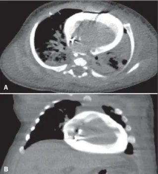
enhanced chest computed tomography. Immediately after iodinated contrast agent infusion (Figure 1), the child presented clinical worsening and was promptly taken back to the intensive care unit. The thoracic surgery team was called.
intensive care unit, with acute renal failure and sepsis with a probable pulmonary fo-cus. Central venous access was not avail-able and was obtained by the intensivist physician by means of puncture of the right internal jugular vein. Subsequently, the patient was referred to undergo contrast INTRODUCTION
Generally, the discussions about com-plications from central venous catheteriza-tion in children concentrate on infectious complications and on the most common me-chanical complications such as pneumotho-rax, hemothorax and venous thrombosis(1).
Nonetheless, pericardial effusion has al-ready been reported by international stud-ies, most of them describing cases of neo-nates submitted to umbilical catheteriza-tion(2,3) and or catheterization through other
central venous lines(4,5).
CASE REPORT
The authors report the case of a three-month-old, preterm child admitted to infant
Figure 1. Contrast enhanced chest computed tomography images. A: Axial section – presence of contrast in the pericardial sac (effusion) and bilateral pneumonia. B: Coronal reconstruction – presence of con-trast in the pericardial sac (effu-sion).
A
386
Daud DF et al. Cardiac tamponade at chest CT in an infant
Radiol Bras. 2013 Nov/Dez;46(6):385–386 Considering the clinical signs of
peri-cardial effusion with cardiac tamponade and consequential cardiac arrest, pericar-diocentesis was performed (Marfan punc-ture) with aspiration of 30 ml hematic fluid and cardiorespiratory resuscitation maneu-vers with heart beat recovery.
The patient was taken to the surgical center assisted by one of the thoracic sur-gery team members while another member was already prepared to initiate the thorac-otomy. Right lateral thoracotomy was per-formed once the hypothesis of right atrial perforation by the central venous catheter was raised. The surgical strategy included pericardiotomy and atrial cardiorrhaphy.
The child’s clinical condition worsened, progressing to death seven days after the procedure due to multiple organs failure.
DISCUSSION
Pericardial effusion with cardiac tam-ponade by itself represents a severe prob-lem for any patient. In a frail child admit-ted to an intensive care unit with other dis-eases, such a condition is potentially fatal. It may be caused by umbilical catheteriza-tion, particularly in neonates(2,3), or other
central venous access lines(4,5).
The cause and prompt diagnosis of such an event during contrast enhanced chest computed tomography scan make this case rare. No similar case report has been found in the literature.
The authors conclude that careful inser-tion techniques as well as continuous ob-servation of the correct positioning and function of central venous catheters,
be-sides the utilization of good quality mate-rials, are essential to aid in the prevention of severe complications.
REFERENCES
1. Askegard-Giesmann JR, Caniano DA, Kenney BD. Rare but serious complications of central line in-sertion. Semin Pediatr Surg. 2009;18:73–83. 2. Megha M, Jain N, Pillai R. Pericardial tamponade
in a newborn following umbilical catheter inser-tion. Indian Pediatr. 2011;48:404–5.
3. Traen M, Schepens E, Laroche S, et al. Cardiac tamponade and pericardial effusion due to venous umbilical catheterization. Acta Paediatr. 2005;94: 626–8.