Carlos Eugênio Villaboim de Castro Lima(a)
Jimmy Cavalcanti Calixto(a) Ana Lia Anbinder(b)
(a) Department of Dentistry, UNITAU -
University of Taubaté, Taubaté, SP, Brazil.
(b) Department of Bioscience and Oral
Diagnosis, São José dos Campos School of Dentistry, UNESP - Univ Estadual Paulista, São José dos Campos, SP, Brazil.
Corresponding author: Ana Lia Anbinder
Av. Engenheiro Francisco José Longo, 777, Jardim São Dimas
São José dos Campos - SP - Brazil CEP: 12245-000
E-mail: [email protected]
Received for publication on Oct 06, 2010 Accepted for publication on Dec 02, 2010
Influence of the association between
simvastatin and demineralized bovine
bone matrix on bone repair in rats
Abstract: Simvastatin, a drug used to lower blood cholesterol, has been reported to have an anabolic effect on bone. The purpose of this study was to evaluate the inluence of simvastatin and demineralized bovine bone matrix (DBBM) on the repair of rat calvarial defects. Defects of 5 mm were created in 64 rats, divided into four groups: no local treatment (control); treatment with DBBM (DBBM); treatment with a combination of simvastatin solution (2.2 mg/50 µl) and DBBM (DBBMSIM-1); and treatment with simvastatin solution (0.5 mg/50 µl) and DBBM (DBBM-SIM-2). Animals were sacriiced on postoperative day 30 or 60, after which the calvariae were X-rayed and prepared for histomorphometric evaluation. The data were submitted to statistical analysis (p < 0.05). X-rays revealed that, on postoperative day 30, animals treated with a lower dose of simvastatin presented the lowest bone density, whereas on post-operative day 60 the use of simvastatin, regardless of the dose, resulted in lower density than that observed in control and DBBM group samples. Histomorphometric analysis revealed that, on postoperative day 30, both DBBM and DBBMSIM-1 had a negative impact on bone formation. On postoperative day 60, none of the combinations tested impaired bone re-pair. These results showed that the association between DBBM and sim-vastatin had a negative impact on bone repair.
Descriptors: Simvastatin; Bone Regeneration; Bone Matrix.
Introduction
Autotransplantation is the gold standard for the repair of large bone defects. However, this technique has two major disadvantages: donor-site morbidity and bone resorption. In the 1960s, Urist1 reported that demineralized bone implants in extraosseous sites induced local miner-alization, a process in which mesenchymal cells differentiate and form new bone. Since then, there have been various studies investigating alter-natives to autotransplantation, including the use of demineralized bone matrix for bone repair.
Statins are widely used to lower blood cholesterol levels. Some studies have reported that statins can stimulate bone formation by stimulating the production of bone morphogenetic protein-2.2
those required to reduce cholesterol levels and are associated with unacceptable toxicity.3 The major limitation to the clinical use of statins for bone re-generation is the lack of an effective carrier for the drug,4 since the success of in vivo bone formation depends on high local concentrations of statins.5 Various authors have investigated new methods and carriers for the administration of statins:
• local delivery in methylcellulose gel;6,7
• local delivery in polylactic acid/polyglycolic acid copolymer;5
• local delivery in gelatin sponge;8 and
• statins used in combination with calcium sul-fate.9,10
The results have often been encouraging.
The purpose of the present study was to evaluate the inluence of the association between simvastatin and demineralized bovine bone matrix (DBBM) on the repair of rat calvarial defects.
Material and Methods
A total of 64 adult male Wistar rats (Rattus nor-vegicus) aged 90 days were used. In order to cre-ate the defects, the animals were anesthetized with 13 mg/kg of xylazine (Anasedan-Vetbrands-Sespo, Jacareí, SP, Brazil) and 33 mg/kg of ketamine (Do-palen Vetbrands - Sespo, Jacareí, SP, Brazil). An in-cision was made through the skin and periosteum, and a trephine drill (Neodent, Curitiba, PR, Brazil) was used to create two critical size defects (5 mm in diameter),11 one in each parietal bone.
The simvastatin solution was prepared by mixing simvastatin (lot no. 0707071512; Galena, Campi-nas, SP, Brazil) and distilled water at a proportion of 2.2 mg/50 µl (solution 16) or of 0.5 mg/50 µl (solu-tion 27) and centrifuging the mixture for 1 h. The animals were divided into four groups:
• control, in which the defects were not illed;
• DBBM, in which the defects were illed with 10 mg of DBBM (GenOx Org Cortical Micro; Baumer S.A., Mogi Mirim, SP, Brazil) and 56 µl of distilled water;
• DBBMSIM-1, in which the defects were illed with 10 mg of DBBM and 56 µl of simvastatin solution 1 (Figure 1); and
• DBBMSIM-2, in which the defects were illed
with 10 mg of DBBM and 56 µl of simvastatin solution 2.
After 30 or 60 days, the animals were sacriiced, and the calvariae were removed and ixed. After radiographic examination, right and left defects were separated, and left-side samples were sent for decalciication. Right-side samples were embed-ded in methyl methacrylate resin, cut into sections, and mounted on slides for microscopic examination (data not shown).
The samples were X-rayed using dental X-ray equipment (Spectro 70X; Dabi Atlante, São Paulo, SP, Brazil) with a digital sensor (Trophy RVG, Ko-dak Dental Systems, Rochester, NY, USA), under the following conditions: 8 mA; 70 KVp; focus-ob-ject distance of 30 cm; exposure time of 0.1 s. The radiographic density (gray levels) of the region of in-terest (circular area = 15,380 pixels) was calculated using the ImageJ 1.31p software (National Institutes of Health, Bethesda, MD, USA). For statistical anal-ysis, we considered the mean density of the two de-fects for each animal.
cyte lacunae. Subsequently, image binarization was performed, and the area was calculated using the ImageJ 1.31p software.
In order to evaluate the effect of the differ-ent types of treatmdiffer-ent at each time point (30 or 60 days), the densitometric data were submitted to analysis of variance (ANOVA) and Tukey’s test, whereas the histomorphometric data were submit-ted to the Kruskal-Wallis test, followed by Dunn’s post-test. In order to evaluate the effect of time, the Student’s t-test or the Mann-Whitney test was used. The level of signiicance was set at 5% for all tests. The analyses were performed with GraphPad Prism 4.0 (GraphPad Softwares Inc., San Diego, CA, USA) and Bioestat 2.0 (Sociedade Civil Mamirauá, Belém, PA, Brazil).
The present study was approved by the Animal Research Ethics Committee of the University of Taubaté, Brazil (protocol no. 0016/07).
Results
After the surgical procedure, 6 rats from group DBBMSIM-1 presented scabbing – which persisted for 30 days – at the incision site. Only 1 animal from group DBBMSIM-2 presented scabbing, observed 14 days after surgery. The lesion was approximate-ly half the size of that seen in animals from group DBBMSIM-1 and had disappeared on postoperative day 30. Also in group DBBMSIM-2, 1 animal died from anesthesia-related complications and was not replaced.
X-ray densitometry
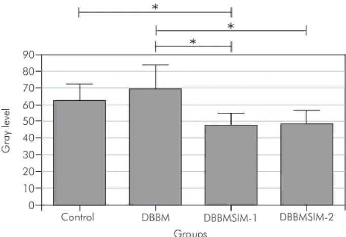
The ANOVA revealed a signiicant difference be-tween the radiographic density values of the samples collected on postoperative day 30 (F = 3.395, df = 3, p = 0.0316) and those of the samples collected on postoperative day 60 (F = 8.574, df = 3, p = 0.0004). Figures 2 and 3 illustrate the differences revealed by Tukey’s test.
The Student’s t-test, used to analyze the effects of time on radiographic density according to the type of treatment, revealed no signiicant differences between the two time points in group DBBMSIM-1 (Figure 4). In the remaining groups, signiicantly greater radiographic density was observed in
ani-Groups 0 10 20 30 40 50 G ray lev e l
Control DBBM DBBMSIM-1 DBBMSIM-2
Figure 2 - Means and standard deviations of radiographic density values on postoperative day 30. * indicates signifi-cant difference (Tukey’s test).
Groups
Control DBBM DBBMSIM-1 DBBMSIM-2 0 10 20 30 40 50 60 70 80 90 G ray lev e l
Figure 3 - Means and standard deviations of radiographic density values on postoperative day 60. * indicates signifi-cant difference (Tukey’s test).
Groups C 3 0 d C 6 0 d D B B M 3 0 d D B B M 6 0 d D B B MSI M-1 3 0 d D B B MSI M-1 6 0 d D B B MSI M-2 3 0 d D B B MSI M-2 6 0 d 0 10 20 30 40 50 60 70 80 90 G ray lev el
mals sacriiced on postoperative day 60.
Histomorphometric analysis
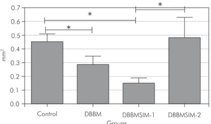
Due to technical dificulties during decalci-ication and slide preparation, one sample col-lected from group DBBM on postoperative day 30 was not used. The Kruskal-Wallis test revealed a signiicant difference between the types of treat-ment, both on postoperative day 30 (H = 13.592; df = 3; p = 0.0035) and on postoperative day 60 (H = 16.828; df = 3; p = 0.0008). Dunn’s post-test revealed that, on postoperative day 30, both DBBM and DBBMSIM-1 had a negative inluence on new bone formation (Figure 5). On postoperative day 60, the two different doses of simvastatin had a negative inluence on bone repair (Figure 6).
The effect of time on bone repair was signiicant only in animals from the control group and in those from group DBBM, the highest density values being observed on postoperative day 60 (Figure 7).
Descriptive histology
On postoperative day 30, the examination of control samples revealed an area of new bone at the edges of the defects. The newly formed bone pre-sented plump osteocytes arranged in a disorganized manner and was covered by lattened osteoblasts. Small bone islands were seen in the center of the defect, which was illed with loose connective tis-sue and mild chronic inlammatory iniltrate. The histological features of group DBBM samples were similar to those of control samples; however, there was less new bone at the edges and in the center of the defect. The connective tissue was thicker in some samples and presented moderate chronic in-lammatory iniltrate. DBBM had been completely reabsorbed in all but 1 of the animals from group DBBM. Group DBBMSIM-1 samples presented few areas of new bone formation, limited to the edges of the defect and composed of highly immature tissue surrounded by plump osteoblasts (Figure 8), as well as regions of old bone resorption. The connective tissue that illed the defect was highly vascularized, thicker than that observed in groups control and DBBM, and presented moderate or severe chronic Figure 5 - Means and standard errors of new bone
forma-tion values on postoperative day 30. * indicates significant difference (Dunn’s post-test).
Groups 0.0
0.1 0.2 0.4
0.3 0.5 0.7
0.6
mm
2
Control DBBM DBBMSIM-1 DBBMSIM-2
Groups 0.00
0.25 0.75
0.50 1.00 1.25
mm
2
Control DBBM DBBMSIM-1 DBBMSIM-2
Figure 6 - Means and standard errors of new bone forma-tion values on postoperative day 60. * indicates significant difference (Dunn’s post-test).
Groups
C
3
0
d
C
6
0
d
D
B
B
M
3
0
d
D
B
B
M
6
0
d
D
B
B
MSI
M-1
3
0
d
D
B
B
MSI
M-1
6
0
d
D
B
B
MSI
M-2
3
0
d
D
B
B
MSI
M-2
6
0
d
0.00 0.50
0.25 0.75 1.00 1.25
mm
2
inlammation. An area of necrosis was observed in 1 animal, and remnants of DBBM were seen amid connective tissue in 50% of group DBBMSIM-1 ani-mals. Group DBBMSIM-2 samples presented few areas of new bone formation, with highly imma-ture tissue. However, there was no resorption of old bone. Remnants of DBBM amid the connective tis-sue that illed the defect were observed in 1 animal. No areas of necrosis were observed.
On postoperative day 60, the newly formed bone at the edges of the defect in control samples was thicker and more mature, containing osteocytes that were smaller and more regularly arranged than those observed on postoperative day 30. In a large number of samples, we observed islands of newly formed bone in the center of the defect. In some samples, linear closure of the defect was nearly com-plete. Areas of mild inlammation were still present. The histological features of group DBBM samples were similar to those of control samples. DBBM-SIM-1 samples once again presented little new bone formation (Figure 9). The connective tissue was slightly thicker and more vascularized than that observed in groups control and DBBM, presenting mild or moderate inlammation. No bone formation was observed in the center of the defect in any of the animals from group DBBMSIM-1. Osteoblasts and osteocytes were plumper in group DBBMSIM-1
samples than in the samples from the remaining groups, and remnants of DBBM were seen in 3 animals. Group DBBMSIM-2 samples once again presented little bone formation. The connective tis-sue that illed the defect was thin, presenting mild chronic inlammatory iniltrate. Neither remnants of DBBM nor necrosis was observed in any of the animals from group DBBMSIM-2.
Discussion
A critical size defect can be deined as the small-est size of an intraosseous wound – in a particular bone and species of animal – that will not heal spon-taneously during the lifetime of the animal12 or over the duration of the study.13 A 5-mm rat calvarial de-fect is considered a critical size dede-fect.11 Our analy-sis of cross-sectional area revealed that none of the samples from the control group presented complete bone regeneration, which allowed us to analyze the osteogenic potential of the treatments tested.
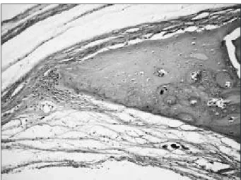
In the present study, X-ray densitometry and histomorphometric analysis were complementary because densitometry was used to evaluate calciied tissue and histomorphometric analysis was used to evaluate decalciied tissue. Although radiographic analysis has certain advantages, such as low cost and rapid results, the accuracy of the method when employed in rat calvarial defect models has been re-Figure 9 - Microscopic appearance of a sample from an animal from group DBBMSIM-1 that was sacrificed on post-operative day 60. Small amount of bone formation at the edges of the defect (H&E; ×200).
ported to be low.14 Therefore, radiographic analysis should not be used as the only method of evalua-tion (i.e., without histological analysis, considered the gold standard). Our results conirmed this. On postoperative day 30, radiographic and histological analyses revealed opposite results for group DBBM-SIM-2. On postoperative day 60, when bone forma-tion was more evident, the two analyses revealed no conlicting results.
Although DBBM has osteoconductive15 and osteoinductive properties, osteoinductivity var-ies according to the commercial preparation.16 We were unable to conirm the beneicial properties of DBBM.15,17 Carneiro et al.18 also investigated the beneicial effects of DBBM and reported no signii-cant differences between the treatment group and the control group. In the present study, DBBM de-layed bone matrix formation in the irst 30 days, exacerbating the inlammatory process. A more in-tense initial inlammation might be related to fac-tors such as incomplete decalciication of the graft, residual lipids or immunopathological features of the recipient.18 In the present study, the association between inlammation and DBBM resorption in the irst 30 days might have affected the osteoconduc-tivity of DBBM. On postoperative day 60, when DBBM was no longer present, no signiicant dif-ferences were observed between groups DBBM and control.
Various authors have investigated local delivery of statins to increase bone formation and reported that statins have a local anabolic effect.5,9,10 Others, however, have found no beneicial effects.9,19,20 The ideal doses and carriers have yet to be determined, and the issue remains controversial. Low doses have no impact on bone repair, whereas high doses can stimulate inlammation. High local concentrations of statins can be cytotoxic due to a drastic reduction in the production of cholesterol, a substance that is important to the integrity of cell membranes.4
In the present study, the use of high local doses of simvastatin caused an intense inlammatory reac-tion, accompanied by tissue necrosis and scabbing. Soft tissue inlammation was also reported by Thy-lin et al.,6 who used 2.2 mg of the drug in
methylcel-lulose gel in rat calvaria; by Nyan et al.,9 who used 1 mg of the drug in calcium sulfate in rat calvaria; and by Stein et al.,7 who used doses of 0.5, 1.0, 1.5, and 2.2 mg of the drug in rat mandible. In the study conducted by Stein et al.,7 the 0.5-mg dose present-ed the best cost-beneit ratio, promoting signiicant bone formation and reduced inlammation. Sato et al.,21 however, used 2 mg of simvastatin in calcium sulfate in rat alveolar bone after incisor extraction and reported no adverse effects. The speed at which the carrier releases the drug also plays a role in the local concentration and effectiveness of simvastatin. Stein et al.7 used 0.1 mg of simvastatin in methylcel-lulose gel and reported no stimulation of bone for-mation, whereas Nyan et al.10 used the same amount of simvastatin in combination with alpha-tricalcium phosphate and reported signiicant new bone for-mation, greater than that obtained using 0.25 and 0.5 mg of the drug.
In the present study, DBBM might not have been an effective carrier of the aqueous solution of sim-vastatin, since the drug delayed DBBM resorption, prolonging the inlammation process. The effects of the association between DBBM and a higher dose of simvastatin were already evident on postopera-tive day 30, when decreased bone formation was observed in group DBBMSIM-1, whereas the ef-fects of the association between DBBM and a lower dose of simvastatin (group DBBMSIM-2) were only evident on postoperative day 60. Despite the evi-dent reduction in inlammation in the DBBMSIM-2 group, the two doses used delayed bone repair. In a study conducted by Ma et al.,20 local delivery of 0.1, 0.9, or 1.7 mg of simvastatin to tibia implants in combination with beta-tricalcium phosphate also had a negative effect on bone repair. Further stud-ies aiming at determining optimal doses and routes of administration of statins are required in order to evaluate more accurately the use of these drugs for bone repair.
Conclusion
treatments tested improved bone regeneration.
Acknowledgements
This research was supported by FAPESP, Grant
12. Hollinger JO, Kleinschmidt MD. The critical size defect as an experimental model to test bone repair materials. J Craniofac Surg. 1990 Jan;1(1);60-8.
13. Gosain AK, Song L, Yu P, Mehara BJ, Maeda CY, Gold LI, et al. Osteogenesis in cranial defects: reassesment of the concept of critical size and the expression of TGF-(beta) isoforms. Plast Reconstr Surg. 2000 Aug;106(2);360-72.
14. Pryor ME, Susin C, Wikesjö UM. Validity of radiographic evaluations of bone formation in a rat calvaria osteotomy defect model. J Clin Periodontol. 2006 Jun;33(6):455-60. 15. Herculiani PP, Cestari TM, Taga EM, Taga R. [Treatment
of perennial bone defect in guinea pigs skull freeze dryed as-sociated or no to bovine bone graft demineralized]. Rev Bras Implant. 2000 Abr-Jun;6(2):7-14
16. Wang JC, Alanay A, Mark D, Kanim LE, Campbell PA, Dawson EG, et al. A comparison of commercially available demineralized bone matrix for spinal fusion. Eur Spine J. 2007 Aug;16(8):1233-40.
17. Marins LV, Cestari TM, Sottovia AD, Granjeiro JM, Taga R. Radiographic and histological study of perennial bone defect repair in rat calvaria after treatment with blocks of porous bovine organic graft material. J Appl Oral Sci. 2004 Jan-Mar;12(1):62-9.
18. Carneiro E, Garcia RB, de Oliveira RC, de Moraes FG, Mene-zes R, Letra A, et al. Microscopic and radiographic analysis of the effect of particle size of demineralized bovine cancellous bone matrix on the repair of bone defects in femurs of rabbits. J Appl Oral Sci. 2005 Apr-Jun;13(2):157-62.
19. Von Stechow D, Fish S, Yahalom D, Bab I, Chorev M, Müller R, et al. Does simvastatin stimulate bone formation in vivo? BMC Musculoskelet Disord. 2003 Apr 28;4:8.
20. Ma B, Clarke SA, Brooks RA, Rushton N. The effect of sim-vastatin on bone formation and ceramic resorption in a peri-implant defect model. Acta Biomater. 2008 Jan;4(1):149-55. 21. Sato D, Nishimura K, Ishioka T, Hondo H, Kuroda S, Ka-sugai S. Local application of simvastatin to rat incisor socket: carrier-dependent effect on bone augmentation. J Oral Tissue Engin. 2005;2(2):81-5.
References
1. Urist MR. Bone: formation by autoinduction. Science. 1965 Nov 12;150 (3698):893-9.
2. Mundy G, Garrett R, Harris S, Chan J, Chen D, Rossini G, et al. Stimulation of bone formation in vitro and in rodents by statins. Science. 1999 Dec 3;286(5446):1946-9.
3. Gutierrez GE, Lalka D, Garrett IR, Rossini G, Mundy GR. Transdermal application of lovastatin to rats causes profound increases in bone and plasma concentrations. Osteoporos Int. 2006;17(7):1033-42.
4. Benoit DS, Nuttelman CR, Collins SD, Anseth KS. Synthesis and characterization of a fluvastatin- releasing hydrogel deliv-ery system to modulate hMSC differentiation and fuction for bone regeneration. Biomaterials. 2006 Dec;27(36):6102-10. 5. Wu Z, Liu C, Zang G, Sun H. The effect of simvastatin on
remodelling of the alveolar bone following tooth extraction. Int J Oral Maxillofac Surg. 2008 Feb;37(2):170-6.
6. Thylin MR, McConnell JC, Schmid MJ, Reckling RR, Ojha J, Bhattacharyya I, et al. Effects of simvastatin gels on murine calvarial bone. J Periodontol. 2002 Oct;73(10):1141-8. 7. Stein D, Lee Y, Schmid MJ, Killpack B, Genrich MA,
Naray-ana N, et al. Local simvastatin effects on mandibular growth and inflammation. J Periodontol. 2005 Nov;76(11):1861-70. 8. Kiliç E, Özeç I, Yeler H, Korkmaz A, Ayas B, Gümüs C. Ef-fects of simvastatin on mandibular distraction osteogenesis. J Oral Maxillofac Surg. 2008 Nov;66(11):2233-8.
9. Nyan M, Sato D, Oda M, Machida T, Kobayashi H, Na-kamura T, et al. Bone formation with the combination of simvastatin and calcium sulfate in critical sized rat calvarial defect. J Pharmacol Sci. 2007 Aug;104(4):384-6.
10. Nyan M, Sato D, Kihara H, Machida T, Ohya K, Kasugai S. Effects of the combination with alpha- tricalcium phosphate and simvastatin on bone regeneration. Clin Oral Implants Res. 2009 Mar;20(3):280-7.
11. Bosch C, Melsen B, Vargervik K. Importance of the critical-size bone defect in testing bone-regenerating materials. J Cra-niofac Surg. 1998 Jul;9(4):310-6.