*Correspondence: Mara Lane Carvalho Cardoso. Departamento de Farmácia, Centro de Ciências da Saúde, Universidade Estadual deMaringá. Avenida Co-lombo, 5790 - BlocoK80. Zona Sete, 87020-900-Maringá -PR, Brasil. E-mail address: [email protected]
A
vol. 50, n. 4, oct./dec., 2014 http://dx.doi.org/10.1590/S1984-82502014000400028
Preparation and characterization of microcapsules of
Pterodon
pubescens
Benth. by using natural polymers
Alexandre Espada Reinas, Jaqueline Hoscheid, Priscila Miyuki Outuki,
Mara Lane Carvalho Cardoso
*Pharmacy Department, State University of Maringá, Maringá, PR, Brazil
An oleaginous fraction obtained from an alcohol extract of the fruit of Pterodon pubescens Benth. (FHPp) was microencapsulated in polymeric systems. These systems were developed using a complex coacervation method and consisted of alginate/medium-molecular-weight chitosan (F1-MC), alginate/ chitosan with greater than 75% deacetylation (F2-MC), and alginate/low-molecular-weight chitosan (F3-MC). These developed systems have the potential to both mask the taste of the extract, and to protect
its constituents against possible chemical degradation. The inluence of the formulation parameters and process were determined by chemical proiling and measurement of the microencapsulation eficiency
of the oleaginous fraction, and by assessment of microcapsule morphology. The obtained formulations were slightly yellow, odorless, and had a pleasant taste. The average diameters of the microcapsules were 0.4679 µm (F2-MC), 0.5885 µm (F3-MC), and 0.9033 µm (F1-MC). The best formulation was
F3-MC, with FHPp microencapsulation eficiency of 61.01 ± 2.00% and an in vitro release proile of 75.88 ± 0.45%; the content of vouacapans 3–4 was 99.49 ± 2.80%. The best model to describe the release
kinetics for F1-MC and F3-MC was that proposed by Higuchi; however, F2-MC release displayed
irst-order kinetics; the release mechanism was of the supercase II type for all formulations.
Uniterms: Pterodon pubescens Benth./pharmacognosy. Sucupira. Microcapsules/preparation. Vouacapans. Alginate. Chitosan. Coacervation method. Natural polymers. Natural products.
Uma fração oleaginosa obtida do extrato etanólico de frutos de Pterodon pubescens Benth (FHPp) foi microencapsulada em um sistema polimérico. Estes sistemas foram desenvolvidos utilizando o método de coacervação complexa, constituído de alginato/quitosana massa molecular média (F1-MC), alginato/ quitosana com desacetilação superior a 75% (F2-MC) e alginato/quitosana de massa molecular baixa (F3-MC). Estes sistemas desenvolvidos têm o potencial tanto de mascarar o sabor do extrato, quanto
de protegê-lo de possível degradação química. A inluência dos parâmetros de formulação e processo foram determinadas por caracterização química, determinação da eiciência de microencapsulação
da fração oleaginosa e por avaliação morfológica da microcápsula. As formulações mostraram-se
ligeiramente amareladas, inodoras e com sabor agradável. Os diâmetros médios das microcápsulas foram
de 0,4679 µm (F2-MC), 0,5885 µm (F3-MC) e 0,9033 µm (F1-MC). A melhor formulação foi F3-MC,
considerando-se que apresentou eiciência de encapsulação de 61,01 ± 2,00%, e peril de liberação in vitro de 75,88 ± 0,45%; o conteúdo dos vouacapanos 3-4 foi 99,49 ± 2,80%. O melhor modelo para descrever a cinética de liberação foi o modelo proposto por Higuchi para F1-MC e F3-MC, entretanto, para F2-MC foi o modelo de primeira ordem, e o mecanismo de liberação foi do tipo super caso II para todas as formulações.
INTRODUCTION
“Sucupira” is the common name given to several species of the genera Pterodon and Bowdichia, both of which belong to the Leguminosae family. The oil extracted from the fruit of the Pterodon sp. has been shown to have several pharmacological properties including anticancer,
antinociceptive, and anti-inlammatory activity, and has
been studied mainly for its effects on arthritis (Carvalho
et al., 1999; Coelho et al., 2005; Vieira et al., 2008; Hoscheid et al., 2013). The raw oil extract is dificult to
use in pharmaceutical preparations without protection of its lipophilic constituents, which are easily oxidized by exposure to air and light.
Natural polymers have potential pharmaceutical applications as protectants because of their low toxicity, biocompatibility, biodegradability, low cost, and natural abundance (Mahkam, 2009). Therefore,the use of both alginate and chitosan has been proposed for drug delivery. Alginates are natural polymers isolated from several species of brown algae and are
composed of 2 monomers, α-l-guluronic acid and β-d -mannuronic acid. Chitosan, another natural polymer derived from chitin, is a linear molecule consisting of d-glucosamine residues with a variable number of randomly located N-acetyl glucosamine groups (Mahkam, 2009).
Microencapsulation is a technique that enables solid or liquid substances to be added to polymeric matrices or to be coated with polymers (Nesterenko et al., 2013; Uddin, Hawlader, Zhu, 2001; Venkatraman
et al., 2000). The oldest, and probably the most widely used, microencapsulation technique involves phase separation by coacervation. This consists of polymer
deposition around the active agent by modiication of
the medium’s physico-chemical properties, such as temperature, ionic strength, pH, and polarity (Suave
et al., 2006). This technique involves the reversible association of two polymers and has some advantages over other techniques, including the possibility of using biopolymers, the absence of an organic solvent, and mild temperature conditions during processing (Kruif, Weinbreck, Vries, 2004; Schmitt et al., 1998).
In light of this knowledge, the objective of the present study was to develop coacervation methods to prepare the oleaginous fraction obtained from an alcohol extract of the fruit of Pterodon pubescens Benth. (FHPp). The microcapsules were prepared using natural polymers and then characterized.
MATERIAL AND METHODS
Material
Alginate with a fraction of 0.28 guluronic acid was obtained from Sigma-Aldrich (São Paulo, Brazil). CaCl2 and Tween 80 were purchased from Vetec (Duque de Caxias, Brazil). Chitosan with an average molecular
weight, chitosan with ≥75% deacetylation, and
low-molecular-weight chitosan were obtained from Sigma-Aldrich (São Paulo, Brazil).
Methods
Plant material and extraction
P. pubescens seeds were obtained from the state of Nossa Senhora do Livramento, Mato Grosso, Brazil. The
taxonomic identity of the respective trees was conirmed
by Dr. Germano Guarim Neto from the Herbarium of the Federal University of Mato Grosso, and a voucher specimen (no. 20502) was deposited at the herbarium of the State University of Maringá.
A crude extract of P. pubescens was obtained and fractionation was performed as previously reported (Hoscheid et al., 2012). Dried fruits of P. pubescens were extracted with ethanol by turbo extraction (Ultra-Turrax UTC115KT, IKA Works, Wilmington, NC, USA) and the extract was filtered. The filtrate was diluted with water and partitioned with hexane. The organic solvent was evaporated in a vacuum evaporator (Büchi® R-114, Flawil, Switzerland) at 40 °C until complete evaporation of the solvent, to obtain the oleaginous fraction (FHPp).
Chemical characterization of FHPp of the alcohol extract of P. pubescens by gas chromatography coupled with mass spectrometry
G a s c h r o m a t o g r a p h y c o u p l e d w i t h m a s s spectrometry (GC-MS) was performed using a system equipped with Thermo Electron Corporation Focus GC, DSQ II (Marietta, USA), with an HP-5 capillary column (30 m × 0.25 mm). The chromatographic conditions and the monitoring of mass fragments (GC-MS-SIM) were carried out in accordance with the methodology previously described by Hoscheid et al. (2012), and vouacapan concentration was monitored using the calibration curve developed and validated by the same author.
Preparation of cross-linked alginate/chitosan microcapsules with CaCl2
0.25% Tween 80 (w/w) and 72.13% FHPp (w/w) was emulsified. This emulsion was atomized using an atomizer with a nozzle diameter of 0.7 mm on a 0.10% chitosan solution (w/w) in acetic acid 1% (v/v)/CaCl2 2.0% (w/v) that was previously homogenized at 4000
rpm. Atomization was performed at a low rate of 1 mL/
min and pressure of 0.5 bar. All tests were performed under the same conditions by changing only the type of chitosan used: F1-MC was prepared using medium-molecular-weight chitosan, F2-MC using chitosan with
≥75% deacetylation, and F3-MC using
low-molecular-weight chitosan. The whole process was conducted in an ice bath maintained at a temperature of -4 °C. The microcapsules were washed with water, coarsely filtered, centrifuged at 3600 rpm, and freeze-dried. Blank microcapsules were prepared without FHPp, but using the same conditions as those used for the FHPp-loaded microcapsules. All formulations were prepared in triplicate.
Morphology and particle size analysis
The microcapsule surfaces were analyzed using scanning electron microscopy (SEM; Shimadzu SS550 Super Scan, Kyoto, Japan) and a metallizer IC 50 ion coater. Particle size analysis was performed using Image Pro Plus software (version 4.5.0.29 for Windows) using Feret’s diameter as a parameter (Barber, 1993). We measured at least 1000 particles and determined their size distribution.
Thermogravimetric analysis and differential scanning calorimetry
Thermogravimetric analysis (TGA) and differential scanning calorimetry (DSC) of the polymers, FHPp, and microcapsules were performed in order to evaluate the variations in the temperature-mass relationship of the materials, and to evaluate possible interactions between the polymers and FHPp. The analyses were carried out using a simultaneous analyzer (NETZSCH STA 409 PC Luxx ®, Witterlsbacherstr, Germany) under a nitrogen
atmosphere, with a low rate of 50 mL/min and heating rate
of 10 °C/min, over a temperature range of 25 to 600 °C.
X-ray diffraction
The X-ray diffraction (XRD) analysis of the polymers, FHPp, and microcapsules was performed using an X-ray diffractometer (Shimadzu LabX XRD-6000,
Kyoto, Japan) at Cukα1, 0.1542 nm, 40 KV, and 40 mA.
Scans were performed between intervals of 2θ; the range
was 5° to 90°, and the speed was set at 0.5 degrees with 2θ/min.
Fourier transform infrared spectroscopy
Infrared spectroscopy analysis of the polymers, FHPp, and microcapsules was carried out using a Fourier transform infrared spectrophotometer (FTIR; BOMEN, MB-100C26, Quebec, Canada) at a range of 400 to 4000 cm-1 and a resolution of 1 cm-1 with samples in KBr pellets.
Fourier transform Raman spectroscopy
Fourier transform Raman (FT-Raman) spectroscopy analysis of the polymers, FHPp, and microcapsules was conducted using an FT-Ram II (Bruker, Vertex 70V, Ettlingen, Germany), with the following parameters: power, 200–250 mW; scans, 100–1500; resolution, 4 cm-1;
crack, 5 mm; and wavelength, 1064 nm.
Microencapsulation efficiency of the oleaginous fraction and content of microencapsulated vouacapans
Loaded microcapsules (200 mg) were extracted with hexane (110 mL) by leaching, over 2 min, at room
temperature, and iltered. The solvent was removed in a
vacuum evaporator, and the extractable core material was dried at 80 °C for 40 min for evaporation of the residual
solvent. The microencapsulation eficiency (ME%) was
calculated using the following formula:
ME%=(Experimental percentage of FHPp content in the microcapsules/Theoretical percentage of FHPp content in the microcapsules) x 100.
The extractable material was resuspended in 2 mL of chloroform for injection and subsequent calculation of the vouacapan concentration (Vouacapan%) present in the loaded microcapsules was determined using GC-MS-SIM analysis.
In vitro release profile
In vitro release proiling was carried out according
to United States Pharmacopeia 32 (2009). Briefly, microcapsules (760 mg) were accurately weighed and placed directly on dissolution test apparatus I (Erweka, DT-800, Heusenstamm, Germany) with a mesh size between 0.36 and 0.44 mm at 100 rpm and 37 ± 0.5 °C. The profiles were determined in sink conditions, with 500 mL of buffer 1.2. After 180 min, the dissolution medium was replaced with 500 mL of buffer 6.8. Samples with a volume of 5.0 mL were collected at 30, 60, 90, 150, 180, 240, 300, 480, 720, and 1440 min, by volume replacement, for calculating the vouacapan concentration in the microencapsulated FHPp, by using GC-MS-SIM. All release tests were performed in triplicate.
the release of the drug from polymeric matrix systems (Korsmeyer et al., 1983; Peppas, 1985).
Statistical analysis
Data are presented as means ± standard deviation (S.D.). Results were analyzed using STATISTICA® 10.0
software (Statsoft, Oklahoma, USA). Analysis of variance (ANOVA) and the post-hoc Tukey’s test were used to
evaluate the encapsulation eficiency. P values of <0.05
were considered statistically signiicant.
RESULTS AND DISCUSSION
Chemical profile
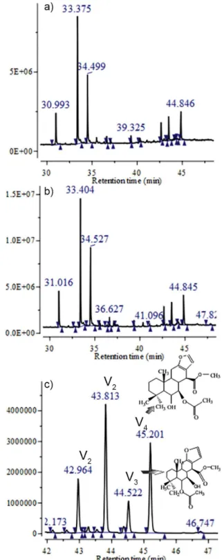
The GC-MS-SIM chemical proile of FHPp after
microencapsulation (Figure 1) remained unchanged when compared with that of FHPp, i.e., the major constituents, geranylgeraniol and vouacapans, remained stable. The transformation of raw plant materials into drugs should preserve the pharmacological and chemical integrity of constituents, ensuring their potential therapeutic value and maintenance of their biological effect and use (Miguel, Miguel, 1999).
Vouacapans are furanoditerpenes involved in the antirheumatic, antinociceptive, and anti-inflammatory activities of the oil extracted from P. pubescens fruit (Carvalho et al., 1999; Nunan, Carvalho, Piloveloso, 1982; Silva et al., 2004; Spindola et al., 2009; Spindola
et al., 2010). Earlier studies by our group have identiied
the presence of 2 vouacapans in FHPp that may be directly involved in their potential therapeutic activity: methyl
6α-acetoxy-7β-hydroxyvouacapan-17β-oate (vouacapan 3) and methyl 6α-hydroxy-7β-acetoxyvouacapan-17β-oate
(vouacapan 4) (Hoscheid et al., 2012). However, the main
compound extracted from FHPp was previously identiied
as 14,15-epoxygeranylgeraniol (retention time 33.38 min) (Hoscheid et al., 2012; Mors et al., 1967) and has been reported to be effective as a chemoprophylactic against schistosomiasis (Mors et al., 1967).
Morphological and particle size analysis
SEM p hoto micr ogr aph y in dicated that the
microcapsules were spherical, lat, and formed clusters
(Figure 2(a), (b), and (c)). This agglomeration is characterized by the residual chitosan in the process of cleaning tools. Similar results were found by Mladenovska
et al. (2007), by analyzing micrographs of alginate/ chitosan particles in 5-aminosalicylic acid. The surface of the microcapsules prepared in the present study appeared
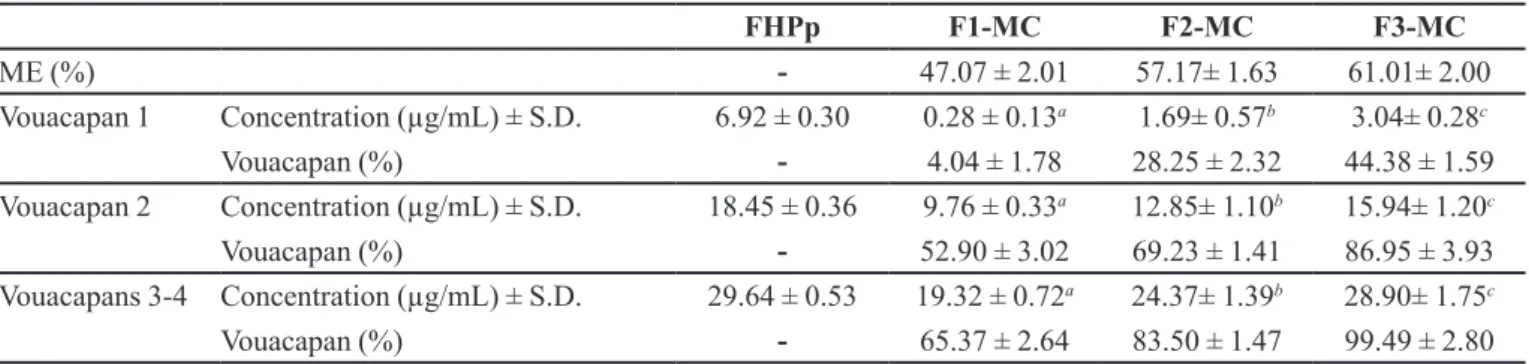
TABLE I -Microencapsulation eficiency of FHPp (ME%) and content of vouacapans microencapsulated (Vouacapan%) obtained by GC-MS-SIM
FHPp F1-MC F2-MC F3-MC
ME (%) - 47.07 ± 2.01 57.17± 1.63 61.01± 2.00
Vouacapan 1 Concentration (µg/mL) ± S.D. 6.92 ± 0.30 0.28 ± 0.13a 1.69± 0.57b 3.04± 0.28c
Vouacapan (%) - 4.04 ± 1.78 28.25 ± 2.32 44.38 ± 1.59
Vouacapan 2 Concentration (µg/mL) ± S.D. 18.45 ± 0.36 9.76 ± 0.33a 12.85± 1.10b 15.94± 1.20c
Vouacapan (%) - 52.90 ± 3.02 69.23 ± 1.41 86.95 ± 3.93
Vouacapans 3-4 Concentration (µg/mL) ± S.D. 29.64 ± 0.53 19.32 ± 0.72a 24.37± 1.39b 28.90± 1.75c
Vouacapan (%) - 65.37 ± 2.64 83.50 ± 1.47 99.49 ± 2.80
a, b and c- One-way ANOVA post-hoc Tukey’s test, specimens differ at p < 0.05.
FIGURE 2 - Photomicrography obtained by scanning electron microscopy of microcapsules obtained from polymeric associations: F1-MC (a); F2-MC (b); F3-MC (c) (2700x).
porous and wrinkled, a feature of the alginate polymer used (Pasparakis, Bouropoulos, 2006).
The particle size analysis revealed that F2-MC had a lower mean diameter than the other formulations. The diameters of F1-MC, F2-MC, and F3-MC were 0.9033 ± 0.48 µm, 0.4679 ± 0.21 µm, and 0.5885 ± 0.33 µm, respectively. Furthermore, the polydispersion index was determined as 0.9300 for F1-MC, 0.7234 for F2-MC, and 0.7467 for F3-MC.
ME% of the oleaginous fraction and Vouacapan%
The results of the encapsulation yield analysis (Table I) showed that the best association was provided
by F3-MC, a fact conirmed by the Vouacapans%. These
results were similar to those obtained by Peniche et al. (2004), who in a study of encapsulation of alginate/ chitosan in shark liver oil showed that increasing the concentration of alginate improves the capsule oil content. However, the present work was limited by the capacity of the peristaltic pump of the spray-dryer that could not atomize concentrations higher than 3% (w/v), which would provide enhanced results according the previous study by Peniche et al. (2004). The alginate reacts with divalent cations such as Ca+2, Sr+2, and
Ba+2, which replace the sodium ions in guluronic acid,
forming a network structure (Shi et al., 2011). Thus, the ratio of guluronic acid should be higher to enhance the encapsulation process.
The content of microencapsulated vouacapans 1 obtained for all formulations was low (<50.00%), indicating that the type of polymer network created by alginate associated with chitosan, was not sufficient for microencapsulation. However, it should be noted that the Vouacapan% of vouacapans 2 and 3-4 showed better results, with a high recovery. The formulation that showed the best Vouacapan% results was F3-MC, which had the highest active concentration, and consequently microencapsulation efficiencies over 44.38%, 86.95%,and 99.49% for vouacapans 1, 2, and 3–4, respectively.
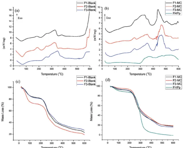
Thermal analysis
By comparing blank microcapsules with FHPp-loaded microcapsules using DSC analysis (Figure 3(a) and (b)), it was possible to detect displacement of the exothermic peak from 578 °C to 467 °C. This shift is caused by a decrease in thermal resistance caused by the interaction of the matrix with FHPp alginate/chitosan, thus demonstrating encapsulation.
microcapsules with those of the microcapsules loaded with FHPp, as illustrated in Figure 3(c) and (d), we observed that the microencapsulation of FHPp resulted in
an increase in water content. The irst mass loss at a low
temperature (<200 °C) corresponds to water desorption (Zohuriaan, Shokrolahi, 2004), and the second mass loss at a higher temperature (>200 °C) is attributed to polymer degradation (Cozic et al., 2009).
It was also observed that the microencapsulation process protected FHPp from degradation, since the residual mass of FHPp alone was 0.11%; however, on F1-MC this increased to 19.60% at the same temperature (500 °C). Similar thermal behavior was observed for the other microcapsules.
The thermal proile of the isolated polymers showed
a broad endothermic peak between 25 and 100 °C, related to polymer dehydration, followed by decomposition. According to Czubenko and Druzynska (2009), the increased water content appears near the endothermic peak and drives the material to higher temperatures with increasing water content.
XRD
In the XRD analysis, the chitosans showed 2 peaks at about 10° and 20°, corresponding to hydrated and anhydrous crystals, respectively (Figure 4a). These results are in agreement with those of Li et al. (2009). After microencapsulation, the typical chitosan peaks disappeared (Figure 4b) and the loaded microcapsules demonstrated an amorphous nature, similar to that of alginate. This suggested that the introduction of alginate on chitosan altered the crystalline structure of chitosan, probably preventing the formation of hydrogen bonds between the amino and hydroxyl groups (Li et al., 2009).
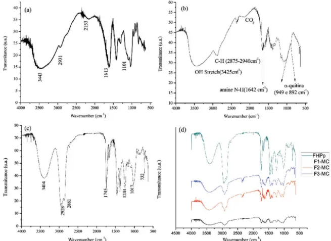
FTIR
The absorption spectrum in the infrared region of alginate (Figure 5a) showed absorption bands at 1613 cm-1
attributed to the stretch of -COO, at 1101 cm-1 because of
the stretch of -C-O-C-, and at 3443 cm-1 because of the
stretch of the hydroxyl group linked by hydrogen bonds.
FIGURE 4 - X-ray diffraction of polymers (a), and from FHPp-loaded microcapsules (b).
Similar results have been observed by other authors (Lawrie
et al., 2007; Pasparakis, Bouropoulos, 2006). According to Czubenko and Druzynska (2009), the vibrations at 2931 cm-1 are attributable to an aliphatic C-H stretch.
The absorption spectrum for low-molecular-weight chitosan (Figure 5b) showed absorption bands at 3425 cm-1
because of stretching of the hydroxyl group (OH). In the region between 2875 and 2940 cm-1, the observed
FIGURE 5 - FT-IR of alginate (a), of low molecular weight chitosan (b), of FHPp (c) and of FHPp-loaded microcapsules (d).
absorption bands were associated with C-H stretching. The band that absorbed at 1642 cm-1 was a result of the
predominance of the amino group (N-H). Similar results were found by Paulino et al. (2009) when they analyzed the infrared spectrum of chitosan.
In the infrared absorption spectrum of FHPp (Figure 5c), we observed absorption bands at 1745 cm-1 attributed
at 1244 cm-1 (indicating the asymmetric C-O stretch of
acetate), at 1017 cm-1 (indicating asymmetric stretch of
the C-O-C ester), and at 732 cm-1 (relecting furan rings).
This trait was also found by Spindola et al. (2009) when
they analyzed 6α-acetoxy,7β-hydroxy-vouacapan.
O n a n a l y s i s o f t h e s a m p l e s o f t h e l o a d e d microcapsules (Figure 5d), we noted that the absorption bands at 3404 cm-1, and those between 2928 and 2861 cm-1
for FHPp were present in varying intensities in the microcapsules, which suggests a variable amount of microencapsulated oil.
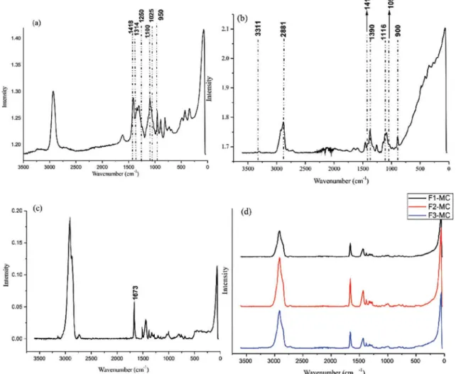
FT-Raman spectroscopy
Characterization of different types of alginate reveals the presence of different proportions of guluronic and mannuronic acid units (Pereira et al., 2003). The presence of these acids can be determined from the characteristic features of their bands. While guluronic acid units originate from a band around 1025 cm-1, mannuronic
acid originates at a band at approximately 1100 cm-1, as can
be seen in Figure 6(a). Thus, the proportion of guluronic/ mannuronic acid concentration can be determined by their relative intensity. These bands were also used by Pereira
et al. (2003) to characterize alginate. In the spectrum of low-molecular-weight chitosan (Figure 6b), we observed bands in the range of 2800-3000 cm-1, which is usually
derived from stretching vibrations of CH groups (Socrates, 2001). Therefore, a band at 2881 cm-1 can be attributed
to these vibrations. Bands at 1419, 1116, and 1094 cm-1
are associated with the vibrations of polysaccharide backbones, and bands at 1116 cm-1 and 1094 cm-1 may
be considered indicative of the symmetric vibration of glycosidic bonds (Zhang et al., 2011). Analyzing the spectra for the FHPp-loaded microcapsules (Figure 6c) and comparing these with the FHPp spectra (Figure 6d), we found that a peak at 1673 cm-1 is characteristic of FHPp,
and is present in all microcapsule samples, indicating that oil was microencapsulated. In general, for esters and monoesters of unsaturated dicarboxylic acids, the C=O bandwidth occurs approximately within the same range (1735-1760 cm-1). With β-keto esters, tautomerism is
possible (Socrates, 2001). In the case of tautomers, a strong band is observed at 1650 cm-1. The peak at 1673 cm-1 is
relective of the ester group (i.e., the vouacapans present
in FHPp), where the effect of hydrogen bonding occurs. When an intramolecular hydrogen bond (internal) is present, the C=O bond shifts to a lower frequency. The two most striking features in the spectrum of a normal ester are the strong C=O band, which appears between 1735 and 1750 cm-1, and stretch C-O bond bands, which
appear between 1300 and 1000 cm-1 (Pavia et al., 2010).
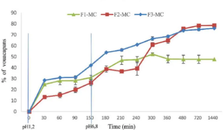
In vitro release profile
The release proile of the FHPp-loaded microcapsules
was generated by measuring vouacapans in FHPp. There
was no statistically signiicant change between the average
percentage of vouacapans released in F2-MC and F3-MC after 24 h (Figure 7); however, it was observed that the release of F3-MC appears to be more accelerated, which could be valuable for oral formulations. We observed that after 3 h (pH 1.2), vouacapan release from F3-MC was 53.85 ± 0.44%, suggesting that the FHPp may be primarily located at the alginate/chitosan interface, a fact made possible by the chitosan-induced relaxation of the polymer mesh.
After 24 h (pH 6.8), the release of vouacapans from F3-MC was 75.88 ± 0.45%, slightly lower than that from F2-MC (78.41 ± 2.23%); in contrast, vouacapan release
from F1-MC was 47.64 ± 2.50%, which was signiicantly
lower than the results obtained with the other two formulations.
According to Ribeiro et al. (2005), there is a lack of knowledge about the behavior of alginate microspheres
in simulated gastric and intestinal luids; however, it is
known that the stability of the alginate/chitosan complex
can be inluenced by environmental parameters such as
pH and ionic strength. Two hours under simulated gastric conditions can lead to the dissociation of the alginate matrix (Shu, Zhu, Song, 2001), providing evidence that this polymers not the limiting factor for the complete liberation of vouacapans.
According to Mi et al. (2002), the ability of alginate/ chitosan microcapsules to swell depends on the pH value. The lower the pH, the greater the swelling, and at pH >6.0, the degree of swelling is reduced. The increased swelling at pH<2.0 is due to the protonation of chitosan, while the slight increase in swelling at pH>5.0 is attributable to the ionization of the carboxylic group of alginate within the alginate/chitosan complex. This is important because the
degree of swelling inluences the process of diffusion, with
increased swelling being associated with a higher diffusion rate. Microcapsules produced with low-molecular-weight chitosan showed less swelling compared to that shown by the microcapsules prepared using larger-molecular-weight chitosans when applied in delivery systems, and were associated with decreased diffusion (Liu et al., 2004).
Several authors have reported a similar percentage release of various drugs from alginate/chitosan beads, demonstrating that these polymer coatings decrease drug release from microcapsules (Anal, Stevens, 2005; González-Rodríguez et al., 2002; Rajendran, Basu, 2009; Yu et al., 2008).
We next examined release kinetics to determine
which model provided the best it for the data. We tested irst-order Q (Mt / M ∞ = Qt = ln ‘. t) and second-order (Mt / M ∞ = Qt = (1 / Q). t) models, as well as the model proposed by Higuchi (Mt / M ∞ = Q0 + KH t1 / 2), for all
formulations.
Following Fick’s law, Mt/M∞ = k.tn and log Mt/M∞
= log k + n log t constant values were obtained: a value of n = 0.45 indicates Case I (Fickian) diffusion or square root of time kinetics, 0.45 >n < 0.89 indicates anomalous (non-Fickian) diffusion, n = 0.89 indicates Case II transport, and n > 0.89 indicates super case II transport (Ritger, Peppas, 1987).
For F1-MC, it was found that the best it was provided
by Higuchi’s model (R = 0.9382 ± 0.0136), by Fick’s law of diffusion, n = 1.5347 (n > 1), r = 0.9664 ± 0.0118, and k = 1.37. However, for F2-MC, the best kinetics model
was irst order (R = 0.9713 ± 0.0290), by Fick’s law of
diffusion n = 1.3733 (n > 1), r = 0.9878 ± 0.0118, and k
= 1.14. It was found that the best it was also provided by
Higuchi’s model with R = 0.8782 ± 0.0136 for F3-MC, with diffusional n = 1.6084, i.e., n>1, r = 0.9525 ± 0.0118, and k = 1.55.
The n value (n>1) indicates that the release mechanism is of the super case II type, in which the simultaneous contribution of processes such as diffusion, swelling, relaxation, and erosion of the polymeric matrix does not obey a Fickian diffusion model (Korsmeyer et al., 1983; Peppas, 1985; Ritger, Peppas, 1987). In the non-Fickian diffusion process, the polymeric chains do not have enough mobility to allow fast penetration of the solvent into the polymer matrix.
CONCLUSIONS
The data revealed that the chemical profile of FHPp was not altered by the microencapsulation process. Thermal and spectroscopic analysis in the infrared region revealed that FHPp was microencapsulated and that the process protected the obtained fraction from degradation. The crystalline chitosan structure was changed with the introduction of alginate, yielding amorphous microcapsules.
Although microcapsules developed with alginate/
chitosan with a deacetylation rate ≥75% (F2-MC) showed
a lower mean diameter, the ME% of the oleaginous fraction and content of microencapsulated vouacapans were greater than when using the combination of alginate/ low-molecular-weight chitosan (F3-MC). The best model to describe the release kinetics was that proposed by Higuchi for F1-MC and F3-MC; however, for F2-MC, the
irst-order model was used, and the release mechanism was
of the supercase II type for all formulations.
The most promising pre-formulation (F3-MC) needs further examination in order to produce a final formulation, and its ex vivo and in vivo stability should also be determined.
ACKNOWLEDGEMENTS
The authors are grateful to Financiadora de Estudos e Projetos (FINEP), Coordenadoria de Aperfeiçoamento de Pessoal do Ensino Superior (CAPES), and Conselho
Nacional de Desenvolvimento Cientíico e Tecnológico (CNPq) for their inancial support. They also acknowledge the Antonio Medina Neto for the DSC and TGA analysis and Edson Marques dos Reis for GC-MS analysis.
REFERENCES
ANAL, A.K.; STEVENS, W.F. Chitosan-alginate multilayer beads for controlled release of ampicillin. Int. J. Pharm., v.290, p.45-54,2005.
BARBER, T.A. Particle population analysis. In: BARBER, T.A. (Ed.). Pharmaceutical particulate matter: analysis and control. Buffalo Grove: Interfarma, 1993. p.266-303.
CARVALHO, J.C.T.; SERTIE, J.A.A.; BARBOSA, M.V.J.; PATRICIO, K.C.M.; CAPUTO, L.R.G.; SARTI, S.J.; FERREIRA, L.P., BASTOS, J.K. Anti-inflammatory activity of the crude extract from the fruits of Pterodon emarginatus Vog. J. Ethnopharmacol., v.64, p.127-133,1999.
COELHO, L.P.; REIS, P.A.; CASTRO, F.L.; GAYER, C.R.M.; LOPES, C.S.; SILVA, M.C.C.; SABINO, K.C.C.; TODESCHINI, A.R.; COELHO, M.G.P. Antinociceptive properties of ethanolic extract and fractions of Pterodon pubescens Benth. seeds. J. Ethnofarmacol., v.98, p.109-116,2005.
COZIC, C.; LICTON, L.; GARDA, M.R.; MARLHOUX, F.; CERF, D.L. Analysis of Arabic gum: Study of degradation and water desorption processes. Food Hydrocolloid, v.23, p.1930-1934, 2009.
CZUBENKO, J.O.; DRUZYNSKA, M.G. Effect of ionic crosslinking on the water state in hydrogel chitosan membranes. Carbohydr.Polym., v.77, p.590-598,2009.
GONZÁLEZ-RODRÍGUEZ, M.L.; HOLGADO, M.A.; SÁNCHEZ-LAFUENTE, C.; RABASCO, A.M.; FINI, A. Alginate/chitosan particulate systems for sodium diclofenac release. Int. J. Pharm., v.232, p.225-234, 2002.
HOSCHEID, J.; REINAS, A.; CORTEZ, D.A.G.; COSTA, W.F.; CARDOSO, M.L.C. Determination by CG-MS-SIM of furanoditerpenes in Pterodon pubescens Benth.: development and validation. Talanta, v.100, p.372-376, 2012.
HOSCHEID, J.; BERSANI-AMADO, C.A.; ROCHA, B.A.; OUTUKI, P.M.; SILVA, M.A.R.C.P.; FROEHLICH, D.L.; CARDOSO, M.L.C. Inhibitory effect of the hexane fraction of the ethanolic extract of the fruits of Pterodon pubescens Benth in acute and chronic inflammation. Evid. Based Compl. Alt. Med., v.2013, p.1-7, art. n. 272795, 2013.
KRUIF, C.G.; WEINBRECK, F.; VRIES, R. Complex coacervation of proteins and anionic polysaccharides. Curr. Opin. Colloid Interface Sci., v.9, p.340-349, 2004.
LAWRIE, G.; KEEN, I.; DREW, B.; CHANDLER-TEMPLE, A.; RINTOUL, L.; FREDERICKS, P.; GRØNDAH, L. Interactions between alginate and chitosan biopolymers characterized using FTIR and XPS. Biomacromolecules, v.8, p.2533-2541, 2007.
LI, X.; XIE, H.; LIN, J.; XIE, W.; MA, X. Characterization and biodegradation of chitosan–alginate polyelectrolyte complexes. Polym. Degrad. Stabil., v.94, p.1-6, 2009.
LIU, X.; XUE, W.; LIU, Q.; YU, W.; FU, Y.; XIONG, X.; MA, X.; YUAN, Q. Swelling behaviour of alginate–chitosan microcapsules prepared by external gelation or internal gelation technology. Carbohydr. Polym., v.56, p.459-464, 2004.
MAHKAM, M. Modiication of nano alginate-chitosan matrix for oral delivery of insulin. Nat. Sci., v.7, p.1-7, 2009.
MI, F.L.; SUNG, H.W.; SHYU, S.S. Drug release from chitosan-alginate complex beads reinforced by a naturally occurring cross-linking agent. Carbohydr. Polym.,v.48, p.61-72, 2002.
MIGUEL, M.D., MIGUEL, O.G. Desenvolvimento de
itoterápicos. São Paulo: Robe, 1999. p.27-41.
MLADENOVSKA, K.; CRUAUD, O.; RICHOMME, P.; BELAMIE, E.; RAICKI, R.S.; VENIER-JULIENNE, M.C.; POPOVSKI, E.; BENOIT, J.P.; GORACINOVA, K. 5-ASA loaded chitosan–Ca–alginate microparticles: Preparation and physicochemical characterization. Int. J. Pharm., v.345, p.59-69, 2007.
MORS, W.B.; FASCIO, M.; MONTEIRO, H.J.; GILBERT, B.; PELLEGRINO, J. Chemoprophylactic agent in schistosomiasis: 14,15-epoxygeranylgeraniol. Science, v.157, p.950-951, 1967.
NESTERENKO, A.; ALRIC, I.; SILVESTRE, F.; DURRIEU, V. Vegetable proteins in microencapsulation: A review of recent interventions and their effectiveness. Ind. Crop. Prod., v.42, p.469-479, 2013.
NUNAN, E.A.; CARVALHO, M.G.; PILOVELOSO, D. Furanediterpenes with anti- and proinlammatory activity. Braz. J. Med. Biol. Res., v.15, p.450-451, 1982.
PASPARAKIS, G.; BOUROPOULOS, N. Swelling studies and in vitro release of verapamil from calcium alginate and calcium alginate–chitosan beads. Int. J. Pharm., v.323, p.34-42, 2006.
PAULINO, A.T.; GUILHERME, M.R.; DE ALMEIDA, E . A . M . S . ; P E R E I R A , A . G . B . ; M U N I Z , E . C . ; TAMBOURGI, E.B. One-pot synthesis of a chitosan-based hydrogel as a potential device for magnetic biomaterial. J. Magn. Magn. Mater., v.17, p.2636-2642, 2009.
PAVIA, D.L.; LAMPMAN, G.M.; KRIZ, G.S.; VYVYAN, J.R. Espectroscopia no infravermelho. In PAVIA, D.L.; LAMPMAN, G.M.; KRIZ, G.S.; VYVYAN, J.R. (Eds.). Introdução à espectroscopia. 4.ed. São Paulo: Cengage Learning Edições Ltda, 2010. p.64-65.
PENICHE, C.; HOWLAND, I.; CARRILLO, O.; ZALDÍVAR, C.; ARGÜELLES-MONAL, W. Formation and stability of shark liver oil loaded chitosan/calcium alginate capsules. Food Hydrocolloid, v.18, p.865-871, 2004.
PEPPAS, N.A. Analysis of Fickian and non-Fickian drug release from polymers. Pharm. Acta Helv.,v.60, p.110-111,1985.
PEREIRA, L.; SOUSA, A.; COELHO, H.; AMADO, A.M.; RIBEIRO-CLARO, P.J.A. Use of FTIR, FT-Raman and 13C-NMR spectroscopy for identiication of some seaweed phycocolloids. Biomol. Enq., v.20, p.223-228, 2003.
RAJENDRAN, A.; BASU, S.K. Alginate-chitosan particulate system for sustained release of nimodipine. Trop. J. Pharm. Res., v.8, p.433-440, 2009.
RIBEIRO, A.J.; SILVA, C.; FERREIRA, D.; VEIGA, F. Chitosan-reinforced alginate microspheres obtained through the emulsification/internal gelation technique. Eur. J. Pharm. Sci., v.25, p.31-40, 2005.
RITGER, P.L.; PEPPAS, N.A. A simple equation for description of solute release I. Fickian and non-ickian release from non-swellable devices in the form of slabs, spheres, cylinders or discs. J. Control. Release, v.5, p.23-36, 1987.
SHI, P.; HE, P.; THE, T.K.H.; MORSI, Y.S.; GOH, J.C.H. Parametric analysis of shape changes of alginate beads. Adv. Powder Technol., v.210, p.60-66, 2011.
SHU, X.Z.; ZHU, K.J.; SONG, W. Novel pH-sensitive citrate cross-linked chitosan ilm for drug controlled release. Int. J. Pharm., v.212, p.19-28, 2001.
SILVA, M.C.C.; GAYER, C.R.M.; LOPES, C.S.; CALIXTO, N.O.; REIS, P.A.; PASSEAS, C.P.B.; PAES, M.C.; DALMAU, S.R.; SABINO, K.C.C.; TODESCHINI, A.R.; COELHO, M.G.P. Acute and topic anti-edematogenic fractions isolated from the seeds of Pterodonpubescens. J. Pharm. Pharmacol., v.55, p.135-141, 2004.
SOCRATES, G. The carbonyl group: C=O. In: SOCRATES, G. (Ed.) Infrared and raman characteristic group frequencies: tables and charts. 3.ed. Chichester: John Wiley, 2001. p.147-148.
SPINDOLA, H.M.; CARVALHO, J.E.; RUIZ, A.L.T.G.; RODRIGUES, A.F.R.; DENNY, C.; SOUSA, I.M.O.; TAMASHIRO, J.Y.; FOGLIO, M.A. Furanoditerpenes from Pterodon pubescens Benth with selective in vitro anticancer activity for prostate cell line. J. Braz. Chem. Soc., v.20, p.569-575, 2009.
SPINDOLA, H.M.; SERVAT, L.; DENNY, C.; RODRIGUES, R.A.F.; EBERLIN, M.N.; CABRAL, E.; SOUSA, I.M.O.; TAMASHIRO, J.Y.; CARVALHO, J.E.; FOGLIO, M.A. Antinociceptive -dihydroxyvouacapan-17β-oate methyl ester isolated from Pterodon pubescens Benth. BMC Pharmacol., v.10, p.1-10, 2010.
SUAVE, J.; DALLAGNOL, E.C.; PEZZIN, A.P.T.; SILVA, D.A.K.; MEIER, M. M.; SOLDI, V. Microencapsulação: inovação em diferentes áreas. Saúde & Amb. Rev., v.7, p.12-20, 2006.
U D D I N , M . S . ; H AW L A D E R , M . N . A . ; Z H U , H . J . Microencapsulation of ascorbic acid: effect of process variables on product characteristics. J. Microencapsul., v.18, p.199-209, 2001.
UNITED STATES PHARMACOPEIA. 32-National Formulary 27. Rockwell, USP, 2009.
VENKATRAMAN, S.; DAVAR, N.; CHESTER, A.; KLEINER, L. An overview of controlled release systems. In: WISE, D.L. (Ed.). Handbook of pharmaceutical controlled release technology. New York: Marcel Dekker, 2000. p.431-463.
VIEIRA, C.R.; MARQUE, M.F.; SOARES, P.R.; MATUDA, L.; OLIVEIRA, C.M.A.; KATO, L.; SILVA, C.C.; GUILLO, L.A. Antiproliferative activity of PterodonpubescensBenth. seed oil and its active principle on human melanoma cells. Phytomedicine, v.15, p.528-532, 2008.
YU, C.Y.; ZHANG, X.C.; ZHOU, F.Z.; ZHANG, X.Z.; CHENG, S.X.; ZHUO, R.X. Sustained release of antineoplastic drugs from chitosan-reinforced alginate microparticle drug delivery systems. Int. J. Pharm., v.357, p.15-21, 2008.
ZHANG, K.; PESCHEL, D.; HELM, J.; GROTH, T.; FISCHER, S. FT Raman investigation of novel chitosan sulfates exhibiting osteogenic capacity. Carbohydr. Polym., v.83, p.60-65, 2011.
ZOHURIAAN, M.J.; SHOKROLAHI, F. Thermal studies on natural and modiied gums. Polym. Test., v.23, p.575-579, 2004.
Received for publication on 17th December 2013