Resorption of roots of different dimension
induced by different types of forces
Abstract: Root resorption is a variable to be considered in induced tooth movement (ITM). It is related to root morphology and alveolar bone crest, and also to the types of forces exerted by mechanotherapy. This histometric study evaluated the predominance of root resorption among roots of different dimensions, following ITM with different types of forces and at different time intervals. The study was conduct-ed on 54 rats dividconduct-ed into three groups, according to the type of force: continuous (CF), continuous interrupted (CIF) and intermittent (IF), at periods of 5, 7 and 9 days. The percentage of resorption between mesio-buccal roots of larger dimension and intermediate roots of smaller di-mension was assessed. The evaluations were performed on the AxioVi-sion software, and the non-parametric analysis of variance for repeated measures in independent groups was further applied, consisting of a
scheme of two factors, and complemented by the Dunn test at a signii -cance level of 5%. The intermediate roots presented a higher percentage of resorption, which was gradual at the periods evaluated for the three types of forces, but mainly for CF. Comparing the intermediate roots
with the mesiobuccal roots, there was a statistically signiicant differ -ence (p < 0.05) in the CF group at day 7 and day 9, and in the FI group,
at day 9. The intragroup analysis evidenced a statistically signiicant
difference (p < 0.05) between the 5th and the 9th day for the intermediate root in the CF group. The intergroup analysis did not reveal any
statis-tically signiicant difference (p > 0.05) in individually analyzed roots.
Keywords: Root Resorption; Tooth Movement; Rats.
Introduction
Orthodontic mechanics involves continuous, continuous interrupted or interrupted and intermittent forces. These forces cause compression and tensile stresses on the periodontal ligament and alveolar bone, inducing morphological and microscopic reactions controlled by chemical media-tors, promoting tooth displacement,1,2,3 which is called orthodontic tooth movement or induced tooth movement (ITM).
The mechanism and biological reaction of ITM depend on the magni-tude, duration and type of force delivered by stainless steel4,5 wires and springs, by superelastics with shape memory, like nickel-titanium alloys (NiTi),5,6,7,8 by ß-titanium or titanium-molybdenum (TMA),9 or by intra- and intermaxillary elastics. These forces can induce variable degrees of root resorption4,5,8,10 in different root morphologies.10,11,12
Osmar Aparecido CUOGHI(a)
Carlos Alberto AIELLO(b)
Alberto CONSOLARO(c)
Pedro Marcelo TONDELLI(d)
Marcos Rogério de MENDONÇA(a)
(a)Department of Pediatric and Community
Dentistry, Faculdade de Odontologia de Araçatuba, Universidade Estadual Paulista – UNESP, Araçatuba, SP, Brazil.
(b)Department of Orthodontics, Hospital de
Reabilitação de Anomalias Craniofaciais – HRAC, Universidade de São Paulo – USP, Bauru, SP, Brazil.
(c)Department of Stomatology, Faculdade de
Odontologia de Bauru, Universidade de São Paulo – USP, Bauru, SP, Brazil.
(d)Department of Oral Medicine and Pediatric
Dentistry, Centro de Ciências da Saúde, Universidade Estadual de Londrina - UEL, Londrina, PR, Brazil.
Declaration of Interests: The authors certify that they have no commercial or associative interest that represents a conflict of interest in connection with the manuscript.
Corresponding Author:
Carlos Alberto Aiello E-mail: [email protected]
DOI: 10.1590/1807-3107BOR-2014.vol28.0013 Epub: Jun 02, 2014
Submitted: May 08, 2013
Root morphology may inluence root resorption;
i.e., teeth with short roots, a triangular shape, pipette-shaped apical thirds or dilaceration pose higher risk of resorption.3,13 Dental traumas,14 as well as the alve-olar crest morphology,11 should also be considered risk factors in root resorption.
The extent of root resorption in ITM can be evalu-ated by different methods and imaging techniques.9,12 The most indicated methods are periapical radio-graphs and cone-beam computed tomography (CBCT), which allow accurate diagnosis in three-dimensional images.11,15,16 Panoramic radiographs or cephalograms have limitations regarding methodology and accu-racy, and thus should be analyzed carefully when assessing their results.
Both the tissue phenomena and the root resorption related to ITM can be investigated in experimental models in rats, by induced movement of the maxillary
irst molar,17,18,19,20,21 and be extrapolated to humans. This study evaluated the predominance of root resorption among roots of different dimensions
dur-ing ITM of the maxillary right irst molar in rats, usdur-ing
different types of forces and time periods.
Methodology
The sample included 54 male young adult Wis-tar rats (Rattus norvegicus albinus). This study was approved by the Institutional Review Board on Ani-mal Studies (Faculdade de Odontologia de Araçatuba, UNESP), under Protocol no. 004938-2009.
The sample was divided into three groups accord-ing to the type of force: continuous (CF), continuous interrupted (CIF) and intermittent (IF). Each group was divided into three subgroups with six animals each, to evaluate changes occurring at periods of 5, 7 and 9 days.
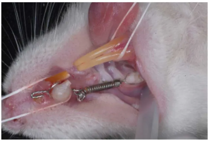
In the CF group (Figure 1A), the springs were maintained without force interruption until the 5th, the 7th and the 9th days. In the CIF group (Figure 1B), the springs were deactivated after three days of con-tinuous force, and maintained in this passive posi-tion until the 5th day, when they were reactivated and maintained in this state until the 7th day, then deac-tivated again until the 9th day. The same procedure was carried out in the IF group (Figure 1C), and in the CIF group, except in regard to the springs, which were removed and replaced.
The experimental procedure was divided into
two stages. In the irst, the murines were 62 days old,
and the procedure comprised of inducing dentoal-Figure 1C. Intermittent force: removed spring
Figure 1B. Continuous interrupted force: non-activated spring in a passive position
veolar ankylosisof maxillary right incisors (Figure 2) to promote absolute anchorage for movement of
the maxillary right irst molar, without inluenc -ing the type of force applied dur-ing movement, especially CIF. This stage was performed accord-ing to a predetermined protocol:22 the anesthetic (Dopalen - ketamine hydrochloride) and the mus-cle relaxant (Anasedan - xylazine hydrochloride) – Vetbrands, Paulínia, Brazil – were mixed and applied by intramuscular injection, followed by luxation and extraction, removal of periradicular tissues and dental papilla, root canal obturation with a mixture of calcium hydroxide and propyl-ene glycol, and root apex sealing with a mixture of grey MTA (Angelus, Londrina, Brazil) and dis-tilled water, and replantation.
A period of two weeks was allowed for formation of ankylosis before the second stage was initiated.
This consisted of ITM of the maxillary right irst
molar when the murines were 76 days old, with a mean weight of 274.75 g, using three different types of forces. The procedure was performed with a 3-mm nickel-titanium closed coil spring Sentalloy® (GAC) (COIL SPRINGS 10-000-25 – GAC Interna-tional Inc., New York, USA), to deliver a continuous force of 50cN, adapted to the cervical region of the
maxillary right irst molar with a 0.20-mm diameter
stainless steel wire, and to the maxillary right inci-sor with a 0.25-mm diameter stainless steel wire. This method followed the principles of Heller and Nanda17 with innovative changes.
The animals were euthanized by injection of an excessive anesthetic dose on completion of each experiment, and histotechnical processing was then performed.
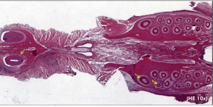
The specimens embedded in parafin blocks were transversely and serially sectioned at 6μm. All roots of the maxillary right irst molar were separated at
the cervical third, immediately below the alveolar bone crest level, where homogeneous structures of alveolar bone tissue and periodontal ligament were observed extending to the incisor roots, and were stained with hematoxylin-eosin (Figure 2).
Histometric analysis
The sections (Figure 2) were examined under a ZEISS Axiophot (Carl Zeiss, Göttingen, Germany) microscope and images of the mesiobuccal and
inter-mediate roots at 25x magniication were captured by
a digital camera connected to the microscope. After acquiring the images, the mesiobuccal and intermediate roots (Figure 3) were visualized using
a microcomputer, demarcated and measured with Imaging Systems - Software Release 4.8.2 – AxioVi-sion (Copyright 1999-2010 by Carl Zeiss, Göttingen, Germany) software to quantify the percentage of resorption in each period of ITM. The greater root perimeter was then contoured at the most external limit of cementum, in red. The resorbed root area was contoured internally in green, and the origi-nal root contour was simulated exterorigi-nally. Then, another contour was made in blue, delimiting the smaller perimeter at the most internal dentin limit, surrounding the pulp externally (Figure 3).
This procedure allowed calculation of the total area of root dentin and cementum, as well as the areas of resorption, expressed in square microme-ters. Accordingly, the percentage of root resorption of the two roots was calculated for each type of force at the different time intervals.
Evaluation of measurement error
For the purpose of method error analysis, 36 sec-tions were randomly selected and the two roots were once again measured after a one-week interval.
The intraexaminer systematic error was calcu-lated by the Wilcoxon Signed Rank Test. The casual error was determined using the error calculation proposed by Dahlberg.23
where d = difference between irst and second
measurements
n = number of repetitions
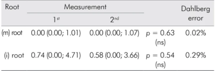
Table 1 shows the method error. The probability values correspond to the systematic error, whereas the values obtained by the Dahlberg formula repre-sent the casual error. No systematic or casual errors were observed, and results considered within accept-able parameters were obtained, thus indicating the reliability of the present conclusions.
Statistical analysis
Comparison of the percentage of root resorption, according to the type of mechanical force, study period and type of root, was made initially applying the Kol-mogorov-Smirnov test to analyze the adherence of data in the normal distribution of probabilities. Since the statistical test revealed abnormal distribution, non-parametric analysis of variance for repeated measures in independent groups was further applied, consist-ing of the two-factor scheme (type of force and study period). The analysis was complemented by the Dunn
multiple comparisons test, at a signiicance level of 5%.24
Results
In Table 2, the intragroup analysis showed that the intermediate root presented a higher percent-age value of resorption for the three types of forces, as compared with the mesiobuccal root, except for day 5 in the intermittent force group. The statisti-cal analysis, represented by Greek letters, indicated
a signiicant difference (p < 0.05) at day 7 and day 9 in the continuous force group, and at day 9 in the intermittent force group.
Table 1. Median, minimum and maximum values, ex-pressed in percentage of measurements of mesiobuccal (m) and intermediate (i) roots.
Root Measurement Dahlberg error 1st 2nd
(m) root 0.00 (0.00; 1.01) 0.00 (0.00; 1.07) p = 0.63 (ns)
0.02%
(i) root 0.74 (0.00; 4.71) 0.58 (0.00; 3.66) p = 0.54 (ns)
0.29%
The comparison among the three study periods for each type of force and the root analyzed, performed for the intragroup analysis and represented by lowercase
Arabic letters, did not reveal any statistically signiicant
result (p > 0.05) for the mesiobuccal root. In regard to the
intermediate root, a statistically signiicant difference
(p < 0.05) was observed for the continuous force (CF) group between the 5th and the 9th days. Even though no statistically signiicant difference was observed for
this root between the 7th and the 9th days, from a practi-cal standpoint, there was an important increase in the numerical values of the medians (0.69 and 1.76),
respec-tively. No statistically signiicant results were observed
for the other two forces (CIF and IF) (p > 0.05). The comparison among the same three study peri-ods for the three types of forces, performed for the inter-group analysis and represented by uppercase Arabic
letters, did not reveal any statistically signiicant dif -ference (p > 0.05) in individually analyzed roots. The highest numerical values of the medians were observed in the intermediate root for the continuous force group.
Discussion
The modiications to the principles of Heller and
Nanda17 in this study allowed absolute anchorage, prevented continuous eruption of incisors and main-tained constant force levels during the ITM of the
maxillary right irst molar. Selection of the trans -verse section at the cervical region of the roots was based on concomitant visualization of the roots of interest, observation of the entire root perimeter and quantity and quality of acellular cementum, which is similar to humans in rats.
Induced tooth movement occurs by application of mechanical forces,1,2,3,4,13,18 which create areas of ten-sile stress and compression. Depending on the type of force, magnitude and duration, cementum resis-tance to resorption may be disrupted by local clast activity, leading to dentin resorption.1
During the application of orthodontic forces,
tri-angular bone crests tend to undergo greater delec -tion, as compared with rectangular- or rhomboi-dal-shaped crests. Since these crests absorb part of this force, they tend to distribute it with lower risk both of lesion to the cementoblast layer and of root resorption. This may justify the greater root resorption percentage in intermediate roots (Table 2), in which the interradicular septa present a rhomboidal or rectangular shape, as compared with mesiobuccal roots, which present a triangular shape on the mesial surface. In addition, because mesiobuccal roots are larger, as compared with intermediate roots, there is better force dissipa-tion, with fewer undesirable effects.
Another aspect refers to the type of movement
generated when the irst molars are moved in an ante -rior or a mesial direction. The trapezoidal shape of these molars causes a tipping movement, with extru-sion in the distal region, thus increasing suscepti-bility to trauma during chewing, as well as greater compression of the mesial periodontal ligament in intermediate and distal roots.
A recent study25 revealed that the periodontal ligament thickness in rats is proportional to the root dimensions, i.e., smaller roots present a smaller thickness of periodontal ligament, as compared with larger roots. This is probably the most important fac-tor explaining the higher susceptibility of interme-Table 2. Median, minimum and maximum values of root
re-sorption expressed in percentage according to type of force, study period and evaluated root.
Force Period Root
Mesiobuccal Intermediate CF 5 days 0.00 (0.00; 0.44) α a A 0.44 (0.00; 2.75) α a A
7 days 0.10 (0.00; 0.35) α a A 0.69 (0.16; 3.38) β ab A 9 days 0.00 (0.00; 0.55) α a A 1.76 (1.06; 7.81) β b A CIF 5 days 0.00 (0.00; 0.38) α a A 0.06 (0.00; 1.20) α a A
7 days 0.00 (0.00; 0.74) α a A 0.36 (0.00; 5.08) α a A 9 days 0.02 (0.00; 0.63) α a A 0.83 (0.00; 7.56) α a A IF 5 days 0.00 (0.00; 0.44) α a A 0.00 (0.00; 2.73) α a A 7 days 0.06 (0.00; 0.46) α a A 0.19 (0.00; 2.80) α a A 9 days 0.00 (0.00; 1.01) α a A 0.41 (0.00; 2.30) β a A The Greek letter enabled interpretation of the intragroup analysis, which compared the mesiobuccal and intermediate roots with each other, according to the type of force and to the different study periods. Two medians followed by the same Greek letter do not present any statistical difference (p > 0.05).
diate roots to resorption, as compared with mesio-buccal roots, considering the same force magnitude of 50cN applied to both roots.
Although some studies show better results in rela-tion to the extent of tooth movement by using NiTi or TMA wires,5,8,9 a continuous interrupted force may be considered the most favorable option, especially in relation to lower predominance of root resorption.1,5,9
Comparing intermittent and continuous forces, Ballard et al.9 reached the same conclusion. That is to say, a continuous force (CF) presented better results in relation to the extent of tooth movement, but induced greater root resorption. This study revealed a tendency for higher percentage values of resorp-tion when CF was used (Table 2); CF presented
sta-tistically signiicant differences between the 5th and the 9th days for intermediate roots.
In relation to continuous interrupted and inter-mittent forces, in this study, a gradual increase was observed in the numerical percentages of resorption in intermediate roots, when compared to the 5th, the 7th and the 9th days. Despite the lack of any statisti-cally signiicant difference, the importance of this
increase should not be ruled out.
Although the results for the intermediate roots (Table 2) in the intergroup analysis indicated no
sta-tistically signiicant difference, they revealed that
continuous force induced the highest percentage of resorption, as compared with the other types of forces, in all the periods.
Thus, notwithstanding the lack of any
statisti-cally signiicant difference, continuous force tends
to be unfavorable as regards risk of root resorption, despite the more favorable results in relation to the extent of orthodontic movement.5,8,9
Another aspect is that the median values in the intermittent force group were the smallest, as com-pared with the other forces. Despite the lower aggres-siveness of this force in regard to root resorption, the results regarding the extent of orthodontic movement for this type of force are less favorable.9,21
This study demonstrated that continuous inter-rupted force promoted intermediate values of root resorption, despite the smaller extent of movement, as compared with only continuous force.5 Therefore, based on the present results, as applied to rat teeth, continuous interrupted force was a good method that could be applied to induce tooth movement in order to lessen the risk of root resorption.
Conclusions
1. The intermediate root presented a higher per-centage of root resorption, as compared with the mesiobuccal root, mainly after application of continuous force.
2. The intermediate root presented a gradual increase in root resorption in the three study periods for the different types of forces.
3. The fact that continuous interrupted force ex-hibited intermediate values among the different types of forces did not rule out the possibility of root resorption.
Acknowledgements
We wish to thank Fundação para o Desenvolvimento da Unesp (Fundunesp) for the inancial support given to this study (Process no. 01214/08).
1. Krishnan V, Davidovitch Z. Cellular, molecular, and
tissue-level reactions to orthodontic force. Am J Orthod Dentofacial
Orthop. 2006 Apr;129(4):469.e1-32.
2. Cattaneo PM, Dalstra M, Melsen B. Strains in periodontal
ligament and alveolar bone associated with orthodontic
tooth movement analyzed by finite element. Orthod
Cra-niofac Res. 2009 May;12(2):120-8.
3. Brezniak N, Wasserstein A. Orthodontically induced in-flammatory root resorption. Part I: the basic science aspects. Angle Orthod. 2002 Apr;72(2):175-9.
4. Reitan K. Initial tissue behavior during apical root resorp-tion. Angle Orthod. 1974 Jan;44(1):68-82.
5. Weiland F. Constant versus dissipating forces in orthodon-tics: the effect on initial tooth movement and root resorption. Eur J Orthod. 2003 Aug;25(4):335-42.
6. Miura F, Mogi M, Ohura Y, Hamanaka H. The super-elastic property of the Japanese NiTi alloy wire for use in ortho-dontics. Am J Orthod Dentofacial Orthop. 1986 Jul;90(1):1-10. 7. Miura F, Mogi M, Ohura Y, Karibe M. The super-elastic Japa-nese NiTi alloy wire for use in orthodontics. Part III. Studies on the Japanese NiTi alloy coil springs. Am J Orthod Dento-facial Orthop. 1988 Aug;94(2):89-96.
8. Owman-Moll P, Kurol J, Lundgren D. Continuous versus in-terrupted continuous orthodontic force related to early tooth movement and root resorption. Angle Orthod 1995;65(6):395-401; discussion 401-2.
9. Ballard DJ, Jones AS, Petocz P, Darendeliler MA. Physical properties of root cementum: Part 11. Continuous vs. inter-mittent controlled orthodontic forces on root resorption. A microcomputed-tomography study. Am J Orthod Dentofacial Orthop. 2009 Jul;136(1):8.e1-8; discussion 8-9.
10. Weltman B, Vig KW, Fields HW, Shanker S, Kaizar EE. Root resorp-tion associated with orthodontic tooth movement: a systematic review. Am J Orthod Dentofacial Orthop. 2010 Apr;137(4):462-76. 11. Brezniak N, Wasserstein A. Orthodontically induced inflam-matory root resorption. Part II: the clinical aspects. Angle Orthod. 2002 Apr;72(2):180-4.
12. Ioannidou-Marathiotou I, Papadopoulos MA, Kokkas A. Orth-odontic treatment and root resorption of teeth: critical analysis of mechanical factors. Hell Orthod Rev 2010:13(1-2):25-42. 13. Roberts-Harry D, Sandy J. Orthodontics. Part 11: orthodontic
tooth movement. Br Dent J. 2004 Apr 10;196(7):391-4; quiz 426. 14. Artun J, Van ‘t Hullenaar R, Doppel D, Kuijpers-Jagtman
AM. Identification of orthodontic patients at risk of severe apical root resorption. Am J Orthod Dentofacial Orthop. 2009 Apr;135(4):448-55.
15. Lund H, Gröndahl K, Gröndahl HG. Cone beam computed tomography for assessment of root length and marginal bone level during orthodontic treatment. Angle Orthod. 2010 May;80(3):466-73.
16. Dudic A, Giannopoulou C, Leuzinger M, Kiliaridis S. Detec-tion of apical root resorpDetec-tion after orthodontic treatment by using panoramic radiography and cone-beam computed tomography of super-high resolution. Am J Orthod Dento-facial Orthop. 2009 Apr;135(4):434-7.
17. Heller IJ, Nanda R. Effect of metabolic alteration of periodon-tal fibers on orthodontic tooth movement. An experimenperiodon-tal study. Am J Orthod. 1979 Mar;75(3):239-58.
18. Verna C, Zaffe D, Siciliani G. Histomorphometric study of
bone reactions during orthodontic tooth movement in rats. Bone. 1999 Apr;24(4):371-9.
19. Miyoshi K, Igarashi K, Saeki S, Shinoda H, Mitani H. Tooth movement and changes in periodontal tissue in response to orthodontic force in rats vary depending on the time of day the force is applied. Eur J Orthod. 2001 Aug;23(4):329-38. 20. Kameyama T, Matsumoto Y, Warita H, Soma K. Inactivated
periods of constant orthodontic forces related to desirable tooth movement in rats. J Orthod. 2003 Mar;30(1):31-7. 21. Hayashi H, Konoo T, Yamaguchi K. Intermittent 8-hour
activation in orthodontic molar movement. Am J Orthod Dentofacial Orthop. 2004 Mar;125(3):302-9.
22. Cuoghi OA, Tondelli PM, Sonoda CK, Aiello CA, Mendon-ça MR, Costa SC. Induction of ankylosis in the incisor for orthodontic tooth movement in rats.Dent Traumatol. 2014 Apr;30(2):112-7. doi: 10.1111/edt.12056. Epub 2013 Jul 7. 23. Houston WJ. The analysis of errors in orthodontic
measure-ments. Am J Orthod. 1983 May;83(5):382-90.
24. Zar JH. Biostatistical analysis. 5th ed. New Jersey: Prentice-Hall; 2009. 994 p.