176 177
176 177
ABSTRACT: The present study assessed condylar displacement between initial maximal habitual intercuspation (MHI) and centric relation (CR), recorded after using a deprogramming occlusal splint for an average period of 7.8 ± 2.1 months prior to any orthodontic treatment. The sample consisted of 22 subjects, 11 male and 11 female, with an average age of 14.2 ± 1.4 years, with Class II malocclusion2 and with no apparent signs or symptoms of
temporomandibular dysfunction (TMD). Condylar displacement was measured using a Panadent axis position indicator in decimal fractions of a millimeter. The original mean vertical displacements and the corresponding standard deviations were 4.24 ± 2.53 mm and 3.86 ± 2.72 mm, respectively, for the right and left sides. Because a significant negative correlation was observed between original condylar displacements and age factors, the dis-placement values were statistically adjusted to 2.74 ± 2.00 mm and 2.44 ± 1.93 mm. On the horizontal plane, the mean displacements measured were –0.72 ± 1.53 mm on the right side and –0.51 ± 1.98 mm on the left. The mean displacement on the transversal plane was 0.03 ± 0.87 mm. A comparison between these values and those observed in non-deprogrammed groups, as well as those published in the related literature, indicates that use of occlusal splints results in greater mean condylar displacement values, especially vertically, between CR and MHI positions, which contributed to a more accurate orthodontic diagnosis.
DESCRIPTORS: Mandibular condyle; Temporomandibular joint; Centric relation.
RESUMO: O presente estudo avaliou os deslocamentos condilares entre a máxima intercuspidação habitual (MIH) inicial e a relação central (RC), registrada após o uso de placa oclusal desprogramadora por período médio de 7,8 ± 2,1 meses antes do tratamento ortodôntico. A amostra consistiu de 22 indivíduos, 11 do gênero masculino e 11 do feminino, com média de idade de 14,2 ± 1,4 anos, com maloclusão de Classe II2, sem sinais e sintomas
aparentes de disfunção temporomandibular (DTM). Os deslocamentos condilares foram medidos com aproximação de décimos de milímetro, por meio do indicador de posição axial Panadent. As médias originais dos deslocamentos verticais dos lados direito e esquerdo e correspondentes desvios padrões mediram 4,24 ± 2,53 e 3,86 ± 2,72 mm, respectivamente. Devido à correlação negativa significante observada entre esses deslocamentos e os fatores relati-vos à idade da amostra, os mesmos foram corrigidos estatisticamente para 2,74 ± 2,00 e 2,44 ± 1,93 mm. No plano horizontal, os deslocamentos médios foram de –0,72 ± 1,53 mm no lado direito e –0,51 ± 1,98 no esquerdo. A mé-dia no plano transversal foi de 0,03 ± 0,87 mm. A comparação destes valores com aqueles observados em grupos não desprogramados e publicados na literatura indica que o uso das referidas placas resulta em deslocamentos condilares médios maiores, especialmente no sentido vertical, entre as posições de RC e de MIH, o que contribuiu para um diagnóstico ortodôntico mais preciso.
DESCRITORES: Côndilo mandibular; Articulação temporomandibular; Relação central.
INTRODUCTION
Since the early 20th century, orthodontists have been subject to criticism for evaluating pa-tients statically, in search for simple morphological malocclusions, while ignoring the possibility of
occasional functional malocclusions. Lauritzen15 (1974) suggests that morphological malocclusions do not necessarily represent any risk of damage to supporting tissues; on the other hand, functional
* PhD, Professors; **Postdoctorate Professor; ***Chair Professor – Department of Orthodontics and Dental Pediatrics, School of Dentistry, University of São Paulo.
Increase of condylar displacement between centric relation and
maximal habitual intercuspation after occlusal splint therapy
Aumento do deslocamento condilar entre relação central e
máxima intercuspidação habitual após terapia com placa oclusal
Solange Mongelli de Fantini* João Batista de Paiva* José Rino Neto*
Gladys Cristina Dominguez* Jorge Abrão**
176 177
176 177
malocclusions are considered triggers of parafunc-tions, and may cause the stomatognathic system self-destruction.
Although the role of occlusion in the etiology of temporomandibular dysfunction (TMD), evaluated in its dental aspects, is still a very controversial issue, Crawford8 (1999) observed a high degree of correlation between signs and symptoms of these dysfunctions and condylar displacement between the positions of centric relation (CR) and maximal habitual intercuspation (MHI).
Centric relation is currently understood as the maxillomandibular relation in which the con-dyles articulate with the thinner and avascular portion of their respective discs; this set occupies an anterosuperior position, against the posterior inclination of the articular eminentia. It is con-sidered a position that does not depend on any dental contact and is clinically discernible when the mandible is positioned superoanteriorly and is restricted to a rotation movement around a trans-verse horizontal axis1. According to Roth21 (1981a), the condyles must also be centralized transversally in the respective mandibular fossae.
On the other hand, MHI corresponds to the position of maximal dental contact established ha-bitually by individuals and does not depend on the condyle-articular fossa relation.
Different studies have shown that, usually, condylar position is different between CR and MHI, giving rise to mandibular discrepancies or displacement between these two positions, which can be measured on a condylar level11,20,24,25,27. These discrepancies may significantly change the characteristics of the malocclusions observed, in-terfering substantially in orthodontic diagnosis and treatment plan6,21,22,24.
According to Roth21,22 (1981a, b), even if asymp-tomatic individuals were submitted to periods of deprogramming with occlusal splints, they would show greater condylar displacement between CR and MHI than originally observed without such deprogramming.
Mindful of the influence of the neuromuscular system on CR registration3-5,7,10,13,14,16-19 and, con-sequently, on measurements of the displacement between CR and MHI positions, the present study proposed to evaluate the possible effects of neuro-muscular deprogramming on these displacement measurements and on their frequency, in a group with similar characteristics to those of another group evaluated earlier11, after a period of using deprogramming occlusal splints.
MATERIAL AND METHODS
Sample
The sample consisted of 22 subjects, eleven male and eleven female, with ages between 11.2 years and 16.8 years (mean of 14.2 ± 1.4 years) (Table 1).
Everyone in the sample was chosen through anamnesis and clinical examination, which were authorized by a parent or guardian, who signed an informed consent form after being duly informed of the procedures.
The following requisites were established as criteria for inclusion: presence of complete perma-nent dentition up to second molars, Angle Class II malocclusion2, identified by analysis of the occlu-sal relations between molars and/or canines, and
TABLE 1 - Gender description, chronological ages, and skeletal maturity index according to the Fishman’s method12 and splint use time.
Patients Gender
Ages at the beginning
of splint therapy (in years)
Skeletal maturity at the beginning
of splint therapy
Splint use time (in months)
1 male 14.4 6.5 6.4
2 male 14.1 8.0 5.5
3 female 12.9 9.0 7.1
4 male 15.0 5.0 5.8
5 male 12.0 4.0 9.9
6 male 14.5 5.0 6.2
7 female 12.1 5.0 5.9
8 male 15.9 7.5 11.6
9 female 13.7 10.5 11.9
10 female 12.6 7.0 7.6
11 male 14.7 7.5 10.6
12 male 15.4 8.0 6.7
13 female 15.4 10.5 6.4
14 female 16.8 11.0 6.9
15 female 15.2 11.0 8.5
16 female 15.1 11.0 6.9
17 male 12.6 – 7.1
18 female 11.2 8.0 6.9
19 male 14.3 8.0 6.2
20 male 13.3 5.0 11.0
21 female 14.4 10.0 10.2
178 179
178 179
lack of clinically perceptible signs or symptoms of temporomandibular dysfunction, such as spon-taneous articular or muscular pain and/or pain during mandibular movements, pain prompted by palpation of the temporomandibular joints (TMJs) and related muscles, restricted mouth opening (maximum opening of less than 40 mm) and lock-ing or dislocated mandible.
This study began after approval of the research project by the Research Ethics Committee, School of Dentistry, University of São Paulo.
Measuring condylar displacement
The displacement between CR – recorded after the neuromuscular deprogramming period with the occlusal splint – and the initial MHI was meas-ured on the three planes of space in decimal frac-tions of a millimeter, based on pairs of casts from each patient, mounted in CR after splint use, in a semiadjustable articulator. Measurements were then taken with the Axis Position Indicator (API) using a millimeter-gauged magnifying glass, all made by the same manufacturer (Panadent Cor-poration, Grand Terrace, CA, USA).
To establish CR after deprogramming, the mandible was manipulated as described by Daw-son9 (1974), and the recording was made in blue wax (Delar Bite Registration Wax, Delar Corp., Lake Oswego, OR, USA) in two stages according to the “Power Centric” technique proposed by Roth (1994 apud Wood et al.26, 1994). On the other hand, the MHI was recorded at the beginning of the study before deprogramming, in Moyco extra-hard pink wax (Moyco Beauty Pink Wax, Moyco Industries, Philadelphia, PA, USA).
The different steps involved in this research were carried out by one single experienced opera-tor, except splint adjustments, which were carried
out by graduate students (master degree), under the orientation and rigorous control of the said operator at each adjustment appointment. Only patients with excellent compliance were included in this study.
Neuromuscular deprogramming
This was achieved using a deprogramming oc-clusal splint built according to criteria proposed by Roth, Rolfs23 (1981). The patients were instructed to wear the splints 24 hours a day, removing them only to enable oral hygiene, until the desired neu-romuscular deprogramming was achieved, as con-firmed by the following criteria: easy mandibular manipulation, mandibular stability, and recur-rence of three consecutive CR records obtained at one-week intervals.
The total splint use time ranged from a mini-mum of 4.8 months to a maximini-mum of 11.9 months. The average time of use was 7.8 ± 2.1 months (Ta-ble 1).
Stages of growth, bone maturity and statistical method
In the selected group, it was possible to iden-tify patients in distinct stages of growth and bone maturity, a process that could explain the differ-ent growth rates while the splint was being used (Table 1).
These different growth rates could, in turn, in-terfere in measurements of condylar displacement under study. After evaluating the stage of skeletal maturity of all the subjects through carpal radi-ography of the left hand, according to the method of Fishman12 (1982), it was possible to establish comparisons (Mann-Whitney U Test) between two sub-groups: with or without accelerated growth (Table 2).
TABLE 2 - Comparisons between groups in accelerated growth period or not, and the studied condylar displace-ments (mm).
Measurement With accelerated growth(n = 15) Without accelerated growth(n = 7) Comparison
Mean ± S.D. Median Mean ± S.D. Median
API vert R 3.07 ± 1.66 2.80 6.73 ± 2.31 6.70 U = 09.5 P = 0.001*
API vert L 2.66 ± 2.08 2.30 6.41 ± 2.15 7.00 U = 08.0 P = 0.001*
API hor R –0.77 ± 1.47 –1.00 –0.60 ± 1.78 –0.20 U = 47.0 P = 0.731
API hor L –0.48 ± 1.67 –0.30 –0.59 ± 2.69 –1.30 U = 46.0 P = 0.680
API transv 0.01 ± 0.81 –0.20 –0.06 ± 1.06 –0.10 U = 48.0 P = 0.783
178 179
178 179
With this in mind, this study proposed to ana-lyze the degree of condylar displacement between original MHI positions and CR positions recorded after neuromuscular deprogramming using oc-clusal splints for an average period of 7.8 ± 2.1 months.
Similarly to other evaluations20,24,27, the re-cords showed great dispersion, accounting for the variability of condylar displacements among individuals. This finding contraindicates the ap-plication of average values observed in studies of this nature to the overall population. Therefore, what is needed to obtain possible displacements between CR and MHI is an examination of each patient individually.
After analyzing the “chronological age” and “skeletal maturity” variables of this group, two subgroups were identified, classified as patients in accelerated growth period or not. Analysis of these two subgroups revealed significant inverse correlations between the variables of age and ver-tical condylar displacement on the right and left sides. It was observed that the older or the more skeletally mature the patient, the less the vertical condylar displacement. These variables, inversely related to the accelerated growth stage, revealed possible influence on the original data, thus jus-tifying the statistical adjustment of these data, based on a multiple linear regression model.
The mean vertical displacements on the right and left sides, both original (4.24 ± 2.53 mm; 3.86 ± 2.72 mm, respectively) and adjusted
TABLE 3 - Spearman’s correlation coefficient between “chronological age” and “skeletal maturity” variables and condylar displacement measurement.
Measurement chronological ageInitial Initial skeletal maturity
API vert R rs = –0.62 rs = –0.68
p = 0.002* p < 0.001*
API vert L rs = –0.54 rs = –0.71
p = 0.009* p < 0.001*
API hor R rs = 0.10 rs = 0.12
p = 0.667 p = 0.533
API hor L rs = 0.08 rs = 0.13
p = 0.732 p = 0.567
API transv rs = 0.33 rs = 0.11
p = 0.137 p = 0.641
*Significant at < 1.0%. API vert: vertical condylar displacement. API hor: horizontal condylar displacement. API transv: trans-versal condylar displacement. R: right. L: left.
An inverse correlation between measurements of age-related factors (bone maturity and chrono-logical age) and vertical condylar displacement on the right and left sides of all participants was ob-served according to Spearman’s rank correlation coefficient (rs) (Table 3).
These comparisons led to the statistical ad-justment of vertical condylar displacements based on a multiple linear regression model according to the following formula:
API vert r = 16.458 – (0.410 × Age) – (0.723 × Maturity) R2 = 0.509
API vert l = 14.047 – (0.223 × Age) – (0.805 × Maturity) R2 = 0.439
Where: API vert: vertical condylar displace-ment; r: right; l: left.
In this model, the discrepancy measurement was used as a dependent variable, and chrono-logical age and skeletal maturity, as independent variables.
The level of significance adopted was 0.05 (α = 5%). Descriptive levels (P) lower than this value were considered significant and represented by “*”.
RESULTS
The original values, corresponding to the vari-ables studied, are presented in Table 4. The result-ing mean values, standard deviations and medians are shown in Table 5.
The original measurements of vertical displace-ment on the right and left sides were statistically adjusted by the variables of age (chronological age and skeletal maturity) and are shown in Table 6. The adjusted mean values, standard deviations and medians are found in Table 7.
DISCUSSION
180 181
180 181
(2.74 ± 2.00 mm; 2.44 ± 1.93 mm, respectively), proved greater than those found previously by Fantini, Abrão11 (2001), in a non-deprogrammed group, with mean values of 1.31 mm on the right side and 1.86 mm on the left side; by Wood, Ko-rne27 (1992), with mean values of 1.24 mm on the right side and 1.13 mm on the left side; by Wood, Elliott25 (1994), with a mean of 1.2 mm on both sides; and by Utt et al.24 (1995), with a mean of 0.91 mm on the right side and 0.73 mm on the left side. In the above-mentioned studies, a simi-lar method was adopted to measure condysimi-lar dis-placement; however, in none of the four mentioned studies was any neuromuscular deprogramming method before recording CR used.
The horizontal displacements observed between CR and MHI, namely, a mean of –0.72 (± 1.53) mm on the right side and –0.51 (± 1.98) mm on the left side, also proved greater than corresponding data found by Fantini, Abrão11 (2001), with mean values
of –0.13 mm on the right side and –0.11 mm on the left side; by Wood, Korne27 (1992), with mean values of –0.32 mm and +0.31 mm on the right and left sides, respectively; and by Wood, Elliott25 (1994), with a mean value of –0.26 mm. Compar-ing the data obtained here with those of Utt et al.24 (1995), the horizontal displacement on the right side observed in this study proved greater than that found by the cited author (0.63 mm); however, horizontal displacement on the left side proved less than the value of 0.64 mm found by the same author.
The mean transversal displacement of 0.03 ± 0.87 mm observed was less than that found by Utt et al.24 (1995), measuring 0.27 mm, and is equal to that found by Fantini, Abrão11 (2001), measuring –0.03 ± 0.30 mm, but in the opposite sense.
Consistently with other authors7,10,13, and based on the data of this study, it was found that deprogramming occlusal splints influence
con-TABLE 4 - Original values, in mm, for vertical condylar displacement on the right side (API vert R) and left side (API vert L); for horizontal condylar displacement on the right side (API hor R) and left side (API hor L); and for trans-versal condylar displacement (API transv).
Patients API vert R API vert L API hor R API hor L API transv
1 2.9 3.8 –0.1 –0.2 –0.1
2 3.9 2.2 –1.6 0.1 –0.4
3 7.0 5.7 –1.7 –1.7 –0.3
4 9.1 8.6 –1.5 –2.0 0.3
5 5.1 3.7 1.3 4.0 –1.0
6 6.7 7.0 –3.7 –3.7 0.5
7 9.3 7.8 –1.4 –1.3 –0.7
8 1.4 0.7 0.6 0.2 0.6
9 2.8 2.2 1.5 –0.3 1.0
10 8.1 8.7 1.4 1.8 –0.7
11 5.9 8.5 –3.4 –3.8 –0.2
12 2.5 2.3 0.5 0.4 1.1
13 2.8 1.5 –1.3 1.4 –0.3
14 1.3 0.9 –0.7 –0.4 0.0
15 1.2 0.2 0.9 1.6 –0.4
16 1.7 1.4 –1.0 –0.8 0.0
17 2.7 2.3 –0.7 2.0 –0.5
18 3.6 3.3 –1.5 –3.5 1.0
19 2.8 3.0 –3.0 –1.7 –1.7
20 5.9 5.3 –0.2 –2.7 2.1
21 4.3 3.2 –1.4 –0.7 –0.8
180 181
180 181
TABLE 5 - Mean values, standard deviations and me-dian, in mm, of vertical condylar displacement on the right and the left sides (API vert R and API vert L, respectively), horizontal condylar displacement on the right and the left sides (API hor R and API hor L, respectively), and transversal condylar displacement (API transv).
Measurement Mean ± S.D. Median
API vert R 4.24 ± 2.53 3.25
API vert L 3.86 ± 2.72 3.10
API hor R –0.72 ± 1.53 –0.85
API hor L –0.51 ± 1.98 –0.35
API transv 0.03 ± 0.87 –0.15
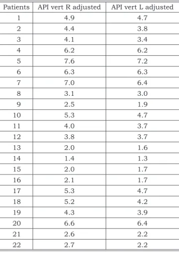
TABLE 6 - Values, in mm, for vertical condylar dis-placement (API vert) on the right (R) and left (L) sides, adjusted by the variables chronological age and stage of skeletal maturity.
Patients API vert R adjusted API vert L adjusted
1 4.9 4.7
2 4.4 3.8
3 4.1 3.4
4 6.2 6.2
5 7.6 7.2
6 6.3 6.3
7 7.0 6.4
8 3.1 3.0
9 2.5 1.9
10 5.3 4.7
11 4.0 3.7
12 3.8 3.7
13 2.0 1.6
14 1.4 1.3
15 2.0 1.7
16 2.1 1.7
17 5.3 4.7
18 5.2 4.2
19 4.3 3.9
20 6.6 6.4
21 2.6 2.2
22 2.7 2.2
TABLE 7 - Mean values, standard deviations and me-dians, in mm, for vertical condylar displacement (API vert) on right (R) and left (L) sides, adjusted by chrono-logical age and skeletal maturity factors.
Measurement Mean ± S.D. Median
API vert R 2.74 ± 2.00 2.20
API vert L 2.44 ± 1.93 1.72
dylar relations, even in asymptomatic patients, since a greater degree of vertical condylar displace-ment was evidenced between CR and MHI. This mentioned displacement brought consequences over the dental, skeletal and soft tissue aspects of malocclusion after such appliance was used. This resulted in a more precise orthodontic diagnosis, with obvious advantages to the patients studied, considering that all of them initiated orthodon-tic treatment immediately after splint therapy. Unfortunately, for practical and economic rea-sons, the recommended use of occlusal splints be-fore orthodontic treatment is traditionally restrict-ed to patients presenting signs and/or symptoms of temporomandibular dysfunction. However, it is noteworthy that even in asymptomatic individuals the period of time during which these deprogram-ming splints are used may influence CR records substantially, and consequentially affect diagnosis and planning of orthodontic corrections. Because impracticality makes it impossible to deprogram all orthodontic patients, it is therefore recommended that CR recording be done carefully, to make sure that such record be as precise as possible. On the other hand, the deprogramming procedure can-not be dismissed, by all means, in symptomatic individuals. This circumstance does not diminish the interest or importance of performing diagnoses in centric relation, a procedure that requires the routine mounting of diagnostic casts in articula-tors for all orthodontic patients.
CONCLUSIONS
The data obtained in the present study leads to the conclusion that:
The neuromuscular deprogramming per-formed in this study, before recording CR, resulted in greater mean condylar
displace-ment between the positions evaluated than that found in similar studies conducted in non-deprogrammed groups.
The displacement between CR and MHI showed frequent incidence, inasmuch as it occurred in 100% of the sample, on at least two planes of space.
182 PB
REFERENCES
1. Academy of Denture Prosthetics. Glossary of prosthodontic terms. 5th ed. St. Louis: Mosby; 1987.
2. Angle EH. Classification of malocclusion. Dent Cosmos 1899;41(2):248-64.
3. Capp NJ, Clayton JA. A technique for evaluation of centric relation tooth contacts. Part I: during normal temporoman-dibular joint function. J Prosthet Dent 1985a;54(4):569-74.
4. Capp NJ, Clayton JA. A technique for evaluation of centric relation tooth contacts. Part II: following use of an occlusal splint for treatment of temporomandibular joint dysfunc-tion. J Prosthet Dent 1985b;54(5):697-705.
5. Carlson N, Moline D, Huber L, Jacobson J. Comparison of muscle activity between conventional and neuromuscular splints. J Prosthet Dent 1993;70(1):39-43.
6. Chang FHF, Chen KC, Shiau YY. The importance of de-termination of jaw position in orthodontic diagnosis and treatment planning for adult patients. Dent Clin North Am 1997;41(1):49-66.
7. Contin I. Estudo comparativo do reposicionamento man-dibular (MIC-RC), frente ao uso do Jig e da placa miorre-laxante em pacientes dentados assintomáticos e com dor miofascial na região da cabeça e pescoço [Tese de Doutora-do]. São Paulo: Faculdade de Odontologia da USP; 1997. 8. Crawford SD. Condylar axis position, as determined by
occlusion and measured by the CPI instrument, and signs and symptoms of temporomandibular dysfunction. Angle Orthod 1999,69(2):103-14.
9. Dawson PE. Evaluation, diagnosis and treatment of oc-clusal problems. St. Louis: Mosby; 1974.
10. Dyer EH. Importance of a stable maxillomandibular relation. J Prosthet Dent 1973;30(3):241-51.
11. Fantini SM, Abrão J. Deslocamentos condilares entre RC e MIH, em jovens assintomáticos, com maloclusão de Cl II. Ortodontia 2001;34(1):28-34.
12. Fishman LS. Radiographic evaluation of skeletal mat-uration. A clinically oriented method based on hand-wrist films. Angle Orthod 1982;52(2):88-112.
13. Kovaleski WC, De Boever J. Influence of occlusal splints on jaw position and musculature in patients with
temporomandibular joint dysfunction. J Prosthet Dent 1975;33(3):321-7.
14. Koyano K, Ogawa T, Sumiyoshi K, Tsukiyama Y, Ichiki R, Suetsugu T. Effect of occlusal splint on masticatory movement in healthy individuals. Cranio 1997;15(2):127-31.
15. Lauritzen A. Atlas of occlusal analysis. Chicago: HAH; 1974.
16. Long JH. Location of the terminal hinge axis by in-traoral means. J Prosthet Dent 1970;23(1):11-24. 17. Lucia VO. A technique of recording centric relation.
J Prosthet Dent 1964;14(3):492-505.
18. Neff P. Trauma from occlusion. Restorative concerns. Dent Clin North Am 1995;39(2):335-54.
19. Nelson SJ. Principles of stabilization bite splint ther-apy. Dent Clin North Am 1995;39(2):403-21.
20. Rosner D, Goldberg GF. Condylar retruded contact position and intercuspal position correlation in dentulous patients. Part I: three-dimensional analysis of condilar registrations. J Prosthet Dent 1986;56(2):230-9.
21. Roth RH. Functional occlusion for the orthodontist. J Clin Orthod 1981a;15(1):32-51.
22. Roth RH. Functional occlusion for the orthodontist. Part III. J Clin Orthod 1981b;15(3):174-98.
23. Roth RH, Rolfs DA. Functional occlusion for the or-thodontist. Part II. J Clin Orthod 1981;15(2):100-23. 24. Utt TW, Meyers CE Jr, Wierzba TF, Hondrum SO. A
three-dimensional comparison of condylar position chang-es between centric relation and centric occlusion using the mandibular position indicator. Am J Orthod Dentofacial Orthop 1995;107(3):298-308.
25. Wood DP, Elliott RW. Reproducibility of the cen-tric relation bite registration technique. Angle Orthod 1994;64(3):211-20.
26. Wood DP, Floreani KJ, Galil KA, Teteruck WR. The effect of incisal bite force on condylar seating. Angle Orthod 1994;64(1):53-61.
27. Wood DP, Korne PH. Estimated and true hinge axis: a comparison of condylar displacements. Angle Orthod 1992;62(3):167-75.