Dispensable for the Formation of the
b
-globin
Active
Chromatin Hub
Ping Kei Chan1, Albert Wai2, Sjaak Philipsen2*, Kian-Cheng Tan-Un1*
1Department of Zoology, Kadoorie Biological Science Building, The University of Hong Kong, Hong Kong Special Administrative Region (SAR), China,2Erasmus MC, Department of Cell Biology, Rotterdam, The Netherlands
Abstract
Hypersensitive site 5 (59HS5) of theb-globinLocus Control Region functions as a developmental stage-specific border in erythroid cells. Here, we have analyzed the role of 59HS5 in the three dimensional organization of theb-genelocus using the Chromatin Conformation Capture (3C) technique. The results show that when 59HS5 is deleted from the locus, both remote and internal regulatory elements are still able to interact with each other in a three-dimensional configuration termed the Active Chromatin Hub. Thus, the absence of 59HS5 does not have an appreciable effect on the three dimensional organization of theb-globinlocus. This rules out models in which 59HS5 nucleates interactions with remote and/or internal regulatory elements. We also determined the binding of CTCF, the only defined insulator protein in mammalian cells, to 59HS5 by using chromatin immunoprecipitation (ChIP) assays. We detect low levels of CTCF binding to 59HS5 in primitive erythroid cells, in which it functions as a border element. Surprisingly, we also observe binding levels of CTCF to 59HS5 in definitive erythroid cells. Thus, binding of CTCF to 59HS5 per sedoes not render it a functional border element. This is consistent with the previous data suggesting that CTCF has dual functionality.
Citation:Chan PK, Wai A, Philipsen S, Tan-Un K-C (2008) 59HS5 of the Humanb-globinLocus Control Region Is Dispensable for the Formation of theb-globin Active Chromatin Hub. PLoS ONE 3(5): e2134. doi:10.1371/journal.pone.0002134
Editor:Axel Imhof, University of Munich and Center of Integrated Protein Science, Germany
ReceivedSeptember 10, 2007;AcceptedMarch 6, 2008;PublishedMay 7, 2008
Copyright:ß2008 Chan et al. This is an open-access article distributed under the terms of the Creative Commons Attribution License, which permits unrestricted use, distribution, and reproduction in any medium, provided the original author and source are credited.
Funding:This work was supported by the HKU School of Professional and Continuing Education (HKU SPACE).
Competing Interests:The authors have declared that no competing interests exist.
* E-mail: [email protected] (SP) ; [email protected] (KT)
Introduction
The humanb-globinlocus contains five genes that are arranged in the same order as their developmental expression pattern: 59-e
(embryonic) Gc Ac(foetal) d b (adult)-39. The major activating region, termed the Locus Control Region (LCR), is located 6 to 22 kb 59to thee-globin gene. The most prominent charactistic of the humanb-globinLCR is that it drives high level, tissue-specific, copy number-dependent and position-independent expression of linked transgenes in mice [1]. The core components of the LCR are the DNase I hypersensitive sites (59HS1-5) [2]. Each of the hypersensitive sites holds a unique array of transcription factor binding sites, including those for GATA1, NF-E2 and EKLF proteins. This suggests that the HS in the human LCR may have different stage-specific activities and cannot be replaced function-ally by each other [3,4]. Transcriptional activation of the b-like globin genes is thought to be a multi-step system. This model includes the active involvement of erythroid transcription factors in the spatial organization of the locus, and increased accessibility to trans-acting factors by recruitment of chromatin remodeling complexes [5]. The resultant transcriptionally active locus is arranged in a three dimensional structure termed the active chromatin hub (ACH), in which the LCR hypersensitive sites interact directly with the transcribed genes through a looping mechanism [6,7]. The deduced spatial arrangement of the regulatory elements and the genes in the ACH offers an explanation for the observation that a gene proximal to the
LCR has a transcriptional advantage over a more distally located gene [8,9]. The mode of action of the LCR to theb-globingenes appears to be orientation dependent as an inverted LCR is incapable of activating downstream globin genes at high levels, and thee-gene can not be activated when placed upstream of the LCR [10]. This property of the LCR may be due to the spatial organization of its hypersensitive sites, but it is also possible that the locus contains elements that block LCR action in one of the directions [11].
Recently, we have shown that human 59HS5 acts as a developmental stage-specific border element in erythroid cells. In that study, we placed a markedb-globingene upstream of the LCR and the genes, using PAC constructs of the humanb-globinlocus in which 59HS5 was flanked by loxP sites [11].
same transgenic lines in which 59HS5 had been removed by Cre-mediated excision.
The first insulator element reported in vertebrates is 59HS4 (cHS4) in the chicken b-globinlocus [12]. Binding of the 11 Zn-finger protein CTCF to the FII region of the cHS4 is thought to play an important role in the activity of cHS4 [13,14]. To investigate the potential activity of CTCF as an enhancer blocking protein on human 59HS5 in vivo, we performed chromatin immunoprecipitation assays using antibodies directed against CTCF.
Results
PAC constructs for 3C analysis of the humanb-globin
locus
To examine the potential conformational changes of the ACH in the presence or absence of 59HS5 in the context of the human
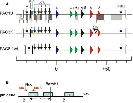
b-globin locus, we employed transgenic mice carrying modified human b-globin PAC transgenes (Fig. 1, [11]). The transgenes were derived from PAC185 which contains the entire humanb -globingene cluster. Homologous recombination inE. coliDH10B cells [15] was used for generation of the constructs. To determine the effect of deletion of 59HS5 on the three dimensional (3D) organization of the locus, 59HS5 was flanked by loxP sites to enable Cre-mediated deletion of 59HS5 (PACD1B). The basal promoter and 59untranslated regions of theb-globin gene, from 2139 to+49 relative to the cap site of the gene, has been removed to generate the b-globin promoter deletion (PAC3K; Fig. 1).
Intactness of the transgenes was carefully mapped by Southern blot analysis using a total of 11 different restriction enzymes and hybridization with cosmid LCRe, -ccdbprobes [16], and smaller probes along theb-globinlocus [11].
3C analysis of the PAC constructs with thebm gene and 59HS5 flanked by loxP sites
Previous studies on human b-globin transgenes in mice have shown that theb-globingene interacts with the ACH when it is transcribed in definitive erythroid cells [7]. In these studies, the Chromosome Conformation Capture (3C) technique [6,7,17] was used to examine the three dimensional organization of theb-globin locus. First, we determined whether introduction of theb-marked (bm) gene, which is an artificially modifiedb-globin gene with part of its exon 2 replaced by equivalent Ac-globin gene [9] (Fig. 1B), and flanking 59HS5 by loxP sites per se caused conformational changes of theb-globinlocus. We performed 3C analysis on the PAC transgenes and compared this directly to the results obtained with the wild type locus (PAC8.1; Fig. 2). In the wild type locus, 59HS1-5 of the LCR and the HS in the 39flanking region (39HS1) interact with each other to form the ACH of theb-globin locus. The results of 3C analysis on the PAC1B transgene show that the spatial structure of the locus is basically unaltered when compared with that of wild type PAC8.1. In brain, a non-erythroid tissue, the PAC transgene shows an essentially linear conformation of theb -globinlocus (data not shown). This is consistent with the notion that the formation of ACH is tissue specific [6].
Effect of the deletion of the 59HS5 on ACH formation To study the effect of 59HS5 on the spatial organization of the
b-globinlocus, we compared the PAC1B line with the PACD1B line, in which 59HS5 had been deleted by the action of Cre recombinase [15].
It has been proposed that transcriptional activation of b-like globin genes at each stage of development is a multi-step process [5]. An LCR holocomplex is formed to allow the accessibility of transcription and chromatin remodeling factors to the locus, thus providing a high local concentration of the relevant trans-acting factors for efficient transcription. The resultant functional active chromatin hub (ACH) comprises the LCR holocomplex interacting directly with the transcribed genes [6,7,18]. This model implies that maintaining the integrity of the ACH is the key to create a chromatin domain permissive for transcriptional regulation.
To examine the possible consequences of deletion of 59HS5 on ACH formation, we compared the 3D structures of the PAC1B and PACD1B transgenes in definitive erythroid cells isolated from 14.5 dpc fetal livers with the 3C technique (Fig. 3). The results show that the overall 3D of theb-globin locus has remained the same when 59HS5 is deleted (PACD1B). However, the relative crosslinking frequency of the 59HS6, the distal hypersensitive site located about 6 kb 59upstream to the HS5 [19], with theb-globin gene is increased by upon the removal of 59HS5 (Fig. 3B, primer 418). This increase in association frequency with the ACH is presumably due to the closer proximity of 59HS6 to the LCR after Cre-mediated excision of 59HS5. 59HS6 is located 800bp closer to the LCR in the PACD1B line. Besides, in the absent of HS5, theb -gene promoter interacts strongly with the A-cpromoter.
globin locus, yolk sac-derived primitive erythroid cells express the human embryonice- and fetalc-globin genes [16].b-like globin gene expression switches to the fetalc- and adultd- andb-globin genes during early definitive erythropoiesis between E12.5 and E14.5. The LCR plays an important role in the regulation of expression of theb-like globins, and the order and distance of the genes relative to the LCR are important parameters for the developmental switch [8,9,20]. These developmental switches of hematopoiesis are accompanied by changes in chromatin structure and spatial organization of regulatory elements throughout the locus [6,7,21]. In a previous study with the humanb-globin PAC transgenes we have shown that 59HS5 functions as an enhancer blocker in embryonic erythroid cells [11]. Therefore, it is interesting to determine the possible conformational changes of the locus without 59HS5 in embryonic erythroid cells.
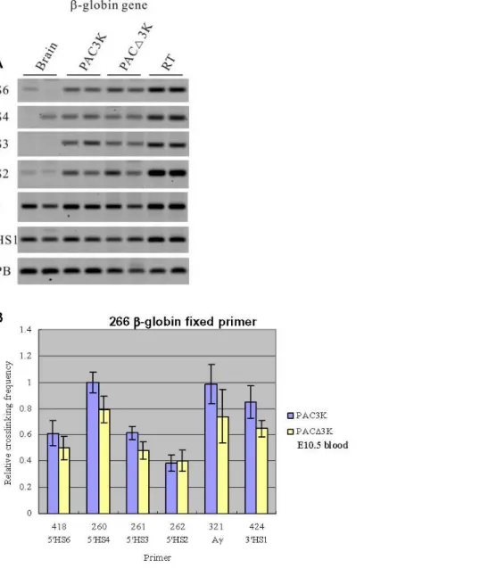
PAC3K transgenic lines, in which the promoter of theb-globin gene in the normal position is deleted, have been used for the 3C analysis. The rationale behind the usage of the PAC3K construct for performing the 3C analysis is that in this configuration, thebm gene located upstream of the LCR is maximally activated in definitive cells, while expression in primitive cells is only detectable after deletion of 59HS5. We therefore compared the locus-wide crosslinking frequencies of the PAC3K and PACD3K (PAC construct with deletion of 59HS5) transgenes at different developmental stages. In the fetal liver, we observed that the 3D conformation of the PAC3K and PACD3K transgenes is virtually identical to that observed for the PAC1B and PACD1B transgenes (data not shown). This implies that neither 59HS5 nor theb-globin promoter is required for structural changes in the ACH occurring during the process ofb-globin gene switching [7,18,22]. The latter Figure 3. 3C analysis of the PAC1B and PACD1B transgenic lines using a primer from the humanb-globingene in combination with primers from other parts of the locus.Fetal livers were collected from E14.5 embryos for this set of 3C experiments. (A) Representative examples of the PCR fragments resulting from the 3C experiments. (B) Histogram of the relative crosslinking efficiencies after quantitation and normalization. The histograms are the average of at least three separate experiments, with each PCR performed in triplicate.
is in agreement with Patrinos et al. [23] who have shown that deletion of theb-globin promoter alone is not sufficient to alter the 3D configuration of the ACH.
Since 59HS5 functions as a border at the primitive stage [11], we next examined the spatial organization of the PAC3K and PACD3K transgenes in embryonic blood samples. The restriction fragment containing 59HS2 [24], a classical enhancer element within the LCR, was used as the fixed fragment for the results presented in Fig. 4. The analysis shows that the spatial organization of the locus is basically unchanged upon deletion of 59HS5 (PACD3K) when compared with the PAC3K transgenic line. In agreement with the notion that thec-globin gene is predominantly expressed in primitive cells, higher relative crosslinking frequencies of the c-globin gene compared to theb-globingene could be observed (Fig. 4). The results imply that in primitive erythroid cells association of the actively transcribed c-globin genes with the ACH principally remains the same in the absence of 59HS5.
For investigating the spatial organization of the chromatin hub (CH), which can be observed in the erythroid progenitor cells withoutb-like globin gene expression [7], we used theb-globingene as the fixed primer in 3C analysis (Fig. 5). Similar results were obtained between the two constructs (PAC3K vs. PACD3K). In conclusion, the results strongly indicate that deletion of 59HS5 of the LCR in the human transgene does not have a measurable effect on the 3 dimensional organization of the b-globin locus, neither in the primitive stage where 59HS5 functions as a border or enhancer blocker, nor in the definitive stage where 59HS5 does not have a known function.
ChIP analysis of CTCF binding to 59HS5 in primitive and definitive erythroid cells
We have shown previously that human 59HS5 has an enhancer blocking function at the primitive stage [11]. However, the mecha-nism by which this function is achieved remains unknown. Although a number of enhancer blocking proteins have been identified in Drosophila[25–27], CTCF is the only protein known so far to mediate enhancer blocking activity in vertebrates [28,29]. Previous studies conducted by Felsenfeld and his colleagues have described a 1.2kb DNA element with strong enhancer blocking activity, located at the 59HS4 of the chickenb-globin gene locus [28,29].
A 250bp core fragment of the cHS4 is fully functional and a single binding site for the protein CTCF, FII, is necessary and sufficient to block the action of an enhancer on a promoter when placed between them [30]. Conserved CTCF binding sites are found at 59HS5 and 39HS1 of both the mouse and human loci.
To investigate whether CTCF binds to 59HS5, ChIP analysis using an antibody to CTCF was performed (Fig. 6). The CTCF ChIP DNA enrichment levels of three fragments were tested, namely, mouse endogenous HS62 (mHS62), human bm (the marked b-globin gene) and human 59HS5. In erythroid tissues, DNA enrichment could be detected by real-time PCR. Our results on mHS62 are in agreement with a previous study [14] and confirm that CTCF binds to the endogenous mouse HS62 fragment. Human 59HS5 exhibits only modest enrichment levels of 1.5 fold at primitive and 2.4 fold at definitive stages. These results support the previousin vitro assay data [13] showing the presence of a CTCF binding site in human 59HS5 [13]. Our data suggest that CTCF also binds to human HS5in vivo.
Simultaneously, thebm gene enrichment level was determined using anti-CTCF ChIP assay. Surprisingly, the DNA enrichment level of bm gene is higher than that of the human 59HS5 and mouse HS62 at both developmental stages (Fig. 6). Further studies are needed to characterize the functional role of CTCF binding to thebm gene.
Discussion
In this study, we have analyzed the role of 59HS5 with respect to the three dimensional configuration of the humanb-globin locus. The transgenic lines carried PAC constructs that were derived from the original 185 kb human b-globin PAC [7]. Transgenic lines carrying single copies of four modified PAC constructs, namely PAC1B, PACD1B, PAC3K and PACD3K, were used [15] to determine possible conformational changes of the locus after deletion of the 59HS5 core region. The 3D conformation of the transgenic loci was determined by 3C analysis. A humanb-globin promoter deletion in the context of the full LCR was used to determine whether or not the interaction between theb-globin gene and the LCR would be perturbed when both 59HS5 and the b -globin promoter (PACD3K) were deleted, as was previously found when both 59HS3 and theb-globin promoter were deleted [23].
structure of theb-globin locus has essentially remained the same when 59HS5 is deleted. Mild increments of the association frequency of 59HS6 to the LCR could be explained by distance effects [9]: 59HS6 interacts more frequently with the ACH once it moves closer to the LCR after Cre-mediated excision of 59HS5. This is consistent with our previous data on humanb-globin transgenic mice which showed that the increase in expression of thebm gene, which is located 59upstream of the LCR, is likely due to the shorter distance of the gene relative to the LCR [11].
Our previous study on the humanb-globin PAC transgenes has shown that 59HS5 is a stage-specific enhancer blocking element that functions at the primitive stage of erythropoiesis [11]. Surprisingly, we did not observe any significant alterations in the structure of the b-globin locus by 3C analysis primitive embryonic blood when 59HS5 was deleted. The results obtained from both the definitive and primitive stages imply that spatial organization of the locus is essentially unchanged upon deletion of 59HS5 (PACD1B) or in the double deletion of HS5 and theb-globin promoter (PACD3K). We
can not completely discount the possible influence of 59HS5 onb -globin locus configuration as this might not be detectable due to limitations of the resolution achieved with the 3C technique. However, we can conclude that the enhancer blocking properties of 59HS5 in primitive erythroid cells is not accompanied by major effects on the spatial organization of theb-globin locus.
Interestingly this study shows that CTCF, the first transcription factor found to have insulating activity [28] and enhancer blocking function [13,14] in mammalian cells, binds to the 59HS5 core region at both the primitive and definitive stages in erythroid cells in vivo. This indicates that CTCF could be involved in the border function of 59HS5 in the primitive cells. Since it also binds to the 59HS5 region in definitive cells, this suggests that CTCF, most likely in conjunction with other proteins, can have opposing effects on gene expression. Filippova et al. [31] proposed that CTCF could be a transcriptional repressor of the c-Myc oncogene in vertebrates. In contrast, the CTCF protein can bind to the promoter of the amyloidbprotein precursor and activate transcription [32]. Figure 5. 3C analysis of the PAC3K and PACD3K transgenes using a primer from the humanb-globin gene as the fixed primer.
Thus, CTCF could be a ‘‘dual functionality’’ protein that has divergent effects on different gene regulation systems. In addition, we show that CTCF binds to the bm gene in both primitive and definitive stage. Three putative CTCF binding sites can be identified at2133 to2149,21005 to2990 and21396 to21412 bp relative to the cap site of thebm gene (Figure S1). Collectively, our results indicate that CTCF may have a more general role in the regulation of theb-globin locus than previously anticipated. Splinteret al. (2006) [33] have recently shown that in CTCF null cells, there were reduced DNA-DNA interaction frequencies between the sites that normally bind CTCF in the mouseb-globin gene locus. Interest-ingly, no changes were seen in the interaction of the surrounding olfactory genes (MOR5B1-3 and MOR3B1-4 [33]), or the expression level of the b-globin genes. It has been proposed previously that CTCF and the chick HS4 insulator sequence are highly concentrated in Matrix Attachment Regions (MARs) which create separate ‘‘loop’’ domains that prevent the interaction between an enhancer and promoter [34]. Collectively, these data suggested that CTCF participates in more than one pathway in the regulation of the globin genes.
Materials and Methods
Transgenic mice
PAC transgenic constructs (Fig. 1) containing theb-globinlocus were generated using homologous recombination inEscherichia coli according to Imanet al.(2000) [15]. Modified PACs were used to generate transgenic mice; these mice have been characterized extensively in Waiet al.(2003) [11].
Tissues used for 3C
Plugged transgenic mice were sacrificed and the embryos were collected at 10.5 and 14.5 dpc (day post coitum) respectively. Fetal liver and brain were collected from 14.5 dpc embryos, and embryonic blood and head were collected at 10.5 dpc. as previously described [6,7,35].
Preparation of 3C templates
The 3C protocol was adapted from Tolhuiset al.(2002) [6]. 16107 cells was defined as 1 unit of sample in a 10ml crosslinking reaction (2% formaldehyde in DMEM with 10% FCS). The samples were cross-linked for 10 minutes at room temperature with gentle mixing. The reaction was quenched by the addition of glycine to 0.125M. Nuclei were harvested by lysis of the cells in 5ml ice-cold lysis buffer (10mM Tris-Cl, 10mM NaCl, 0.2% NP-40 pH8.0) containing
protease inhibitors (pefabloc, Roche) for 10 minutes on ice. Nuclei were resuspended in 0.5ml 16HindIII restriction buffer (Roche) containing 0.3% SDS and incubated for 1 hour at 37uC with agitation. Triton X-100 was added to 2% (v/v), and the nuclei were further incubated for 1 hour at 37uC to sequester the SDS. The crosslinked DNA was digested overnight at 37uC with 400U HindIII. The restriction enzyme was inactivated by the addition of SDS to 1.6% and incubated at 65uC for 20 minutes with agitation. The reaction was diluted with 7ml 16ligation buffer (Promega) and incubated for 1 hour at 37uC. The chromatin was ligated with 100U T4 DNA ligase (Promega) for 4 hours at 16uC followed by 30 minutes at room temperature. 600mg of Proteinase K was added, and samples were incubated overnight at 65uC to reverse the cross-links. Next, the samples were incubated for 30 minutes at 37uC with 1500mg of DNase-free RNaseA, and the DNA was purified by phenol extraction and ethanol precipitation. The DNA pellet was resuspended in 150ml 10 mM Tris-Cl (pH7.5).
Preparation of Random Template control
In order to normalize the amplification efficiency of different primer sets, the PAC185 [15] plasmid containing the humanb -globin locus was used for preparation of the random template control (RTC). Theoretically, this random template control contains all possible ligation products in equimolar amounts. Wild-type mouse genomic DNA was mixed with PAC185 plasmid and a 60–70kb PAC containing the mouseXPBlocus (PAC Clone
#443-C18, MRC gene services). The genomic DNA/PAC clones mixture was processed according to the 3C technique [6].
The processed products were serially diluted with wild-type mouse genomic DNA until the PAC DNA was present in a molar ratio similar to single-copy b-globin locus transgenic DNA (line PAC8.1), as assessed by quantitative PCR [6,7]. PCR products were run on a 2% agarose gel and quantified with a Typhoon 9200 imager (Amersham). Typically 300ng of DNA was subsequently used for each 3C-PCR reaction. Each PCR reaction was performed in triplicate and repeated at least three times. Sequences of the 3C PCR primers are shown in Table 1.
Anti-CTCF ChIP assays
16107 cells were defined as 1 unit of sample for 10ml ChIP crosslinking reaction (2% formaldehyde and 40mM HEPES pH7.9 in DMEM with 10% FCS). The samples were crosslinked for 20 minutes at room temperature with gentle mixing. The reaction was quenched by the addition of glycine to 0.125M. The cells were harvested by centrifugation at 1500rpm for 5 minutes at 4uC. The cells were first washed with PBS buffer containing protease inhibitors (1 mM phenylmethylsulfonyl fluoride (PMSF), 1mg/ml aprotinin and 1mg/ml pepstatin A) and centrifuged at 1500rpm for 5 minutes. 10 ml of Triton-wash buffer (0.25% Triton-X 100, 10mM Na-EDTA, 0.5 mM Na-EGTA and 10 mM Tris-Cl pH8.0) was added and the samples were incubated at room temperature for 10 minutes. The cells were pelleted by centrifugation at 1500rpm for 5 minutes and they were further washed with 10ml NaCl-wash (200mM NaCl, 1 mM Na-EDTA, 0.5mM Na-EGTA and 10mM Tris-Cl pH8.0) buffer at room temperature for 10 minutes. After centrifugation at 1500rpm for 5 minutes, the pellet was resuspended in 500ml sonication buffer (1mM Na-EDTA, 0.5mM Na-EGTA and 10mM Tris-Cl pH8.0) and kept on ice for 5 minutes. DNA was sheared by sonication to lengths between 200 and 1000bp, keeping the sample cold on ice. Cell debris was removed by centrifugation. ChIP reactions were performed according to the Upstate Biotechnology chromatin immunoprecipitation protocol with 1mg of anti-CTCF antibody (Upstate Biotechnology) per IP reaction. Enrichment of specific Figure 6. CTCF-ChIP assay analysis of the binding of CTCF
protein to the mouse HS62 (mHS62),bm and human 59HS5 in the primitive (A) and definitive (B) stage.Fold enrichment of the test sequence in bound with CTCF versus input/starting material is shown on the y axis.
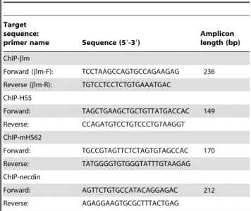
immunoprecipitated sequences, including 59HS5,bm (markedb -globingene), mHS62 (mouse HS62) and a fragment of the mouse Necdin gene (control), were determined by real-time PCR. Fold enrichment was calculated by comparing the ratio toNecdinin pull-down-eluate against the value from input-starting material. The sequences of the ChIP PCR primers are shown in Table 2. The location of thebm gene primers were shown in Fig. 1B.
Supporting Information
Figure S1 Alignment of the cHS4 CTCF binding site with thebm promoter. Alignment of the cHS4 CTCF binding site with thebm promoter (1). Thebm gene cap site (+1) are indicated with a blue
letters. The proposed CTCF binding sites in thebm sequence are indicated with red letters and the cHS4 CTCF binding sequence is shown on top of each proposed binding site. Conserved binding sequences are highlighted with a gray background. The alignment was performed with the ClustalX program (2) Reference List 1. Farrell CM, West AG, Felsenfeld G (2002) Conserved CTCF insulator elements flank the mouse and human beta-globin loci. Mol Cell Biol 22: 3820-3831. 2. Thompson JD, Gibson TJ, Plewniak F, Jeanmougin F, Higgins DG (1997) The CLUSTAL_X windows interface: flexible strategies for multiple sequence alignment aided by quality analysis tools. Nucleic Acids Res 25: 4876-4882.
Found at: doi:10.1371/journal.pone.0002134.s001 (0.04 MB DOC)
Acknowledgments
We would like to thank Dr. Roy Drissen and Dr. Laura Gutierrez for their assistance throughout this study.
Author Contributions
Conceived and designed the experiments: SP PC KT. Performed the experiments: PC. Analyzed the data: SP PC KT. Contributed reagents/ materials/analysis tools: SP KT AW. Wrote the paper: SP PC KT.
References
1. Grosveld F, Antoniou M, van Assendelft GB, de Boer E, Hurst J, et al. (1987) The regulation of expression of human beta-globin genes. Prog Clin Biol Res 251: 133–144.
2. Forrester WC, Takegawa S, Papayannopoulou T, Stamatoyannopoulos G, Groudine M (1987) Evidence for a locus activation region: the formation of developmentally stable hypersensitive sites in globin-expressing hybrids. Nucleic Acids Res 15: 10159–10177.
3. Fraser P, Pruzina S, Antoniou M, Grosveld F (1993) Each hypersensitive site of the human beta-globin locus control region confers a different developmental pattern of expression on the globin genes. Genes Dev 7: 106–113.
4. Bungert J, Tanimoto K, Patel S, Liu Q, Fear M, et al. (1999) Hypersensitive site 2 specifies a unique function within the human beta-globin locus control region to stimulate globin gene transcription. Mol Cell Biol 19: 3062–3072.
5. Levings PP, Bungert J (2002) The human beta-globin locus control region. Eur J Biochem 269: 1589–1599.
6. Tolhuis B, Palstra RJ, Splinter E, Grosveld F, de Laat W (2002) Looping and interaction between hypersensitive sites in the active beta-globin locus. Mol Cell 10: 1453–1465.
7. Palstra RJ, Tolhuis B, Splinter E, Nijmeijer R, Grosveld F, et al. (2003) The beta-globin nuclear compartment in development and erythroid differentiation. Nat Genet 35: 190–194.
8. Hanscombe O, Whyatt D, Fraser P, Yannoutsos N, Greaves D, et al. (1991) Importance of globin gene order for correct developmental expression. Genes Dev 5: 1387–1394.
9. Dillon N, Trimborn T, Strouboulis J, Fraser P, Grosveld F (1997) The effect of distance on long-range chromatin interactions. Mol Cell 1: 131–139. 10. Tanimoto K, Liu Q, Bungert J, Engel JD (1999) Effects of altered gene order or
orientation of the locus control region on human beta-globin gene expression in mice. Nature 398: 344–348.
11. Wai AW, Gillemans N, Raguz-Bolognesi S, Pruzina S, Zafarana G, et al. (2003) HS5 of the human beta-globin locus control region: a developmental stage-specific border in erythroid cells. EMBO J 22: 4489–4500.
12. Hebbes TR, Clayton AL, Thorne AW, Crane-Robinson C (1994) Core histone hyperacetylation co-maps with generalized DNase I sensitivity in the chicken beta-globin chromosomal domain. EMBO J 13: 1823–1830.
13. Farrell CM, West AG, Felsenfeld G (2002) Conserved CTCF insulator elements flank the mouse and human beta-globin loci. Mol Cell Biol 22: 3820–3831.
Table 1.Sequence of 3C-PCR primers.
Target gene locus
Target sequence:
primer# Sequence (59-39)
Humanb-globin 59Olfactory Receptor gene:
318
CAGCTTGGGTCAAATACAG
Humanb-globin Markedb-globin gene (bm):
474
ACCACCCTTTCCACAGTC
Humanb-globin HS5: 442
CTGCACACTTTCAGTCCG
Humanb-globin HS4 260
CAAATGGGTGACTGTAGGG
Humanb-globin HS3 261
GCCTGCATTTATTGTTGTG
Humanb-globin HS2 262
GAACTGCTCATGCTTGGAC
Humanb-globin Ac-globin gene (Ac): 321
GAAGAAAGACCTCTATAGGACAGG
Humanb-globin b-globin gene (b): 266
TGGTTATGGTCAGAGCCTC
Humanb-globin 39HS1: 424
ACATGGATAATACTGTTCCCC
Mouse XPB XPB 341&343
Forward: TGACCTCCACACTCCTGAC
Reverse: ATGCGCAATTAGAAACTGC
doi:10.1371/journal.pone.0002134.t001
Table 2.PCR primers used in the Anti-CTCF ChIP Real-time PCR experiments.
Target sequence:
primer name Sequence (59-39)
Amplicon length (bp)
ChIP-bm
Forward (bm-F): TCCTAAGCCAGTGCCAGAAGAG 236
Reverse (bm-R): TGTCCTCCTCTGTGAAATGAC
ChIP-HS5
Forward: TAGCTGAAGCTGCTGTTATGACCAC 149
Reverse: CCAGATGTCCTGTCCCTGTAAGGT
ChIP-mHS62
Forward: TGCCGTAGTTCTCTAGTGTAGCCAC 170
Reverse: TATGGGGTGTGGGTATTTGTAAGAG
ChIP-necdin
Forward: AGTTCTGTGCCATACAGGAGAC 212
Reverse: AGAGGAAGTGCGCTTTACTGAG
14. Bulger M, Schubeler D, Bender MA, Hamilton J, Farrell CM, et al. (2003) A complex chromatin landscape revealed by patterns of nuclease sensitivity and histone modification within the mouse beta-globin locus. Mol Cell Biol 23: 5234–5244.
15. Imam AM, Patrinos GP, de Krom M, Bottardi S, Janssens RJ, et al. (2000) Modification of human beta-globin locus PAC clones by homologous recombination in Escherichia coli. Nucleic Acids Res 28: E65.
16. Strouboulis J, Dillon N, Grosveld F (1992) Developmental regulation of a complete 70-kb human beta-globin locus in transgenic mice. Genes Dev 6: 1857–1864.
17. Dekker J, Rippe K, Dekker M, Kleckner N (2002) Capturing chromosome conformation. Science 295: 1306–1311.
18. Wijgerde M, Grosveld F, Fraser P (1995) Transcription complex stability and chromatin dynamics in vivo. Nature 377: 209–213.
19. Bulger M, van Doorninck JH, Saitoh N, Telling A, Farrell C, et al. (1999) Conservation of sequence and structure flanking the mouse and human beta-globin loci: the beta-beta-globin genes are embedded within an array of odorant receptor genes. Proc Natl Acad Sci U S A 96: 5129–5134.
20. Grosveld F, van Assendelft GB, Greaves DR, Kollias G (1987) Position-independent, high-level expression of the human beta-globin gene in transgenic mice. Cell 51: 975–985.
21. Gribnau J, Diderich K, Pruzina S, Calzolari R, Fraser P (2000) Intergenic transcription and developmental remodeling of chromatin subdomains in the human beta-globin locus. Mol Cell 5: 377–386.
22. Gribnau J, de Boer E, Trimborn T, Wijgerde M, Milot E, et al. (1998) Chromatin interaction mechanism of transcriptional control in vivo. EMBO J 17: 6020–6027.
23. Patrinos GP, de Krom M, de Boer E, Langeveld A, Imam AM, et al. (2004) Multiple interactions between regulatory regions are required to stabilize an active chromatin hub. Genes Dev 18: 1495–1509.
24. Tuan DY, Solomon WB, London IM, Lee DP (1989) An erythroid-specific, developmental-stage-independent enhancer far upstream of the human ‘‘beta-like globin’’ genes. Proc Natl Acad Sci U S A 86: 2554–2558.
25. Roseman RR, Pirrotta V, Geyer PK (1993) The su(Hw) protein insulates expression of the Drosophila melanogaster white gene from chromosomal position-effects. EMBO J 12: 435–442.
26. Zhou J, Barolo S, Szymanski P, Levine M (1996) The Fab-7 element of the bithorax complex attenuates enhancer-promoter interactions in the Drosophila embryo. Genes Dev 10: 3195–3201.
27. Capelson M, Corces VG (2006) SUMO conjugation attenuates the activity of the gypsy chromatin insulator. EMBO J 25: 1906–1914.
28. Chung JH, Whiteley M, Felsenfeld G (1993) A 59element of the chicken beta-globin domain serves as an insulator in human erythroid cells and protects against position effect in Drosophila. Cell 74: 505–514.
29. Chung JH, Bell AC, Felsenfeld G (1997) Characterization of the chicken beta-globin insulator. Proc Natl Acad Sci U S A 94: 575–580.
30. Bell AC, West AG, Felsenfeld G (1999) The protein CTCF is required for the enhancer blocking activity of vertebrate insulators. Cell 98: 387–396. 31. Filippova GN, Fagerlie S, Klenova EM, Myers C, Dehner Y, et al. (1996) An
exceptionally conserved transcriptional repressor, CTCF, employs different combinations of zinc fingers to bind diverged promoter sequences of avian and mammalian c-myc oncogenes. Mol Cell Biol 16: 2802–2813.
32. Vostrov AA, Quitschke WW (1997) The zinc finger protein CTCF binds to the APBbeta domain of the amyloid beta-protein precursor promoter. Evidence for a role in transcriptional activation. J Biol Chem 272: 33353–33359. 33. Splinter E, Heath H, Kooren J, Palstra RJ, Klous P, et al. (2006) CTCF mediates
long-range chromatin looping and local histone modification in the beta-globin locus. Genes Dev 20: 2349–2354.
34. Yusufzai TM, Felsenfeld G (2004) The 59-HS4 chicken beta-globin insulator is a CTCF-dependent nuclear matrix-associated element. Proc Natl Acad Sci U S A 101: 8620–8624.