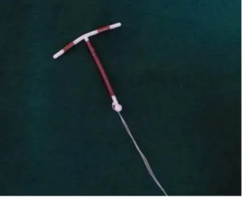
A CASE OF MISSING IUCD
Texto
Imagem
Documentos relacionados
We report a case of Aspergillus meningitis in an immunocompetent patient successfully treated with fluconazole..
There are two previous reports, one of an endometrioid ovarian carcinoma, and the other of a mucinous cystadenocarcinoma, of cases that presented with rupture during pregnancy 4,5 ,
Risk factors for the uterine perforation occurrence are the following: clinician’s inexperience, nulliparity, unfa- vourable uterine position, uterine scars, changes and defor-
We report the case of a patient in whom bilateral uterine artery embolization, followed by curettage of cervical canal were required in a patient with cervical pregnancy who
a case of intravenous leiomyomatosis with invasion of the inferior vena cava and extension to the right atrium, successfully treated with surgical approach.. Case report: Female
The surgical complication in this case was treated successfully, and there was resolution of the profile on postoperative day 4 following the second intervention, as
We report a case of pseudoaneurysm following PCNL in supine position assessed by angiotomography with a 3-D reconstruction, managed successfully by endovascular occlusion
Delayed presentation of an isolated gallbladder rupture following blunt abdominal trauma: a case report. Isolated perforation of
