Duodenum of AIDS Patients Despite Antiretroviral
Treatment
Dag E. Nilssen1,2, Per Brandtzaeg1*
1Laboratory for Immunohistochemistry and Immunopathology (LIIPAT), Centre for Immune Regulation (CIR), University of Oslo, and Department of Pathology, Oslo University Hospital, Rikshospitalet, Oslo, Norway,2Department of Medicine, Akershus University Hospital, Lørenskog, Norway
Abstract
Intraepithelial lymphocytes (IELs) bearing the cd T-cell receptor are a unique intestinal subset whose function remains elusive. Here, we examine how they behave in AIDS and during various regimens of antiretroviral treatment in order to obtain mechanistic insight into their adaptive or innate functionalin vivoproperties. IELs were studied by multimarker two-colour immunofluorescencein situstaining. Consecutive duodenal biopsies were obtained from advanced infection-prone HIV+ patients (n= 30). The systemic adaptive immune status was monitored by determining T-cell subsets and immunoglobulins in peripheral blood. ThecdIEL ratio (median 14.5%, range 1.5–56.3%) was significantly increased (p,0.02) compared with that in clinically healthy HIV2control subjects (n= 11, median 2.8%; range 0.3–38%), although the number ofcdIELs per mucosal length unit (U) only tended to be increased (4.0/U in HIV+versus3.2/U in HIV2subjects). Notably, the total number of CD3+IELs was significantly reduced in AIDS (p
,0.0001, 39.6/U in HIV+versus86.4/U in HIV2 subjects). Almost 100% of thecd IELs were CD82 and they often expressed the Vd1/Jd1-encoded epitope (median 65.2%). HIV+ patients on highly active antiretroviral therapy only tended to have a lower ratio ofcdIELs (median 12.8%) than those receiving no treatment (median 14.3%) or 1 nucleoside analogue (NA) (median 23.5%) or 2 NAs (median 13.0%). This minimal variation among therapy groups, contrasting the treatment response of systemic and local adaptive immunity, harmonizes with the novel idea derived from animal experiments thatcdT cells are largely innate cells in first-line microbial defence.
Citation:Nilssen DE, Brandtzaeg P (2012) IntraepithelialcdT Cells Remain Increased in the Duodenum of AIDS Patients Despite Antiretroviral Treatment. PLoS ONE 7(1): e29066. doi:10.1371/journal.pone.0029066
Editor:Douglas F. Nixon, University of California San Francisco, United States of America
ReceivedAugust 8, 2011;AcceptedNovember 20, 2011;PublishedJanuary 4, 2012
Copyright:ß2012 Brandtzaeg, Nilssen. This is an open-access article distributed under the terms of the Creative Commons Attribution License, which permits unrestricted use, distribution, and reproduction in any medium, provided the original author and source are credited.
Funding:This study was supported by grants from the Norwegian Cancer Society, the Research Council of Norway, Anders Jahre’s Foundation for the Promotion of Science, University of Oslo, and Medinnova, Oslo University Hospital, Rikshospitalet. The funders had no role in study design, data collection and analysis, decision to publish, or preparation of the manuscript.
Competing Interests:Medinnova is a limited company owned by Oslo University Hospital and was recently merged with a similar service unit of the University of Oslo into a new company named Inven2. As of January 1, 2011, the two service units are co-located at Gaustadalle´en 21, Oslo, Norway in Oslo Innovation Center. Medinnova continues to deliver competitive services adapted for biomedical research environment needs for health enterprises, hospitals, universities and colleges or individuals. It manages all categories of research collaboration and promotes clinical research, laboratory research and animal experimental studies. Medinnova thus contributes to high-quality research and acts in accordance with agreements. Support of the authors’ study from Medinnova and any other source does not alter the authors’ adherence to all the PLoS ONE policies on sharing data and materials.
* E-mail: per.brandtzaeg@medisin.uio.no
Introduction
There is currently consensus that developing a human immunodeficiency (HIV) vaccine will be essential to stop the global acquired immunodeficiency syndrome (AIDS) epidemic [1], but human trials based on parenteral immunization have yielded disappointing results. Therefore, the general opinion is that more basic science studies of HIV cell entry and mucosal immunology are required to boost the development of an efficacious vaccine [2,3]. Perhaps induction of a mucosal secretory immunoglobulin A (IgA) antibodies together with a cytotoxic response in mucosal and systemic CD8+
T cells is what novel efforts should aim at [4,5]. In addition, it may be possible to reinforce innate immune mechanisms to enhance mucosal protection.
T cells expressing thecdT-cell receptor (TCR) are believed to be critical in immune regulation, tumour surveillance and primary immune responses. Studies of TCR-mediated selection of T cells
in mice support the view that somecdsubsets are unconventional and positively rather than negatively selected on cognate self antigen [6,7]. However, recent findings have revealed effector functions apparently reflecting a mix of innate programming and acquired plasticity [8]. More than 20 years ago we reported a striking increase of duodenalcdintraepithelial lymphocytes (IELs) in coeliac disease [9], and acdCD8+
IEL subset in such patients has recently been shown to have attributes of regulatory cells – at least partly by secreting TGF-b upon NKG2A– HLA-E interaction with intestinal epithelial cells [10].
We have also reported an increased proportion of duodenalcd
IELs in patients with hypogammaglobulinaemia associated with mild to moderate intestinal villous atrophy [11] and in selectively IgA-deficient subjects without infections [12]. We found in similar studies of HIV+patients that the duodenalcdIEL proportion was
Here, we retrospectively studied alterations incdIELs by two-colour immunofluorescence staining in duodenal tissue sections from patients with late-stage HIV type 1-infection. We related the distribution of these IELs to the number of B cells (CD19+
), T cells (CD4+
and CD8+
) and b2-microglobulin (b2-M) in peripheral blood as well as to clinical manifestations, treatment combinations, and various stages of disease progression. We had the opportunity to study immune alterations in severely immunodeficient patients during periods of simple treatment (1991–96), often with 1 nucleoside analogue (NA) or 2 NAs, compared with highly active antiretroviral therapy (HAART) of more recent years. By observing the response of IEL subsets – particularly the cd T cells – compared with adaptive immunity markers, we hoped to obtain more mechanistic insight into theirin vivofunctional nature in humans as most of our current understanding of these unique immune cells come from studies in mice.
Results
Distribution ofcdintraepithelial lymphocytes
The total number of CD3+
IELs per mucosal U was significantly lower (p,0.0001) in the HIV+
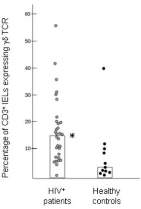
patients than in the controls (39.6/U versus 86.4/U). There was a striking variability among the patients as to the density ofcd IELs (Fig. 1) but the total number per mucosal length unit (U) tended to be increased (median 4.0/Uversus3.2/U). Therefore, the averagecdIEL ratio (Fig. 2) in the 30 HIV+
patients (median 14.5%, range 1.5–56.3%) was significantly increased (p,0.02) compared with values in the healthy controls (median 2.8%, range 0.3–38%). Autofluorescence in some of the old biopsies limited countable areas, and two patients were excluded from IEL counts/U. However, the ratios and numbers of cd IELs were well correlated (r= 0.74), substantiating the validity of the recording methods.
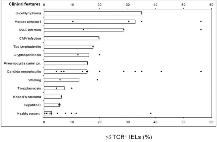
Regarding the clinical subgroups of HIV+
patients, those with B-cell lymphoma, herpes simplex II infection, or with Mycobacte-rium avium intracellularecomplex (MAC) infection had particularly high ratios ofcdIELs (Fig. 3). We also observed relatively high ratios in two patients with cytomegalovirus (CMV) infection or tuberculous lymphadenitis, whereas those with Candida infection showed a wide range (4.5%–56.3%). The lowest cd IEL ratios were seen in HIV+patients suffering from toxoplasmosis, Kaposi’s
sarcoma, or hepatitis C virus infection.
When we related thecdIELs to different antiretroviral therapy, the highest ratios were seen in HIV+
patients on single medication (1 NA, median 23.5%; p,0.04versuscontrols); these patients were the earliest included ones (before April, 1996) and often had intercurrent infections. Thecd IEL ratios were also increased in untreated patients (median 14.3%; p,0.008) and in those on double nucleoside analogues (2 NAs, median 13.0%; p,0.04), whereas those on triple combination therapy (HAART, median 12.8%) did not reach significance (p = 0.08) due to widely scattered ratios (Fig. 4).
As mentioned above, the total number ofcdIELs tended to be increased despite a highly significant decrease of the total number of CD3+
IEL/U in the HIV+
patients as a group (medians 31.7– 45.7/U) compared with healthy controls (Fig. 4). In our previous studies we classificated three HIV+
subgroups according to length of survival after biopsy [13,14], but this clinical grouping did not provide a better cd IEL discrimination. It should be noted, however, that the 7 premortal patients died of diseases unrelated to AIDS, e.g. cerebral haemorrhagia, cardiac left ventricle dysfunc-tion, aortic valve stenosis and neurolues.
Two-colour staining for CD8 revealed that almost allcd IELs were CD82 (median 100%, range 85–100%), as previously
reported in coeliac disease (median 90%) [9] and primary immunodeficiencies (median 94–99%) [11,12] and also in healthy
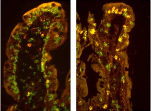
Figure 1. Paired immunofluorescence staining (merges) for CD3 (green) andcdTCR (red).The left panel shows a sectioned duodenal villus from an HIV+patient who had retained an abundance of intraepithelial lymphocytes (IELs) which are mainly CD3+cdTCR2(presumablyabTCR+CD8+)
and only few are CD3+
cdTCR+
(yellow; median 14%). The right panel shows comparable field from an HIV+
patient with depletion of IELs but with highly increased CD3+
cdTCR+
ratios (yellow; median 60%). In both sections some nonspecifically stained deposits are seen in the lamina propria, while the epithelium is visualized due to autofluorescence (which increased during storage of the frozen tissue samples).
controls (median 75%) [11]. A large fraction of cd IELs in the HIV+
patients (median 65.2%, range 21.7–100%) expressed the Vd1/Jd1-encoded epitope revealed by mAb dTCS-1 – almost identical to that in patients with coeliac disease (67%) [9], primary immunodeficiencies (63–84%) [11,12], and healthy controls (75%) [9,11]. No difference was seen among the subgroups of HIV+
patients.
Infection parameters in peripheral blood related tocd IELs
When circulating T-cell subsets in HIV+ patients were
compared with healthy blood donors [15], a tendency to reduced median numbers of CD8+ cells (0.36
6109/l; range, 0.057–
1.676109/l versus 0.4056109/l; range, 0.18–1.236109/l) was
observed while the CD4 counts were, as expected, significantly (p,0.0001) reduced (0.0206109/l; range, 0.0012–0.256109/l
versus0.8156109/l; range, 0.5–1.586109/l) – particularly in late
stage (Group II, 0.0196109/l) and preterminal (Group III,
0.0156109/l) AIDS patients.
Plasma HIV RNA (n= 12) increased as expected towards the end of life: Group I, 57000 copies/ml; Group II, 70000 copies/ml; and Group III, 191000 copies/ml. We observed a trend towards a higher CD4 count (0.0606109/l,n= 9versus0.0226109/l) and a
lower median plasma HIV RNA level (76000 copies/ml) in patients receiving HAART.
No relationship was seen between the relative (Fig. 2) or absolute (Fig. 4) numbers ofcdIELs and the level of circulating CD4+
or CD8+
T cells, the CD4:CD8 ratio or the B-cell number in peripheral blood.
Serum immunoglobulins andb2microglobulin
Hypergammaglobulinaemia was seen in 16 of the HIV+
patients, mostly IgG (18.3 g/l; range, 8.7–44.6 g/l) dominated by IgG1 (median, 11.2 g/l) and IgG3 (median, 1.8 g/l). There was only a slight increase of IgA (median 3.5 g/l) and also IgM (median, 1.5 g/l) was within normal limits. Compared with IgA in untreated HIV+patients (median, 4.2 g/l), the level tended to be
decreased in patients on HAART (median, 3.1 g/l), similarly to what we observed for duodenal IgA-producing plasma cells [14]. Theb2-M level in HIV+patients (median, 3.7 mg/l;n= 28) was significantly increased (p,0.0001) compared with healthy controls (median, 1.2 mg/l). No relationship was observed between serum
b2-M and circulating CD4+or CD8+cells, duodenalcdIELs or any of the other immunological variables.
Long-time survivors
Three of the ten ARC/AIDS patients were so-called long-time survivors (LTSs), living for 15–17 years after becoming HIV+.
Only one of them received HAART and one was not treated at all. Two of them were IVDAs and one was homosexual. Compared with the HIV+
patients as a group, they had lower median ratios and numbers of duodenalcdIELs (7.1%, number 1.6/U), average levels of circulating CD4+
T cells, and mostly low serum IgG1. One 38-year old female IVDA received stavudine (d4T) and methadone. She suffered from wasting syndrome, AIDS dementia, and chronic hepatitis B and C virus infection, and had remarkably elevated serum immunoglobulins (especially IgG, 33.6 g/l) and relatively many duodenal IgG-producing plasma cells (15.2%), mainly of the IgG1 subclass [14]. We found elevated duodenalcd
IELs in this LTS (19.2%, number 5.5/U). All other immunological variables were near the average values.
Discussion
More information is needed about the compartmentalized distribution of lymphoid and accessory immune cells in human intestinal mucosa to enable modulation of local immunity in a rational manner [16]. This has been emphasized in face of the failure of parenteral vaccines to reinforce immunity at the site of HIV entry [1–5]. However, ethical restrictions in sampling mucosal tissue from AIDS patients prohibit functional studies of local immune cells ex vivo, thus limiting our efforts to obtain mechanistic insight to observing their treatment responsein vivo.
Here, we confirmed the relative increase of duodenalcdIELs in HIV patients, as seen in our previous study of patients included before 1996 [13]. A concurrent study did not find elevatedcdIELs in HIV+
patients, whether they had diarrhoea or not [17]. This discrepancy might have a technical origin as the study was partly based on flow-cytometric analyses of dispersed cells. We performed quantitativein situcell phenotyping and enumeration with a careful morphometric approach. In our HIV+
patients especially highcdIEL ratios were associated with intercurrent B cell lymphoma, herpes simplex II infection, MAC infection, CMV infection, and tuberculous lymphadenitis.
Our results suggested that a relative expansion of thecd IEL fraction occurs in advanced AIDS at the same time as the total IEL population is reduced. Most human IELs are memoryab
CD8+T cells [18,19]; based on recently published immunization
experiments (both in humans and mice) [20], their long-term residence at body surfaces, including the gut epithelium, may depend on repetitive inductive activity in peripheral lymphoid organs. It has been reported that viral control and early HIV disease progression is associated with a synchronous differentiation of the HIV-specific and total CD8+
memory cell populations [21].
Figure 2. Scatter diagram of the percentage of duodenal cd
IELs in HIV+ patients compared with HIV2 control subjects.
Median ratios are represented by vertical columns (*: p,0.02, Mann-Whitney two-tailed test).
However, as the peripheral immune organs with their CD4+
helper T cells deteriorate in AIDS, chronic infectious stimuli are apparently unable to maintain the dominating ab CD8+
IEL
population due to lack of T-cell homing to the intestinal mucosa [22]. Thus, there must be other rules for cd IELs which are maintained or expanded in response to opportunistic
micro-Figure 4. ThecdIEL subset remains elevated in AIDS patients despite antiretroviral treatment.Scatter diagram showscd:CD3 ratios for duodenal IELs,cdIEL numbers per mucosal unit (above 1 mm of muscularis mucosae), and numbers of CD3+
IELs per mucosal unit in HIV+
patients – all variables displayed according to administered antiretroviral treatment compared with no treatment and with data from HIV2control subjects.
Medians are represented by vertical columns. NA, nucleoside analogue; HAART, highly active antiretroviral therapy. doi:10.1371/journal.pone.0029066.g004
Figure 3. Scatter diagram of the percentage of duodenalcdIELs in relation to clinical features of individual HIV+patients.
Median ratios are represented by horizontal columns. CMV, cytomegalovirus infection; MAC,Mycobacterium avium intracellularecomplex.
organisms, apparently with no direct HIV effect as antiretroviral treatment did not reduce the response. This was in contrast to the expanded IgA+
duodenal plasma cell population that was significantly decreased by HAART in virtually the same patient material [14].
The function of cd IELs is probably not limited to first-line defence. In mice the activation ofcdT cells sometimes follows that ofabT cells, and thecdT cells may mainly contribute to immune regulation and tissue repair [23]. The common overlap of cd
TCR-directed pathogen-encoded epitopes with molecules ex-pressed by ‘‘stressed’’ host cells has fuelled the view thatcdT-cell responses are driven by lack of tissue homeostasis (inflammation, cell damage or transformation) rather than by microbial challenges [23]. In mice, it has been shown thatcdT cells express the toll-like receptors TLR1 and TLR2, as well as dectin-1, and respond to microbial products [24]. In addition, they express the aryl hydrocarbon receptor (AhR) and can therefore receive other environmental signals that may shape their functional capacity, including the production of the IL-17 cytokine [24]. Thus, these cells do not necessarily need TCR engagement but can be triggered by their innate receptors or, alternatively, by the cytokines IL-1 or IL-23 derived from activated macrophages or dendritic cells [25].
If it can be shown that human cd IELs have immune modulating properties similar to murinecdT cells, this would fit with the fact that they are not negatively affected by deterioration of the adaptive immune system in advanced AIDS, but rather expand in such HIV+
patients. Interestingly, it was recently shown that murine cd IELs are a self-renewing population with little migration and recirculation out of their specialized niche, whereas peripheralcdIELs are highly motile [26].
We have earlier reported a marked decrease of duodenalcd
IELs in premortal AIDS patients [13]. A combined deficiency of
cdIELs and IgA-producing plasma cells at the end stage of AIDS could in part be responsible for the fatal result. In the present study we could not show this; but it should be noted that most of the preterminal patients included here died of diseases unrelated to AIDS. There was a strikingly variable positive effect of HAART on the cd IEL counts in the AIDS patients, although immune reconstitution was usually confirmed by favourable CD4-cell counts and plasma HIV RNA levels in peripheral blood.
Serum levels ofb2-M and neopterin (an intermediate in the biosynthesis of tetrahydro-biopterin), which are markers of immunostimulation [27], showed an inverse correlation with duodenalcdIELs in end-stage AIDS in our previous study [13]. A similar strong tendency of increasingb2-M concentrations towards the end of life was not observed in the present study. This could reflect the beneficial effect of HAART in advanced AIDS. One of the three LTSs showed particularly good prognosis (undetectable HIV RNA level) and had elevated serum IgG and duodenal IgG+
plasma cells (especially IgG1). This suggested complement-activating local immunity – a response also noted in other studies [28,29]. Additionally, we found raised ratio and number of cd
IELs (19.2%, 5.5/U) in this patient, contributing to the idea that these cells exert immunoregulatory functions.
There is a recent focus on the intestinal epithelial barrier as an important variable in HIV infection. Pigtail macaques (PTMs) rapidly progress to AIDS after simian immunodeficiency virus (SIV) infection. The explanation seems to be that uninfected PTMs commonly show damage to their intestinal barrier [30]. This results in microbial translocation and deleterious immune activation both locally and systemically; a higher frequency of CD4+
memory/effector T cells ensues, and these cells are the primary target for SIV/HIV infection.
Th17 cells are important in microbial defence by attracting neutrophils and maintaining the mucosal barrier; but these CD4+
cells are preferentially lost during HIV infection [31], although they may be restored by HAART [32]. Importantly, however,cd
T cells may respond more rapidly with IL-17 secretion because of their innate nature [24,25,32], and therefore be compensating for the Th17 cell loss. Moreover, there is experimental evidence in mice thatcdT cells may amplify the function of Th17 cells [25] and inhibit regulatory cells T (Treg) cells [33]. Putative Tregcells (CD4+CD25+FOXP3+) in peripheral blood may be increased in
HIV+
patients and dampen various defences against the virus; but decrease of Tregcells has been reported in chronic HIV infection [34,35]. Thus, it remains unresolved whether Treg cells are protective or detrimental in HIV+
patients. Also the balance between Th17 cells,cdT cells and Tregcells shows a complexity afflicted with many uncertainties [36]. Nevertheless, a recent study reported fewer Tregcells in rectosigmoid mucosal biopsies of HIV long-term ‘‘controllers’’ than ‘‘non-controllers’’ [37].
Taken together, it remains unknown to what extent these immunological variables influence the course of HIV infection. However, cd T cells with specificity for Candida albicans, and bearing the Vd1 TCR chain as well as possessing cytoplasmic IL-17, have been identified in peripheral blood of HIV+
patients [38]. Whether these cells have similar functional properties ascdIELs remains uncertain. We observed a relative expansion of duodenal Vd1+ cd IELs in AIDS patients, even in those receiving
antiretroviral therapy. This probably reflected an intensified innate response to an increased luminal load of microbial components. Secondary intestinal infections are common in AIDS, and it is important to define how the mucosal defence behaves in these patients. A better delineation of intestinal HIV immunology, including the late stage-induced local response, is crucial to this end.
Materials and Methods
Biological samples
Duodenal biopsy specimens and peripheral blood were collected consecutively during the period 1991–98 from 30 patients infected with HIV type 1 (5 women: median age, 33 years and range, 26– 40 years; and 25 men: median age, 39 years and range, 26–52 years); 18 were homosexual men, 7 intravenous drug abusers (IVDAs), and 5 had experienced heterosexual virus transmission in Africa (Table S1). Informed verbal consent was obtained from all patients. The biopsy material was derived from the Diagnostics and Treatment Biobank of the Oslo University Hospital. This collection of biological material intended for clinical examination, diagnostics and treatment does not require explicit informed consent according to The Norwegian Treatment Biobank Act 2003.
In addition, similar biopsy and blood samples were obtained from 11 immunologically intact HIV2controls (seven females and four males; median age 21 years and range, 4.5–76 years) with no histological abnormalities in duodenal mucosa. Dyspepsia, growth retardation, irritable bowel syndrome, and hypothyroid disease were the reasons for endoscopic examination in a minority of the subjects as we also included volunteers (health workers) with no symptoms. Part of the same control material has been used in previous studies with informed written consent [11–13], and the Regional Ethics Committee approved the inclusion of additional controls to replace exhausted material.
All HIV+patients had serum IgG antibodies to HIV type 1 as
confirmed by Western blotting (DuPont, Wilmington, DE). When clinically classified according to the criteria of the Centers for Disease Control and Prevention (CDC), Atlanta (GO, USA), 5 subjects were staged in group CDC IVA/B, 23 in group CDC IVC1, and 2 in group CDC IVD – the latter 25 fulfilling diagnostic criteria for full-blown AIDS. Because most of the patients were classified as having late-stage AIDS, CD4+T-cell
counts did not differ much. Median survival time after the first positive HIV test was 81 months (range 11–156 months;n= 20). Seven patients could retrospectively be defined as premortal due to death within 7 months (six homosexual men, one raped female refugee). However, they died mainly of diseases unrelated to AIDS, e.g. cerebral haemorrhagia, cardiac left ventricle dysfunc-tion, aortic valve stenosis and neurolues.
Clinical features
All HIV+
patients suffered from different on-going opportunistic and/or non-opportunistic infections; only one of them had skin or mucosal lesions consistent with Kaposi’s sarcoma, and one had non-Hodgkin malignant lymphoma. Eight of the patients had chronic intermittent diarrhoea, of which two had cryptosporidi-osis, one systemic CMV infection, and four herpes simplex type II infection. Altogether twelve patients had oral and/or oesophageal candidiasis, three suffered from MAC infection and two from wasting (.10% unwanted weight loss). Only one of these patients had duodenitis with histological signs of moderate, chronic inflammation. Other serious infections such asPneumocystis carinii
pneumonia was found in one patient, two had toxoplasmosis (encephalitis), two chronic hepatitis C virus infection and one tuberculous lymphadenitis (Table S1).
Four patients still received antiretroviral monotherapy with zidovudine (ZDV) at the time of sampling (1995–96) while eight used two other NAs such as didanosine (DDI) and/or stavudine (d4T) as well (Table S1). Ten patients tolerated satisfactorily combination therapy with one of these drugs together with lamivudine (3TC) and HIV protease inhibitors (PI) such as indinavir (Crixivan) or nelfinavir (Viracept), constituting HAART, whereas eight others had discon-tinued all antiretroviral treatment because of intolerable side effects. None received immunoglobulin replacement therapy.
Quantification of Ig isotypes, B cells, T-cell subsets,b2 -microglobulin and HIV RNA in peripheral blood
Measurements of serum IgG, IgA, and IgM were performed by nephelometry with commercial standards (Behringwerke, Mar-burg, Germany), while quantification of serum IgG subclasses was performed with monoclonal antibodies (mAbs) in commercial kits (The Binding Site, Birmingham, UK). The blood samples were collected as near the time of biopsy as possible (usually within 1 to 2 hours before) and analysed on the same day, or frozen at220uC or270uC for later analyses.
CD19+ B cells and CD4+/CD8+ T cells in peripheral blood
were enumerated by a standard flow cytometric method. b2-M was measured by a microparticle enzyme immunoassay (Abbott IMX; Abbott laboratories, Abbott Park, IL, USA). HIV RNA was measured by a reverse transcriptase-polymerase chain reaction (RT-PCR) assay (Amplicor HIV-1 Monitor, Roche Diagnostic System, Brachburg, NJ, USA). The detection limit of the kit was 400 HIV-1 RNA copies/ml; lower values were also included. This methodology was only available after August 1996 in our hospital.
Tissue sample processing
Multiple mucosal biopsy specimens (n= 8–10) were obtained endoscopically (Olympus GIF K10) from the distal duodenum.
Endoscopical signs of chronic inflammation were occasionally observed in the distal oesophageal (n= 3), prepyloric gastric (n= 1) or duodenal mucosa (n= 2). Two specimens from each subject were processed for conventional histological evaluation. Other tissue specimens were fixed in 2% (w/v) paraformaldehyde for electron-microscopic evaluation or processed by snap-freezing in liquid nitrogen for immunohistochemistry (see below). All gastrointestinal endoscopies were performed in the HIV+
patients solely for clinical indications, mostly to evaluate their symptoms such as dysphagia, diarrhoea or cachexia. Ethical reasons prevented us from obtaining additional biopsies.
Histological evaluation
A tissue section from each biopsy series was stained with haematoxylin and eosin for histological evaluation. In addition, scanning electron microscopy was performed to classify villus changes as previously described [11]. Such combined evaluation showed normal villi in all 30 HIV+
patients. Only two of them had histological signs of moderate, chronic duodenal inflammation. All control subjects had histologically normal duodenal biopsies.
Two- or three-colour immunohistochemical staining
Serial cryosections were cut at 6mm and incubated for 1 hour at ambient temperature with pairs of unlabelled murine mAbs of the following human specificities:d chain of cd TCR (TCRd1, IgG1, 1/20; T Cell Sciences Inc., Cambridge, MA, USA) and CD3 (clone RIV9, IgG3, 1/10; Sanbio, Am Uden, The Nether-lands);dchain ofcd TCR and theachain of CD8 (BMA-081, IgG2a, 1/40; Behringwerke, Marburg, Germany); CD4 (anti-Leu-3a and -3b, IgG1, 1/40; Becton Dickinson, Mountain View, CA, USA) and CD3 (RIV9, IgG3, 1/10; Sanbio); and CD8 (anti-Leu-2b, IgG2a, 1/20; Becton Dickinson) mixed with BMA-081 (IgG2a, 1/80) and CD3 (anti-Leu-4, IgG1, 1/80; Becton Dickinson). Secondary antibody reagents were various combinations of biotinylated and fluorescein isothiocyanate-conjugated goat IgG (0.01 to 0.05 g/litre) with specificity for mouse IgG1, IgG2a, or IgG3 (Southern Biotechnology, Birmingham, ALA, USA). These reagents were applied for 1.5 hour, and then streptavidin-Texas red (0.0025 g/litre; Bethesda Research Laboratories, Gaithers-burg, MD, USA) was applied for 30 min. Human IgG at 0.8 g/ litre (Kabi Vitrum, Stockholm, Sweden) was added to the goat reagents to block species cross-reactivity [9]. Because mAb TCRd1 reacts with all cd T cells, sequential staining was performed as detailed elsewhere [9] to examine the subset expressing the Vd1/ Jd1-encoded epitope for mAbdTCS-1 (IgG1; T Cell Sciences).
Fluorescence microscopy and cell counting
Immunofluorescence was examined at a magnification of6300 in a Nikon microscope (Eclipse E600; ocular grid; Nikon, CFI) equipped with a Ploem-type vertical illuminator with interference filters for selective observation of red, green, or blue emission. The results were recorded on Ectachrome professional 800/1600 ASA daylight film. All sections were examined blinded by the same investigator. The percentage distribution of cd IELs was determined by evaluating every CD3+
IEL forcdTCR expression (Fig. 1). Also coexpression of CD8 and Vd1/Jd1 oncdIELs was similarly examined.
The total number of CD3+
andcdIEL was related to a 316-mm grid width, including all parts of the epithelium from the luminal face to the muscularis mucosae. The number of CD3+
100) [39] was followed by 7-amino-4-methylcoumarin-3-acetic acid-conjugated goat anti-rabbit IgG (1/20; Vector Laboratories, Burlingame, CA, USA).
Control reference data
The ratios ofcdIELs as well as the numbers of CD3+andcd
IELs per mucosal U were determined as described above in histologically normal duodenal mucosa of the eleven HIV2
control subjects. Reference data were available in our laboratory for the normal expression of the Vd1/Jd1-encoded epitope oncd
IELs [9]. Normal CD4:CD8 T-cell ratios for duodenal lamina propria and epithelial compartments were based on published data [40].
As normal reference data for circulating lymphocyte subsets, we used results from healthy (HIV2) blood donors (n= 60) [15] recorded as described previously [41]. Reference data from blood donors were also available for serum concentrations of b2-M (n= 18). The HIV RNA level in normal controls was set at,400 copies/ml plasma.
Statistical analysis
Statistical calculations were based on the software program SPSS (Chicago, IL) while the Mann-Whitney 2-tailed test for unpaired samples was used for comparison between various categories of HIV+
patients and control subjects. Results are presented as median and observed range. Differences between groups were considered statistically significant at p,0.05. Correlations between mucosal CD3+
IELs or cd IELs and circulating T-cell subsets orb2-M in the HIV+ patients and the
HIV2controls were calculated by the Spearman rank correlation
test (SPSS), as was the relationship between the proportions and numbers of cd IELs. The reproducibility of in situ T-cell enumerations by paired immunofluorescence staining was excel-lent as documented in our previous studies [9,11,12].
Ethical considerations
The study was approved by the Committee for Medical Research Ethics (Health Region South-East, Oslo, Norway).
Supporting Information
Table S1 Clinical and immunological features of the HIV-1-infected patients in relation to treatment regi-men.
(DOC)
Acknowledgments
We thank the technical staff at LIIPAT, Oslo University Hospital, Rikshospitalet, and the Department of Infectious Diseases, the Department of Clinical Chemistry and the Section of Gastroenterology, Oslo University Hospital, Ullevaal, Oslo, Norway, for valuable contributions. Hege Eliassen at the Department of Pathology is gratefully acknowledged for excellent assistance with the manuscript preparation.
Author Contributions
Conceived and designed the experiments: DEN PB. Performed the experiments: DEN. Analyzed the data: DEN. Contributed reagents/ materials/analysis tools: DEN PB. Wrote the paper: DEN PB.
References
1. Virgin HW, Walker BD (2010) Immunology and the elusive AIDS vaccine. Nature 464: 224–231.
2. Broliden K, Haase AT, Ahuja SK, Shearer GM, Andersson J (2009) Introduction: Back to basics: mucosal immunity and novel HIV vaccine concepts. J Intern Med 265: 5–17.
3. Shacklett BL, Critchfield JW, Ferre AL, Hayes TL (2009) Mucosal T-cell responses to HIV: responding at the front lines. J Intern Med 265: 58–66. 4. Hirbod T, Kaul R, Reichard C, Kimani J, Ngugi E, et al. (2008)
HIV-neutralizing immunoglobulin A and HIV-specific proliferation are indepen-dently associated with reduced HIV acquisition in Kenyan sex workers. AIDS 22: 727–735.
5. Masopust D (2009) Developing an HIV cytotoxic T-lymphocyte vaccine: issues of CD8 T-cell quantity, quality and location. J Intern Med 265: 125–137. 6. Pennington DJ, Silva-Santos B, Hayday AC (2005)c/dT cell development –
having the strength to get there. Curr Opin Immunol 17: 108–115. 7. Ciofani M, Zu´n˜iga-Pflu¨cker JC (2010) Determining cd versus aß T cell
development. Nat Rev Immunol 10: 657–663.
8. Bonneville M, O’Brien RL, Born WK (2010)cdT cell effector functions: a blend of innate programming and acquired plasticity. Nat Rev Immunol 10: 467–478. 9. Halstensen TS, Scott H, Brandtzaeg P (1989) Intraepithelial T cells of the
TCRc/d+
CD82and Vd1/Jd1+
phenotypes are increased in coeliac disease. Scand J Immunol 30: 665–672.
10. Bhagat G, Naiyer AJ, Shah JG, Harper J, Jabri B, et al. (2008) Small intestinal CD8+
TCRcd+
NKG2A+
intraepithelial lymphocytes have attributes of regula-tory cells in patients with celiac disease. J Clin Invest 118: 281–293. 11. Nilssen DE, Halstensen TS, Frøland SS, Fausa O, Brandtzaeg P (1993)
Distribution and phenotypes of duodenal intraepithelialc/dT cells in patients with various types of primary B-cell deficiency. Clin Immunol Immunopathol 68: 301–310.
12. Nilssen DE, Aukrust P, Frøland SS, Mu¨ller F, Fausa O, et al. (1993) Duodenal intraepithelialc/dT cells and soluble CD8, neopterin andb2-microglobulin in serum of IgA-deficient subjects with or without IgG subclass deficiency. Clin Exp Immunol 94: 91–98.
13. Nilssen DE, Mu¨ller F, Øktedalen O, Frøland SS, Fausa O, et al. (1996) Intraepithelialc/dT cells in duodenal mucosa are related to the immune state and survival time in AIDS. J Virol 70: 3545–3550.
14. Nilssen DE, Øktedalen O, Brandtzaeg P (2004) Intestinal B cell hyperactivity in AIDS is controlled by highly active antiretroviral therapy. Gut 53: 487–493. 15. Brinchmann JE, Vartdal F, Thorsby E (1989) T lymphocyte subset changes in
human immunodeficiency virus infection. J Acquired Immune Defic Syndr 2: 398–403.
16. Pabst R, Russel MW, Brandtzaeg P (2008) Tissue distribution of lymphocytes and plasma cells and the role of the gut. Trends Immunol 29: 206–208. 17. Snijders F, Meenan J, van den Blink B, van Deventer SJ, ten Kate FJ (1996)
Duodenal intraepithelial and lamina propria T lymphocytes in human immunodeficiency virus-infected patients with and without diarrhoea. Scand J Gastroenterol 31: 1176–1181.
18. Brandtzaeg P, Halstensen TS, Scott H, Sollid LM, Valnes K, et al. (1989) Epithelial homing ofcdT cells? Nature 341: 113–114.
19. Brandtzaeg P, Farstad IN, Helgeland L (1998) Phenotypes of T cells in the gut. In: Mucosal T cells, MacDonald TT, ed. Chemical Immunology 71: 1–26. 20. Masopust D, Choo D, Vezys V, Wherry EJ, Duraiswamy J, et al. (2010)
Dynamic T cell migration program provides resident memory within intestinal epithelium. J Exp Med 207: 553–564.
21. Burgers WA, Riou C, Mlotshwa M, Maenetje P, de Assis Rosa D, et al. (2009) Association of HIV-specific and total CD8+
T memory phenotypes in subtype C HIV-1 infection with viral set point. J Immunol 182: 4751–4761.
22. Ciccone EJ, Read SW, Mannon PJ, Yao MD, Hodge JN, et al. (2010) Cycling of gut mucosal CD4+
T cells decreases after prolonged anti-retroviral therapy and is associated with plasma LPS levels. Mucosal Immunol 3: 172–181. 23. Hayday AC (2009)c/dcells and the lymphoid stress-surveillance response.
Immunity 31: 184–196.
24. Martin B, Hirota K, Cua DJ, Stockinger B, Veldhoen M (2009) Interleukin-17-producingcdT cells selectively expand in response to pathogen products and environmental signals. Immunity 31: 321–330.
25. Sutton CE, Lalor SJ, Sweeney CM, Brereton CF, Lavelle EC, et al. (2009) Interleukin-1 and IL-23 induce innate IL-17 production from cd T cells, amplifying Th17 responses and autoimmunity. Immunity 31: 331–341. 26. Chennupati V, Worbs T, Liu X, Malinarich FH, Schmitz S, et al. (2010)
Intra-and intercompartmental movement ofcdT cells: intestinal intraepithelial and peripheral cd T cells represent exclusive nonoverlapping populations with distinct migration characteristics. J Immunol 185: 5160–5168.
27. Bhalla RB, Safai B, Mertelsmann R, Schwartz MK (1983) Abnormally high concentrations of b2-microglobulin in acquired immunodeficiency syndrome (AIDS) patients. Clin Chem 29: 1560.
28. Schneider T, Zippel T, Schmidt W, Pauli G, Heise W, et al. (1997) Abnormal predominance of IgG in HIV-spesific antibodies produced by short-term cultured duodenal biopsy specimens from HIV-infected patients. J AIDS Hum Retrovir 16: 333–339.
30. Klatt NR, Harris LD, Vinton CL, Sung H, Briant JA, et al. (2010) Compromised gastrointestinal integrity in pigtail macaques is associated with increased microbial translocation, immune activation, and IL-17 production in the absence of SIV infection. Mucosal Immunol 3: 387–398.
31. Prendergast A, Prado JG, Kang YH, Chen F, Riddell LA, et al. (2010) HIV-1 infection is characterized by profound depletion of CD161+
Th17 cells and gradual decline in regulatory T cells. AIDS 24: 491–502.
32. Klatt NR, Brenchley JM (2010) Th17 cell dynamics in HIV infection. Curr Opin HIV AIDS 5: 135–140.
33. Petermann F, Rothhammer V, Claussen MC, Haas JD, Blanco LR, et al. (2010)
cdT cells enhance autoimmunity by restraining regulatory T cell responses via an interleukin-23-dependent mechanism. Immunity 33: 351–363.
34. Bernardes SS, Borges IK, Lima JE, Milanez Pde A, Conchon-Costa I, et al. (2010) Involvement of regulatory T cells in HIV immunopathogenesis. Curr HIV Res 8: 340–346.
35. Sachdeva M, Fischl MA, Pahwa R, Sachdeva N, Pahwa S (2010) Immune exhaustion occurs concomitantly with immune activation and decrease in regulatory T cells in viremic chronically HIV-1-infected patients. J Acquir Immune Defic Syndr 54: 447–454.
36. Kanwar B, Favre D, McCune JM (2010) Th17 and regulatory T cells: implications for AIDS pathogenesis. Curr Opin HIV AIDS 5: 151–157. 37. Loke P, Favre D, Hunt PW, Leung JM, Kanwar B, et al. (2010) Correlating
cellular and molecular signatures of mucosal immunity that distinguish HIV controllers from noncontrollers. Blood 115: e20–32 (erratum in: Blood 2010, 116: 2402).
38. Fenoglio D, Poggi A, Catellani S, Battaglia F, Ferrera A, et al. (2009) Vd1 T lymphocytes producing IFN-c and IL-17 are expanded in HIV-1-infected patients and respond to Candida albicans. Blood 113: 6611–6618.
39. Huitfeldt H, Brandtzaeg P (1985) Various keratin antibodies produce immunohistochemical staining of human myocardium and myometrium. Histochemistry 83: 381–389.
40. Selby WS, Janossy G, Bofill M, Jewell DP (1983) Lymphocyte subpopulations in the human small intestine. The findings in normal mucosa and in the mucosa of patients with adult coeliac disease. Clin Exp Immunol 52: 219–228. 41. Brinchmann JE, Vartdal F, Gaudernack G, Markussen G, Funderud S, et al.