Dopaminergic Modulation of the
Voltage-Gated Sodium Current in the Cochlear
Afferent Neurons of the Rat
Catalina Valdés-Baizabal, Enrique Soto, Rosario Vega*
Instituto de Fisiología, Benemérita Universidad Autónoma de Puebla, Puebla, México
Abstract
The cochlear inner hair cells synapse onto type I afferent terminal dendrites, constituting the main afferent pathway for auditory information flow. This pathway receives central control input from the lateral olivocochlear efferent neurons that release various neurotransmitters, among which dopamine (DA) plays a salient role. DA receptors activation exert a protective role in the over activation of the afferent glutamatergic synapses, which occurs when an ani-mal is exposed to intense sound stimuli or during hypoxic events. However, the mechanism of action of DA at the cellular level is still not completely understood. In this work, we studied the actions of DA and its receptor agonists and antagonists on the voltage-gated sodium current (INa) in isolated cochlear afferent neurons of the rat to define the mechanisms of do-paminergic control of the afferent input in the cochlear pathway. Experiments were per-formed using the voltage and current clamp techniques in the whole-cell configuration in primary cultures of cochlear spiral ganglion neurons (SGNs). Recordings of the INashowed that DA receptor activation induced a significant inhibition of the peak current amplitude, leading to a significant decrease in cell excitability. Inhibition of the INawas produced by a phosphorylation of the sodium channels as shown by the use of phosphatase inhibitor that produced an inhibition analogous to that caused by DA receptor activation. Use of specific agonists and antagonists showed that inhibitory action of DA was mediated both by activa-tion of D1- and D2-like DA receptors. The acactiva-tion of the D1- and D2-like receptors was shown to be mediated by a Gαs/AC/cAMP/PKA and Gαq/PLC/PKC pathways respectively.
These results showed that DA receptor activation constitutes a significant modulatory input to SGNs, effectively modulating their excitability and information flow in the auditory pathway.
Introduction
The organ of Corti consists of several cells types that perform auditory functions harmoniously. The hair cells are responsible for the sensory transduction and synaptic activation of the affer-ent neurons. The outer hair cells (OHC) play a role mainly related to the cochlear amplifier,
OPEN ACCESS
Citation:Valdés-Baizabal C, Soto E, Vega R (2015)
Dopaminergic Modulation of the Voltage-Gated Sodium Current in the Cochlear Afferent Neurons of the Rat. PLoS ONE 10(3): e0120808. doi:10.1371/ journal.pone.0120808
Academic Editor:Saobo Lei, University of North
Dakota, UNITED STATES
Received:July 8, 2014
Accepted:February 6, 2015
Published:March 13, 2015
Copyright:© 2015 Valdés-Baizabal et al. This is an
open access article distributed under the terms of the
Creative Commons Attribution License, which permits unrestricted use, distribution, and reproduction in any medium, provided the original author and source are credited.
Data Availability Statement:All relevant data are
within the paper.
Funding:This work was supported by a grant from
Consejo Nacional de Ciencia y Tecnología de México (CONACyT) grant 167052 to ES, grants VIEP-BUAP to RV and ES, and PIFI 2012-2013 grant. CVB was supported by CONACyT fellowship 216652. The funders had no role in study design, data collection and analysis, decision to publish, or preparation of the manuscript.
Competing Interests:The authors have declared
while the inner hair cells (IHC) fundamentally function in the detection of auditory stimuli. The IHC receive afferent innervation from type I spiral ganglion neurons (SGNs), which com-prise approximately 95% of the cochlear afferents [1], while the OHC receive afferent innerva-tion through the type II SGNs, which correspond to approximately 5% of the cochlear afferents.
The efferent neurons originating from the lateral superior olivary complex (LOC) make syn-aptic contacts with the afferent neurons innervating the IHC [2]. These neurons have been found to release various neurotransmitters including acetylcholine, dynorphin [3], encephalin [4], calcitonin gene-related peptide (CGRP) [5], GABA [6], adenylate cyclase-activating poly-peptide [7], and dopamine (DA) [8]. Another group of efferent neurons originates from the medial superior olivary complex (MOC) and synapse directly onto the OHC [9]. The MOC ef-ferents release acetylcholine [3], GABA [6] and CGRP [10]. A particular case consists of a group of neurons originating from the periolivary nucleus, which form part of the efferent sys-tem innervating both the OHC and IHC that release serotonin as a neurotransmitter [11].
The type I SGNs express D1, D2, D4 and D5 dopaminergic receptor subtypes [12,13,14], which belong to the large family of G-protein coupled receptors that have seven transmem-brane segments. Based on their pharmacological properties and structural homology, dopami-nergic receptors are classified into two families consisting of D1-like receptors, which include the D1- and D5-receptors, and D2-like receptors, which include the D2-, D3- and D4-receptors [15].
The olivocochlear efferent neurons in guinea pig show tyrosine hydroxylase immunoreac-tivity, which is the enzyme that catalyzes the synthesis of DA [16]. DA is present in the cochlea at birth in the rat and its concentration increases with age to approximately 5-fold by 30 days after birth [17]. Sound conditioning triggers an up-regulation of tyrosine hydroxylase both in the lateral efferent of cochlea and in the lateral superior olivary complex and acoustic trauma reduced these levels [18].
DA modulates the sound evoked compound action potential (CAP) of auditory nerve with no effects on cochlear microphonic, summating or endocochlear potentials [19], indicating that DA action is taken place at postsynaptic level upon the terminals of afferent dendrites. The action of DA in the cochlea has been associated with a neuroprotective mechanism in afferent neurons, and the CAP modulation depends on the subtype(s) of DA receptors activated [20]. DA was shown to reduce the action potential amplitude in isolated SGNs of the mouse [21]. In the guinea pig, DA decreased the action potential discharge of afferent neurons that is induced by glutamatergic agonists in a dose-dependent manner [22]. The D1 receptor was localized at the spiral ganglia neurons and at the base of the IHC. The amplitude of the CAP was enhanced by D1 receptor agonists an effect that was abolished by a protein kinase A (PKA) inhibitor [23], and the level of glutamate receptor phosphorylation was increased by D1 receptor activa-tion indicating that it is mediated by PKA signal transducactiva-tion pathway [23]. Studies in mice have shown that D1 and D5 deletions reduce the response threshold to high frequency stimula-tion and that D2 receptor delestimula-tion increases the threshold for all frequencies. Mice with com-bined deletions of D2, D4 and D5 receptors show increased noise vulnerability [14].
In this work, we studied the effect of DA receptor pharmacology on the voltage gated INaof
cochlear afferent neurons. We showed that down regulation of voltage-gated Na+current by a second messenger cascade activated by DA receptors involves G proteins. The action of D1-and D2-like DA receptors was shown to be mediated by a Gαs/AC/cAMP/PKA and Gαq/PLC/
PKC pathways respectively and may significantly contribute to neuroprotective action by regu-lating the gain of the afferent neurons.
Material and Methods
The study was performed in strict accordance with the recommendations in theGuiding Prin-ciples in the Care and Use of Vertebrate Animals in Research and Trainingof the American Physiological Society and with the regulations of theGeneral Health Law Research Subject Healthof the Ministry of health of México. The animal protocol was reviewed and approved by the Institutional Animal Care and Use Committee (IACUC) of the Autonomous University of Puebla (VIEP-BUAP). All efforts were made to minimize animal suffering and to reduce the number of animals used. The animals were provided by the 'Claude Bernard' animal facility of the Autonomous University of Puebla.
Isolation and culture of SGNs
For the animal experiments, Long Evans rats (postnatal day 8–9) were killed by decapitation. The upper part of the skull and the brain were removed, and the temporal bones were dissected from the cranium under a stereoscopic microscope (Nikon, Tokyo, Japan) and placed in L-15 medium (GIBCO, Grand Island, NY, USA). The bony shell of the cochlea, the stria vascularis and the organ of Corti were removed. The spiral ganglion was then carefully extracted from the cochlear modiolus and the bony spiral lamina. The spiral ganglion was incubated with 1.25 mg/ ml collagenase IA and 1.25 mg/ml porcine trypsin dissolved in L-15 culture medium for 30 min at 37°C. The ganglia were then rinsed with fresh culture medium, triturated with a fire-polished Pasteur pipette, and centrifuged at 4,000 rpm for 5 min. The supernatant was discarded, and this procedure was repeated three times. Isolated neurons were plated on cover slides pretreated with 100μg/ml poly-D-lysine (Sigma-Aldrich, St. Louis, MO, USA) in 35-mm petri dishes (Corning,
Lowell, MA, USA) with 4 ml of modified L-15 medium (supplemented with 10% fetal bovine serum, 500 IU penicillin, 15.7 mM NaHCO3, 15.8 mM HEPES and pH adjusted to 7.4). The
neu-rons were maintained in an atmosphere of 95% air and 5% CO2at 37°C for 18–24 hours until
re-cording, at which time the cover slides were mounted on the stage of an inverted phase-contrast microscope (TMS, Nikon Co. Tokyo, Japan) [24].
Drugs
Drug perfusion was made with a gravity-driven flow system (flow rate of 0.5 ml/s) consisting of three square perfusion tubes coupled to a step motor (SF-77B; Warner Instruments, Ham-den, CT, USA) for rapid solution change. DA, A-68930 (D1-like selective agonist),SCH-23390
(D1 selective antagonist), quinpirole (D2-like agonist), eticlopride (D2-like antagonist), 8-Br-cAMP (8-Br-cAMP analog), H-89 (PKA inhibitor), NPC-15437 (selective protein kinase C inhibi-tor), GDP-β-S (G protein unspecific blocker), U-73122 (Phospholipase C-PLC- inhibitor), for-skolin (adenylyl cyclase activator), IBMX (cAMP phosphodiesterase inhibitor), Rp-cAMP (specific PKA inhibitor), NiCl2(T type Ca2+channel blocker), nifedipine (L type Ca2+channel
blocker), pertussis toxin (PTx, Gi/oprotein irreversible inhibitor), and TTX (Na+channel
blocker) were all purchased from Sigma-Aldrich (St Louis, MO, USA). Dihydrexidine (D1 se-lective agonist) and okadaic acid (phosphatase 1 and 2A inhibitor) were purchase from Tocris Bioscience, (Ellisville, MO, USA) and Santa Cruz Biochemicals respectively. All drugs were prepared according to specifications. DA was always added with 100μM of ascorbic acid. With
some drugs, the experiments were performed in low light conditions.
to block the action of an agonist was evaluated. Some drugs (GDP-β-S, H89, NPC-15437 and okadaic acid) were dissolved in the intracellular solution (2 min were allowed after establishing the whole cell for the drug to dialyze into the cell). In the experiments in which Rp-cAMP or U-73122 were used the cells were incubated with the drug for 30–60 min before the recording. For the experiments using PTx cells were incubated for 24 hrs with the toxin before recording.
Electrophysiological recording
For electrophysiological recordings, the voltage and current clamp techniques in the whole-cell configuration were used. Experiments were performed at room temperature (23–25°C). For the voltage clamp, the cells were bathed with an external solution containing (in mM): 1.8 CaCl2, 1 MgCl2, 10 HEPES, 90 NaCl, 45 TEA-Cl, 10 4-AP and 10 glucose (at pH 7.4). For
electrophysiological recordings, glass pipettes with a resistance of 2 to 5 MΩwere used, and the pipettes were pulled from borosilicate glass capillaries using a laser puller (P 2000, Sutter Inst. Novato, CA, USA). The pipette solution contained (in mM): 5 HEPES, 8 EGTA, 10 NaCl, 10 TEA-Cl, 30 CsCl, 100 CsF, 2 ATPMg and 1 GTPNa (at pH 7.2). Ionic currents from SGNs were recorded with an Axopatch 200B amplifier (Molecular Devices, Union City, CA). Com-mand pulse generation and data sampling were performed with a 16-bit data acquisition sys-tem (Digidata 1440A, Molecular Devices, Union City, CA, USA) controlled by pCLAMP 10 software (Molecular Devices). Data were sampled at 10 kHz and low-pass filtered at 20 kHz. The passive properties of the cell, membrane capacitance (Cm), membrane resistance (Rm), ac-cess resistance (Ra) and time constants (Ƭ), were measured online with the pCLAMP program at−70 mV, with a 10 mV hyperpolarizing pulse. Capacitance and series resistance (80%) were
compensated electronically.
Current-voltage relationships and availability curves were constructed using a standard double-pulse protocol; from a holding potential of−100 mV, a series of 50 ms prepulses
be-tween−110 and 50 mV were followed by a 50 ms test pulse to−20 mV (time interval between
sweeps was 6 s). In all cases, the amplitude of the current, the half activation or inactivation voltage, and the respective slopes were evaluated by fits to a Boltzmann equation:
f xð Þ ¼ A1A2 1þ eðVV1=2Þ=dx
þ A2
whereV1/2is the half-maximum voltage,dxis the slope factor,A1is the minimum value,A2is
the maximum value, andf(x)is the probability.
The concentration-response curve was fit using a Hill equation:
E ¼ EmaxC n
ECn
50þCn
where E is the predicted effect of the drug,Emaxis the maximum effect,Cis the drug
concentra-tion,nis the Hill coefficient andEC50is the drug concentration producing half of the
maximum effect.
For the current clamp experiments, a normal external solution of the following composition was used (in mM): 140 C2H5O4SNa, 1.8 CaCl2, 5.4 K-Gluconate, 1.2 MgCl2and 10 HEPES.
The internal solution contained (in mM): 10 C2H5O4SNa, 0.134 CaCl2, 125 K-Gluconate, 5
HEPES and 10 EGTA. Square current pulse series from−0.1 to 0.6 nA with 0.1 nA steps and
100 ms pulse lengths was used to determine the action potential threshold. The characteristics of the action potentials, elicited by 3 ms suprathreshold pulses, were analyzed off-line using Clampfit in the pClamp 10.2 bundle (Molecular Devices) and Origin 8.0 (Microcal Software, Northampton, MA, USA) software. The phase-plane plot of the response to current clamp
pulses was constructed by plotting the first derivative of the membrane voltage with respect to the time (dV/dt) of the first action potential versus the membrane potential [25]. The threshold of the action potential was defined as the voltage at which dV/dt increases suddenly, and its amplitude was defined as the voltage between the maximum peak and the resting potential. The duration of the action potential was measured at 50% of the spike amplitude. The maxi-mum depolarization rate (MDR) and maximaxi-mum repolarization rate (MRR) were measured in the phase plane as the maximum and minimum dV/dt values, respectively. The afterhyperpo-larization (AHP) was defined as the difference between the minimum voltage following the ac-tion potential and the membrane potential. Significant differences between the means were determined using Student’s t-test, and a value ofP<0.05 was considered statistically significant.
To study the drug effects in a more dynamical condition, sinusoidal current injection was used. The frequency of the stimuli was 10 or 20 Hz and cells were stimulated by sinusoidal cur-rent injection during 9 s, the amplitude was suprathreshold, based on the threshold value de-fined for each neuron by square current pulse injection described above. Values of current amplitude injection ranged from 150 pA to 600 pA.
Recordings were analyzed off-line using Clampfit 10 (Molecular Devices) and OriginPro 8 software (Microcal Software, Northampton, MA, USA). Statistical significance was determined using a paired Student’st-test withP<0.05. Numerical data are presented as the mean ± S.E. M. For comparisons between groups of different experimental series, unpairedt-test was used.
Results
DA effect on the I
Naamplitude of SGNs
The average capacitance of SGNs (n= 380) was 9 ± 0.5 pF. Some cells were identified as basal or apical cells, and no difference in membrane capacitance was found between cells from basal (8 ± 0.4 pF,n= 52) or apical portion of the cochlea (8 ± 0.3 pF,n= 85). The INawas blocked by
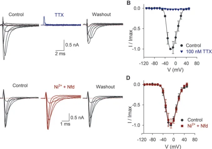
TTX (Fig. 1A-B) and unaltered by the calcium channel antagonists nifedipine (10μM) and
nickel (100μM) (Fig. 1C-D). The average density of the sodium current was 216 ± 13 pA/pF
(n= 97). No correlation between the current amplitude and the cell membrane capacitance was found (R2= 0.01).
Perfusion of DA reduced the INapeak amplitude, an effect that took place during the first
min-ute of its application (Fig. 2A-B). Comparing the values of the current amplitude at−10 mV, DA
significantly decreased the Na+current at 3μM, 10μM and 100μM by 36 ± 12%, 40 ± 9% and
49 ± 5%, respectively, and shifted the V1/2towards more negative potentials. When 10μM of DA
(n= 9) was perfused, the maximum INaamplitude decreased 43 ± 8%,P =0.001 at−20 mV
(Fig. 2C). DA caused a significant 7 mV, 12 mV, 8 mV, 9 mV and 14 mV hyperpolarizing shift in
the V½of the inactivation curve at 1 nM (P= 0.03), 100 nM (P= 0.001), 1μM (P= 0.02), 3μM
(P= 0.01) and 100μM (P<0.001), respectively. With 10μM DA non-significant changes of the
activation or inactivation curves was found (n= 9;P>0.05) (Fig. 2D). The concentration-re-sponse curve of the effect of DA on the INashowed that DA inhibits the INain a concentration
de-pendent manner (Fig. 2E). Data were fitted (solid line) by a dose-response function with an IC50
of 2.5μM.
The effect of 100μM of DA on the peak INaamplitude was blocked by the co-application of
100μM SCH-23390 and 1μM eticlopride (D1- and D2-like antagonists). In control DA
pro-duced an inhibition of the INaof 49 ± 5% (n= 6;P<0.001), and after the coapplication of
G protein and Na
+channel phosphorylation
To determine the participation of G-proteins in the DA action, GDP-β-S (500μM), which is a
non-hydrolyzable GDP analog was used to block G proteins (GDP-β-S was dissolved in the in-tracellular solution,n= 5). In this condition, DA (100μM) perfusion did not produce any
sig-nificant effect on the INaamplitude (current increased 8 ± 5%,P= 0.265), nor significant
changes in the activation or inactivation curves or voltage sensitivity of the INa, indicating that
DA receptor activation implies the activation of a G-protein coupled receptor (Fig. 3).
When the inhibitor of type 1 and 2A protein phosphatases, okadaic acid (100 nM), was per-fused INapeak amplitude decreased 43 ± 2% (n= 5,P<0.001) and shifted the half inactivation
voltage towards more negative potentials (from−70 ± 1 mV to−80 ± 1 mV,P= 0.003) with
not change of the slope. Non-significant changes of the activation curve were found (Fig. 3). Since okadaic acid promotes the phosphorylated state of the sodium channels (assuming that, in the cells, the basal concentration of cAMP activates the PKA that phosphorylate them and PKA autophosphorylation on Thr197site maintain the PKA in active state) the inclusion of 100 nM okadaic acid dissolved in the intracellular solution was tested (n= 5). Okadaic acid re-duced the inhibition prore-duced by 100μM DA from 49 ± 5% under control conditions to 16 ±
7% (P= 0.126). With okadaic acid non-significant changes in the activation or inactivation
Fig 1. Effects of TTX, nickel and nifedipine on the INa.A) INaproduced by voltage pulses from−110 mV to 50 mV in control conditions, during 100 nM TTX
perfusion and 2 min after washout. In this and the following figures, only representative traces are shown. B) Current-voltage relationship in control conditions (black) and with TTX perfusion (blue). C) Current recording in control conditions and after coapplication of 10μM nifedipine (Nfd) and 100μM nickel. D) Current-voltage relationship in control conditions (black) and with Ni2+plus nifedipine perfusion (red). In all traces, the dotted line indicates zero current.
doi:10.1371/journal.pone.0120808.g001
curves or voltage sensitivity of the INawas produced by DA. These indicate that
phosphoryla-tion of the Na+channel is the result of DA receptor activation (Fig. 3).
Functional expression of D1-like receptors
Two D1-like agonists were studied A-68930 and dihydrexidine. The perfusion of the D1-like agonist 300 nM A-68930 (n= 14) decreased the INaamplitude 29 ± 4% (P<0.001) (Fig. 4A-B)
and shifted the V½of the inactivation curve 7 mV leftward (P<0.001), with no change in the
slope of inactivation (Fig. 4C). Experiments using the D1 agonist dihydrexidine (100 nM) shown that it also inhibits INa23 ± 6% (P= 0.009,n= 5;Fig. 4D) and displaced the inactivation
V½10 mV to more negative potentials (from−71 mV in control to-81 mV;P= 0.01).
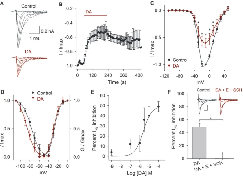
Fig 2. Effects of DA on the INa.A) Representative experiment showing the effect of 10μM DA after control perfusion. B) Temporal course of the inhibitory
action of DA 100μM on the INaamplitude. Bar indicates the DA perfusion. C) Current-voltage relationship in control and with 10μM DA. DA decreased the INa
by 43±8% at−20 mV (P= 0.001;n= 9). D) Activation and inactivation curves in control and after DA application. DA caused a hyperpolarizing shift of the V½
of the inactivation curve of 8 mV at 10μM (P= 0.8). In this and following activation and inactivation curves the data were fitted with a Boltzmann function (solid lines). E) Concentration-response relationship of the effect of DA (1 nM to 100μM), with at leastn= 6 for each point. The data were fitted with a concentration
response curve (solid line) with an IC50of 2.5 x 10⁻⁶M and a Hill coefficient of 1. F) Bar graph shows that a mixture of D1 and D2 antagonists (100μM
SCH-23390 + 1μM eticlopride) completely blocks DA action (P= 0.006). Inset show a representative recording of the SCH-23390 (SCH) and eticlopride (E) actions. Calibration bars 0.2 nA and 1 ms. Asterisks denote a significant effectP<0.05.
Experiments in which the D1 antagonist SCH-23390 (100μM) was perfused shown that
this drug blocked the action of 300 nM A-68930 (n= 5) reducing its effect to a non-significant 12 ± 5% inhibition of the INa(P= 0.073) indicating that agonist action takes place by specific
D1 receptor activation (Fig. 4E-G). Paradoxically, we found that the application of 300 nM of SCH-23390 significantly decreased the INa(between−30 mV and 40 mV) with a maximum
ef-fect of 31 ± 3% (n= 5,P= 0.001). The coapplication of A-68930 with SCH-23390 produced an additional inhibition of 29 ± 3% (P<0.001). With 300 nM of SCH-23390, the V½ of the inac-tivation curve shifted 10 mV leftward (P<0.001), and co-application with 300 nM of A-68930 shifted the inactivation curve an additional 4 mV leftward (P= 0.016) (Data not shown).
Ketanserin, a serotonin receptor antagonist, also behaves as a D1 and D5 receptors antago-nist [26]. In our experiments when ketanserin (1μM) was co-applied with A-68930 300 nM
(n= 5), the agonist no longer produced a significant effect on the INapeak amplitude, the V1/2
of the inactivation curve was shifted from−66 ± 1 mV to−76 ± 1 mV (P= 0.01) without
modi-fication of the activation curve (Data not shown).
Signaling pathway activated by D1-like receptors
To determine the D1 signaling mechanisms two pathways were considered: D1 coupled to Gαs
protein and D1 coupled to Gαqprotein.
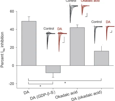
Fig 3. Intracellular mechanism inherent to D1- and D2-like receptors.Bars indicate the percent INa
inhibition produced by 100μM DA and compared with its effect when 500μM GDP-β-S (P<0.001) were added intracellularly. The perfusion of 100 nM okadaic acid decreased the INaamplitude in a similar
percentage as 100μM DA (P= 0.3 unpairedt-test). While 100 nM okadaic acid added intracellularly occluded the DA action (P= 0.01). Insets above bars show representative recordings in control conditions and after drug application. Asterisks denote a significant difference (P<0.05 unpairedt-test). Calibration bars are 1 ms and 0.5 nA for all recordings.
doi:10.1371/journal.pone.0120808.g003
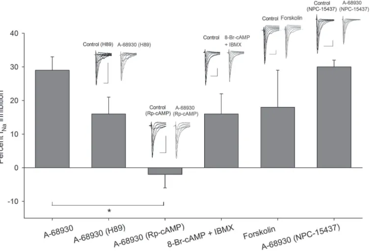
Inclusion of a selective inhibitor of PKA, H89 (1μM), in the intracellular solution (n= 6)
produced a decrease in the effect of 300 nM of A-68930 on the INafrom 29 ± 4% in control
condition to 16 ± 5% (at−10 mV) (P= 0.027). No changes in the V½or the slope of activation
or inactivation curves were found (Fig. 5).
Preincubation of the cells with Rp-cAMP (50μM, during 30–60 min), which is one of the
most specific PKA inhibitors available, completely blocked the action of 300 nM A-68930 on the INa(current increased 2 ± 4%,P= 0.644). No changes in the V½or the slope of activation
or inactivation curves were observed. These results show that PKA participate in the signaling pathway activated by D1-like receptor (Fig. 5).
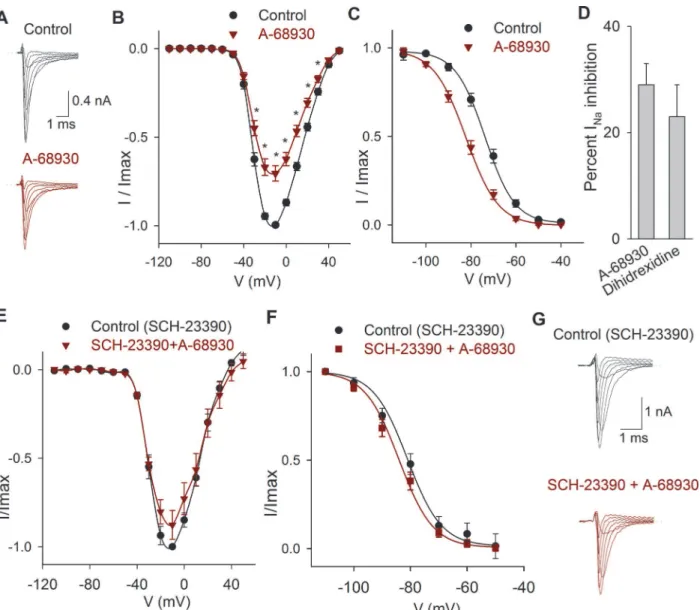
Fig 4. Effects of D1 related drugs on the INa.A) Recordings of the INain control condition and after perfusion of A-68930 (300 nM). B) Current-voltage
relationship of the INaunder control conditions and after 300 nM A-68930. The maximum decrease of the current was 29±4% at−10 mV. C) Steady state
inactivation of the INain control conditions and after 300 nM A-68930, which caused a leftward shift of the inactivation. D). Bar graph comparing the inhibitory
effect of 100 nM dihidrexidine with A-68930 effect. Non-significant difference was found (P= 0.445; unpairedt-test). E) Current-voltage relationship of the INa
in control conditions (with 100μM SCH-23390), and after the co-application of 300 nM A-68930. F) Steady state inactivation of the INain control (with 100μM
SCH-23390) and after A-68930 + SCH-23390, which caused non-significant changes. G) Typical recordings of the INashowing that SCH-23390 significantly
reduced the inhibitory action of 300 nM A-68930.
An experimental series using 8-Br-cAMP (500μM), along with cAMP phosphodiesterase
inhibitor IBMX (100μM), produced a 16 ± 6% (P= 0.04,n= 5) inhibition of the INa, with
non-significant changes in the activation or inactivation curves or voltage sensitivity of the INa
(Fig. 5). Furthermore, to determine the adenylyl cyclase (AC) participation in the pathway
acti-vated by D1-like receptors, forskolin (AC activator) was used. Forskolin (10μM) decreased the
INa18 ± 11% (n= 5:P= 0.04). The V½of the inactivation curve shifted 8 mV leftward (P=
0.02;Fig. 5). Together, these results indicate that intracellular increments of cAMP produce by itself a significant decrease of the INamimicking the D1-like receptor activation.
The use of 500 nM NPC-15437 (selective PKC inhibitor) in the pipette produced non-sig-nificant changes in the inhibitory action of 300 nM A-68930 (n= 8), which decreased the INa
30 ± 2 at−20 mV (P<0.001) (Fig. 5). The V½and slope of the activation curve did not change,
and the V½of the inactivation curve shifted 12 mV leftward (P<0.001) with no change of the
slope (Fig. 5). This result indicates that PKC is not significantly participating in the D1-like signaling pathway.
Fig 5. Transducer mechanisms activated by D1-like receptors.Bars show the effect of A-68930 in control condition in comparison with its action while other drugs were used. The use of H89 in the intracellular solution decreased the inhibitory effect of A-68930 from 29±4% to 16±5% at−10 mV (P= 0.08).
When cells were preincubated with 50μM Rp-cAMP, A-68930 effect was completely blocked (P<0.001). Coapplication of 8-Br-cAMP and IBMX mixture caused a decrease in the INacurrent which mimicked the effect of A-68930 (P= 0.114). Forskolin also mimicked the effect of D1-like agonist decreasing the
INa18±11% (P= 0.27). The NPC-15437 in the intracellular solution did not significantly modify the A-68930 effect (P= 0.92). Insets above bars show typical
recordings of the INaunder control conditions and after drug application. Calibration bars are 2 ms and 0.5 nA for all recordings.
doi:10.1371/journal.pone.0120808.g005
Functional expression of D2-like receptors
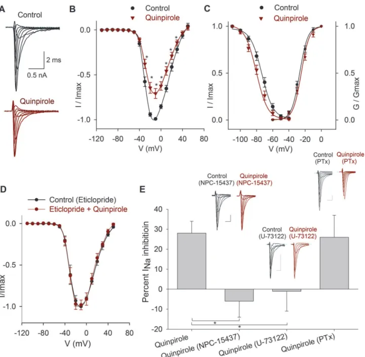
The application of the D2-like receptor agonist quinpirole (1μM) significantly decreased the INa
peak amplitude at all the voltages (n= 10) (Fig. 6A-B). At−10 mV, the INapeak amplitude was
28 ± 6% (P= 0.001). The V½of the inactivation curve was shifted 5 mV leftward (P<0.001),
with no changes in the slope (Fig. 6C). The effects of quinpirole were blocked by 1μM eticlopride
(D2-like receptor antagonist), which reduced quinpirole action to 2 ± 6% (n= 4;P= 0.76,
Fig. 6D).
Signaling pathway activated by D2-like receptors
To study the signaling mechanisms of D2-like receptors two possibilities were considered: D2 receptors coupled to Gαq/PLC/PKC protein or D2 receptors coupled to Gi/oprotein.
Initially, the selective PKC inhibitor NPC-15437 (500 nM) dissolved into the pipette solu-tion was tested when activating the D2 receptors with quinpirole (1μM,n= 5). NPC-15437
produced a significant block of D2 agonist effect whose action becomes under NPC-15437 a non-significant increase of 6 ± 8% of the INa(P= 0.5); thus indicating that D2 receptors are
coupled to a signaling pathway that leads to PKC activation (Fig. 6E). In cells that were prein-cubated for 30–60 min with 10μM U-73122 (PLC blocker), the subsequent perfusion of 1μM
quinpirole produced a non-significant 1 ± 10% increase of the INa(n= 6,P= 0.91) (Fig. 6E).
When cells were pretreated (for 24 hrs) with 400μM of PTx, quinpirole inhibition of the INa
was 26 ± 11% (n= 5,P= 0.027) which is similar to the inhibition of 28 ± 6% produced under control condition (P= 0.001) (Fig. 6E). Together these results indicate that D2-like receptor ac-tivation involves Gαq/PLC/PKC pathway.
Current Clamp Experiments
Current clamp recordings of SGNs were also performed to examine voltage responses to current pulse injection. The cell voltage was fixed at−80 mV, and at more depolarized membrane voltages,
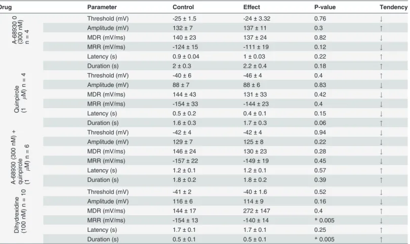
no action potential discharge was produced even by highly depolarizing (>50 mV) current pulse injection. Action potential waveform parameters under control conditions were (n= 24): ampli-tude = 117 ± 5 mV, duration = 1 ± 0.2 ms, latency = 4 ± 1 ms, threshold = -38 ± 2 mV, MDR = 144 ± 11 mV/ms, MRR-150 ± 9 mV/ms. The use of DA agonists in current clamp experiments re-vealed that action potential parameters showed various modifications, summarized inTable 1. DA 100μM decreased the action potential amplitude 10 ± 3%, (n= 4,P= 0.04) with
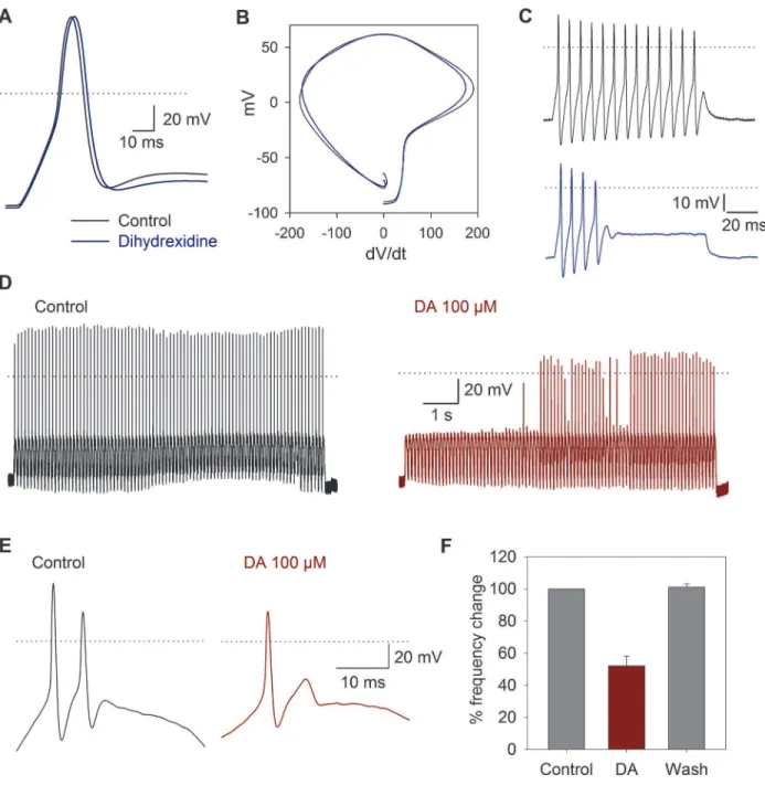
non-signifi-cant changes in other action potential parameters. Under 100 ms current injection, 90% (n= 46) of the cells produced a single action potential when stimulated with suprathreshold depolarizing current pulses. The other 8% slowly adapted producing between 3–7 action potentials, and only 2% of the cells produced sustained, non-adapting, repetitive spiking. In neurons with repetitive spike activity under square current pulse injection, the D1 agonist dihydrexidine (100 nM,n= 10) produced a significant decrease of the MRR from−154 to−140 mV/ms (P= 0.005). The duration
at 50% of the action potential was increased by 7 ± 0.1% (P= 0.005), and in two cells that showed repetitive spike activity, a reversible (44%) reduction in the number of action potentials produced by the current pulse injection was induced (Fig. 7A-C).
To study the dynamics of the action potential discharge suprathreshold sinusoidal current pulses were used. The sinusoidal stimulation showed that application of 100μM DA (n= 7)
Discussion
The SGN consist of 95% type I, and 5% type II cells [27], thence it is 19 times more likely to en-counter a type I cell. Additionally, the capacitance of the cells in our experiments coincided with the value reported for type I cells (9 ± 0.5 pF, n = 380). The type II neurons have lower
Fig 6. Effects of quinpirole on the INa.A) Typical recordings of the INaunder control conditions and after 1μM quinpirole perfusion. B) Current- voltage
relationship of the INain control conditions and after 1μM quinpirole. The maximum inhibition was 28±6% at−10 mV. C) Conductance activation and steady
state inactivation curves of the INain control conditions and after 1μM quinpirole perfusion. Quinpirole caused a 5 mV hyperpolarizing shift in the V½of the
inactivation curve. D) Current-voltage relationship of the INain control condition (with 1μM eticlopride) and after coapplication of 1μM quinpirole. E) Bar graph
of the percent inhibition of the INaby quinpirole in control, with NPC-15437 in the intracellular solution (P= 0.004) and when the cells were preincubated with
U-73122 (P= 0.02) or PTx (P= 0.82). Insets above bars show typical recordings of the INain control and after drug application. Calibration bars 0.5 nA, 1 ms
for all recordings.
doi:10.1371/journal.pone.0120808.g006
capacitanceffi6 pF [28], and they are not innervated by dopaminergic fibers [29]; thus, alto-gether these data support the notion that we were recording essentially from type I neurons.
DA-receptor gene expression studies in the cochlea have shown significant interspecies dif-ferences. Transcripts of the genes for D2 (long) and D3 receptors were found in SGNs from the mouse cochlea (14–18 days old), while those of the D2 (Short) and D4 receptors were not am-plified [12]. In contrast, all DA receptor subtypes were found in rat SGNs [13]. A recent report using immunohistochemistry and RT-PCR in postnatal day 10–13 mice described the expres-sion of D1, D2 and D5 receptors in SGNs and OHC, and D4 receptor was found exclusively in SGN. With no evidence of D3 receptor expression in mice cochlear tissues [14].
Our results show that DA, A-68930 and quinpirole, cause a decrease of the INain SGNs.
The D2-like antagonist eticlopride blocks the effect of quinpirole, indicating the specific action of quinpirole and the functional expression of D2-like receptors. The D1-like antagonist SCH-23390 blocks the action of A-68930, indicating the functional expression of D1-like receptors in the spiral ganglion neurons. The use of ketanserin shows that it blocks the A-68930 action on the INaamplitude, thus behaving as a D1-like receptor antagonist. It is worth noting that
SCH-23390 and eticlopride have an inhibitory effect on the INaby itself. A similar paradoxical
effect of these antagonists was observed in the auditory activity of guinea pig [19]. The intrinsic inhibitory action of the antagonist may be produced because DA receptors may have constitu-tive activity similar to that found in histamine receptors [30] and in D1/D2 receptor chimeras [31]. There is also a group of D1-like receptors that are insensitive to the antagonism of
SCH-Table 1. Action potential parameters in control conditions and after drug use.
Drug Parameter Control Effect P-value Tendency
A-68 930 0 (30 0 nM) n= 4
Threshold (mV) -25±1.5 -24±3.32 0.76 #
Amplitude (mV) 132±7 137±11 0.3 "
MDR (mV/ms) 140±23 137±24 0.82 #
MRR (mV/ms) -124±15 -111±19 0.12 #
Latency (s) 0.9±0.04 1±0.03 0.22 "
Duration (s) 2±0.3 2.2±0.4 0.18 "
Quinpi role (1 m M) n =
4 Threshold (mV) -40±6 -46±4 0.4 "
Amplitude (mV) 88±7 88±6 0.83 #
MDR (mV/ms) 144±43 131±33 0.42 #
MRR (mV/ms) -154±33 -144±23 0.4 #
Latency (s) 0.5±0.2 0.4±0.1 0.15 #
Duration (s) 1.6±0.3 1.7±0.3 0.06 "
A-689 30 (30 0 nM) + quinpi role (1 m M) n = 6
Threshold (mV) -42±4 -42±4 0.94 #
Amplitude (mV) 129±7 125±8 0.22 #
MDR (mV/ms) 146±24 130±23 0.28 #
MRR (mV/ms) -157±22 -149±19 0.45 #
Latency (s) 1.2±0.1 1.2±0.1 0.57 "
Duration (s) 1.8±0.2 1.8±0.2 0.39 "
Dihydr exidi ne (100 nM ) n =
10 Threshold (mV) -41±2 -40±1.6 0.52 #
Amplitude (mV) 116±6 114±9 0.16 #
MDR (mV/ms) 144±17 272±147 0.4 "
MRR (mV/ms) -154±13 -140±14 *0.005 #
Latency (s) 1.7±0.1 1.7±0.1 0.25 "
Duration (s) 0.5±0.1 0.5±0.1 *0.005 "
23390 [32]. The inhibitory effect of SCH-23390 could also be due to an unspecific interaction with other non-dopaminergic receptors or due to a direct effect on ionic channels.
To determine the intracellular pathways participating in the dopaminergic action in the SGN, we employed a pharmacological approach. Use of GDP-β-S decreased the DA effect, in-dicating that DA receptor actions are mediated by a G protein. In most cell types in which a G protein modulation of the INahas been described, the most common mechanism was a
Fig 7. Current clamp response to sinusoidal stimulation and DA receptor activation.A) The use of 100 nM dihydrexidine (blue traces) shifted the maximum repolarization rate and increased the action potential duration. B) Phase plane plot of action potentials under control conditions and after dihydrexidine application. C) Action potentials produced by a current pulse injection of 150 pA were reduced from 13 under control conditions to 4 after dihydrexidine (100 nM). D) Typical response to sinusoidal current injection (10 Hz, 150 pA). Before the stimuli, the cells were held at−80 mV. E) In a cell
discharging in a 2:1 phase lock to sinusoidal stimuli, the use of DA reduced action potential discharges per cycle to a phase lock of 1:1 F) Bar graph showing the percent change in the number of action potentials in the control, after 100μM DA application and after washout of the drug.
doi:10.1371/journal.pone.0120808.g007
reduction of the current amplitude due to a decrease in the Na+channel availability [33], al-though also a negative adjustment of the voltage dependence of rapid inactivation has been de-scribed [33]. In our experiments, both mechanisms seem to be occurring since we found current amplitude reduction and modifications of the inactivation V½with most of the drugs
tested. Considering that okadaic acid reproduces and occludes the DA effect, we concluded that Na+channel phosphorylation and subsequent modification of the current amplitude is es-sential for the DA action.
The D1-like and D2-like receptors act through different pathways. In our system, blocking PKA with H89 or Rp-cAMP significantly reduced the action of the D1-like agonist, indicating that PKA participates in its signal transduction. Previous studies using H89 and forskolin have also shown that the dopaminergic modulation of the CAP in the auditory system implies the PKA participation [23].
Our results show that increasing the cAMP levels (using a cAMP analog plus a phosphodi-esterase inhibitor or an AC activator) mimics the DA effect. Thus indicating that increase in the cAMP secondary to adenylyl cyclase activation leads to PKA activation. Together these re-sults showed that D1-like dopamine receptors activate a Gαs/AC/cAMP/PKA pathway (Fig. 8).
However, we cannot rule out the involvement of additional signaling pathways. The D1-like re-ceptors have been shown to be coupled to three different signaling mechanisms: SCH-23390 sensitive Gαs/olf/AC/PKA, adenosine A2A receptor-dependent Gαs/olf/AC/PKA and Gαq/PLC
[32]. In our experiments no significant effect was produced by PKC inhibition on D1-like re-ceptor actions, indicating that Gαq protein seems not to participate in the SGNs D1 receptors response. In neostriatal neurons, even a marginal activation of PKC potentiates the effects of PKA [32], and in striato-nigral neurons (P25 rats), D1-like receptors decrease the INavia PKA,
but PKC is also involved in the INamodulation [34].
In addition, protein-protein interactions of the carboxy terminal intracellular sites of the D1 receptor with N-methyl-D-aspartic acid (NMDA) receptor subunits [35,36], and GABA-A in-teractions with the D5 receptor [37] have been described. The D1 and D2 heteromeric recep-tors may interact with A1 and A2A adenosine receprecep-tors [38]. Heterodimers of metabotropic glutamate receptors mGlu(5) and D2 receptors and oligomeric receptors containing more than two protomers, including mGlu (5), D2 and A2A adenosine receptors, are found in rat striatum homogenates [39]. Also heteromers of D1 with type 3 histamine receptor (H3) have been shown in transfected mammalian cells and confirmed by bioluminescence resonance energy transfer (BRET) and binding assays [40]. Thus a complex pharmacology of the D1 agonists and antagonists is not an unexpected phenomena given the complexity of receptor interactions with other membrane proteins.
The D2-like receptors may be coupled to Gαi/oor Gαqproteins. In striatal medium spiny
neurons and cholinergic interneurons, the activation of D2 receptors leads to a potent decrease of the INathrough a PKC-dependent mechanism [41]. In our system blockade of PKC
elimi-nates the effect of the D2-like receptor activation. Additionally, block of Phospholipase C (PLC); which inhibits the hydrolysis of phosphatidylinositol 4,5-bisphosphate (PIP2) to inositol
trisphosphate (IP3) and diacyl-glycerol (DAG), decreasing the cytosolic free calcium necessary
for PKC activation, also produced a complete block of the effects due to D2-like receptor acti-vation (Fig. 8). Since no significant effect of PTx was found, we concluded that in our system, the Gαi/odid not to have a role in reference to the INacurrent modulation and most probably
D2-like signaling pathway is through Gαq/PLC/PKC pathway activation.
The activation of D1- or D2-like receptors exerts similar action on the INain SGNs. It is
different from those of their constituent receptors [42,43,44]. In striatal medium spiny neu-rons, the D1 and D2 receptors are segregated into discrete populations, but there is increasing functional and anatomical evidence that they may be co-expressed in a subpopulation of neu-rons. A new complex receptor in the striatum, the D1-D2 heteromeric receptor, with unique functional features would constitute a third neuronal population with physiological relevance [45].
In the inner ear, the complexity of the sound stimuli make it important to have a finely tuned system capable of segregating the features of each sound. Such process requires very pre-cise differential modulation of the afferent neuron gain. In the cochlear efferent system, preci-sion seems to be obtained thanks to the participation of various neurotransmitters including ACh, GABA, CGRP, enkephalins and DA, among others. We propose that the function of the release of multiple neurotransmitters from the efferent terminals and the expression of their re-ceptors in SGNs is that all neurotransmitters act in a synergistic form, carrying specific func-tions to establish their final integrated effects
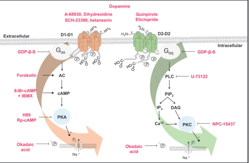
Fig 8. Scheme of the signaling pathways activated by D1- and D2-like receptors in the spiral ganglion neurons.Receptors are shown as
homodimers. Phosphorylation and glycosylation sites are indicated (P and Y). The drugs used in this work are shown in red. Lines with transverse-endings indicate blockade and those with circle-endings stimulation. D1-like receptors are coupled to a Gαsprotein leading to AC stimulation, thus increasing cAMP
levels and subsequent PKA activation. PKA phosphorylates the Na+channels thus decreasing the INa. D2-like receptors are coupled to a Gαqprotein whose
activation stimulates the PLC, which cleaves PIP2into IP3and DAG, the IP3increases the Ca2+concentration, and both Ca2+and DAG activates PKC
leading also to a Na+channel phosphorylation thus decreasing the I
Na. In both cases, phosphorylation was prevented by okadaic acid.
doi:10.1371/journal.pone.0120808.g008
We found that under current pulse stimulation most of the SGN adapted quickly and dis-charged a single action potential. These results coincide with those described by other authors [46,47]. Applying DA to cells stimulated with sinusoidal current injection allowed us to show the inhibitory action of DA in the action potential discharge of SGNs. The use of dihydrexidine in cells with repetitive discharge showed that there is an increase in the duration of the action potential in the cells that discharged throughout the pulse. This change in the AP duration is reflected by a change in the number of action potentials throughout the current pulse. These results support the notion that DA released from the olivocochlear efferent neurons depresses the discharge of the afferent neurons, constituting a protective mechanism in conditions of is-chemia or acoustic overstimulation, which may lead to an excitotoxic damage from excess glu-tamate release and sustained action potential discharge [48,49]. The DA neurotransmission enhancer rasagiline (a monoamine oxidase type B inhibitor) has been approved as an otopro-tectant, showing that DA release from lateral olivocochlear fibers exert a protective action against excitotoxicity, a pathological factor in the aminoglycoside-induced sensorineural hear-ing loss [50]. Also pramipexole (a D2/D3 receptor agonist) is an effective agent against subjec-tive tinnitus, action that may involve both central and peripheral targets in the auditory system [51].
In the SGNs, INahas a fundamental role in the spike generation [52]. Previous works have
shown that DA decreases spike frequency of afferent neurons only in cases in which they are overstimulated by glutamate [22]. This is analogous with what we found. Because in isolated SGNs dopamine exerted a clear cut inhibitory effect only in those cells that were subjected to a continuous activation by sinusoidal stimuli. In cells with very low or no activity the mild effects seen in AP waveform parameters should be due to the fact that INaavailable for an AP
genera-tion is exceeding causing that AP generagenera-tion has a large security factor. Thus, modulatory changes of the INacould not block action potential generation but modulate the discharge rate.
Although DA uptake block has been shown to reduce spontaneous activity and sound-evoked compound action potential of the auditory nerve in a dose-dependent manner [53]. We found that the DA receptor activation may produce a fine adjustment of afferent neuron gain, without producing a complete block of the afferent input, thus constituting a functionally secure and ef-ficient mechanism for gain control of the cochlear afferent activity.
Acknowledgments
Proof reading of the English manuscript was performed by Nature Publishing Group Language Editing.
Author Contributions
Conceived and designed the experiments: CVB ES RV. Performed the experiments: CVB. Ana-lyzed the data: CVB ES RV. Contributed reagents/materials/analysis tools: CVB ES RV. Wrote the paper: CVB ES RV. Supervised the project: RV ES.
References
1. Pujol R. Lateral and medial efferents: a double neurochemical mechanism to protect and regulate inner and outer hair cell function in the cochlea. Br J Audiol. 1994; 28: 185–191. PMID:7735146
2. Warr WB, Guinan JJ Jr. Efferent innervation of the organ of corti: two separate systems. Brain Res. 1979; 173: 152–155. PMID:487078
3. Altschuler RA, Kachar B, Rubio JA, Parakkal MH, Fex J. Immunocytochemical localization of choline acetyltransferase-like immunoreactivity in the guinea pig cochlea. Brain Res. 1985; 338: 1–11. PMID:
4. Eybalin M, Pujol R. Immunofluorescence with Met-enkephalin and Leu-enkephalin antibodies in the guinea pig cochlea. Hear Res. 1984; 13: 135–140. PMID:6370942
5. Lu SM, Schweitzer L, Cant NB, Dawbarn D. Immunoreactivity to calcitonin gene-related peptide in the superior olivary complex and cochlea of cat and rat. Hear Res. 1987; 31: 137–46. PMID:3502259
6. Eybalin M, Parnaud C, Geffard M, Pujol R. Immunoelectron microscopy identifies several types of GABA-containing efferent synapses in the guinea-pig organ of Corti. Neuroscience. 1988; 24: 29–38. PMID:3285238
7. Drescher MJ, Drescher DG, Khan KM, Hatfield JS, Ramakrishnan NA, Abu-Hamdan MD, et al. Pituitary adenylyl cyclase-activating polypeptide (PACAP) and its receptor (PAC1-R) are positioned to modulate afferent signaling in the cochlea. Neuroscience. 2006; 142: 139–164. PMID:16876955
8. Altschuler RA, Hoffman DW, Wenthold RJ. Neurotransmitters of the cochlea and cochlear nucleus: immunocytochemical evidence. Am J Otolaryngol. 1986; 7: 100–106. PMID:2870655
9. Guinan JJ Jr. Cochlear efferent innervation and function. Curr Opin Otolaryngol Head Neck Surg. 2010; 18: 447–453. doi:10.1097/MOO.0b013e32833e05d6PMID:20717032
10. Sliwinska-Kowalska M, Parakkal M, Schneider ME, Fex J. CGRP-like immunoreactivity in the guinea pig organ of Corti: a light and electron microscopy study. Hear Res. 1989; 42: 83–95. PMID:2584160
11. Bartolomé MV, Gil-Loyzaga P. Serotonergic innervation of the inner ear: is it involved in the general physiological control of the auditory receptor?. Int Tinnitus J. 2005; 11: 119–125. PMID:16639911
12. Karadaghy AA, Lasak JM, Chomchai JS, Khan KM, Drescher MJ, Drescher DG. Quantitative analysis of DA receptor messages in the mouse cochlea. Brain Res. 1997; 44: 151–156.
13. Inoue T, Matsubara A, Maruya S, Yamamoto Y, Namba A, Sasaki A, et al. Localization of dopamine re-ceptor subtypes in the rat spiral ganglion. Neurosci Lett. 2006; 399: 226–229. PMID:16490310
14. Maison SF,Liu XP, Eatock RA, Sibley DR, Grandy DK, Liberman MC. Dopaminergic signaling in the co-chlea: receptor expression patterns and deletion phenotypes. J Neurosci. 2012; 32: 344–355. doi:10. 1523/JNEUROSCI.4720-11.2012PMID:22219295
15. Missale C, Nash SR, Robinson SW, Jaber M, Garon MG. Dopamine receptors: from structure to func-tion. Physiol Rev. 1998; 78: 189–225. PMID:9457173
16. Niu X, Bogdanovic N, Canlon B. The distribution and the modulation of tyrosine hydroxylase immunore-activity in the lateral olivocochlear system of the guinea-pig. Neuroscience. 2004; 125: 725–733. PMID:
15099686
17. Gil-Loyzaga P, Parés-Herbute N. HPLC detection of dopamine and noradrenaline in the cochlea of adult and developing rats. Dev Brain Res. 1989; 48: 157–160. PMID:2752573
18. Niu X, Tahera Y, Canlon B. Environmental enrichment to sound activates dopaminergic pathways in the auditory system. Physiol Behav. 2007; 92: 34–39. PMID:17631367
19. Ruel J, Nouvian R, Gervais d’Aldin C, Pujol R, Eybalin M, Puel JL. Dopamine inhibition of auditory nerve activity in the adult mammalian cochlea. Eur J Neurosci. 2001; 14: 977–986. PMID:11595036
20. Garrett AR, Robertson D, Sellick PM, Mulders WH. The actions of dopamine receptors in the guinea pig cochlea. Audiol Neurootol. 2010; 16: 145–157. doi:10.1159/000316674PMID:20668375
21. Sun W, Salvi RJ. Dopamine modulates sodium currents in cochlear spiral ganglion neurons. Neurore-port. 2001; 26: 803–807.
22. Oestreicher E, Arnold W, Ehrenberger K, Felix D. Dopamine regulates the glutamatergic inner hair cell activity in guinea pigs. Hear Res. 1997; 107: 46–52. PMID:9165346
23. Niu X,Canlon B. The signal transduction pathway for the dopamine D1 receptor in the guinea-pig co-chlea. Neuroscience. 2006; 137: 981–990. PMID:16330149
24. Limón A, Pérez C, Vega R, Soto E. Ca2+-activated K+current density is correlated with soma size in rat vestibular-afferent neurons in culture. J Neurophysiol. 2005; 94: 3751–3761. PMID:16107534
25. Bean BP. The action potential in mammalian central neurons. Nat Rev Neurosci. 2007; 8: 451–465. PMID:17514198
26. Sunahara RK, Guan HC, O'Dowd BF, Seeman P, Laurier LG, Ng G, et al. Cloning of the gene for a human dopamine D5 receptor with higher affinity for dopamine than D1. Nature. 1991; 350: 614–619 PMID:1826762
27. Spoendlin H. Anatomy of cochlear innervation. Am J Otolaryngol. 1985; 6: 453–467. PMID:3909832
28. Szabó ZS, Harasztosi CS, Sziclai I, Szucs G, Rusznak Z. Ionic currents determining the membrane characteristics of type I spiral ganglion neurons of the guinea pigs. J Neurosci. 2002; 16: 1887–1895.
29. Darrow KN, Simons EJ, Dodds L, Liberman MC. Dopaminergic innervations of the mouse inner ear: ev-idence for a separate cytochemical group of cochlear efferent fibers. J Comp Neurol. 2006; 498: 403– 414. PMID:16871528
30. Arrang JM, Morisset S, Gbahou F. Constitutive activity of the histamine H3 receptor. Trends Pharmacol Sci. 2007; 28: 350–357. PMID:17573125
31. Kozell LB, Neve KA. Constitutive activity of a chimeric D2/D1 dopamine receptor. Mol Pharmacol. 1997; 52: 1137–1149. PMID:9396784
32. Kuroiwa M, Bateup HS, Shuto T, Higashi H, Tanaka M, Nishi A. Regulation of DARPP-32 phosphoryla-tion by three distinct dopamine D1-like receptor signaling pathways in the neostriatum. J Neurochem. 2008; 107: 1014–1026. doi:10.1111/j.1471-4159.2008.05702.xPMID:18823371
33. Carr DB, Day M, Cantrell AR, Held J, Scheuer T, Catterall WA, et al. Transmitter modulation of slow, ac-tivity-dependent alterations in sodium channel availability endows neurons with a novel form of cellular plasticity. Neuron. 2003; 39: 793–806. PMID:12948446
34. Cantrell AR, Scheuer T, Catterall WA. Voltage-dependent neuromodulation of Na+channels by D1-like
dopamine receptors in rat hippocampal neurons. J Neurosci. 1999; 19: 5301–5310. PMID:10377341
35. Lee FJ, Xue S, Pei L, Vukusic B, Chéry N, Wang Y, et al. Dual regulation of NMDA receptor functions by direct protein-protein interactions with the dopamine D1 receptor. Cell. 2002; 111: 219–230. PMID:
12408866
36. Pei L, Lee FJ, Moszczynska A, Vukusic B, Liu F. Regulation of dopamine D1 receptor function by physi-cal interaction with the NMDA receptors. J Neurosci. 2004; 24: 1149–1158. PMID:14762133
37. Liu F, Wan Q, Pristupa ZB, Yu XM, Wang YT, Niznik HB. Direct protein-protein coupling enables cross-talk between dopamine D5 and gamma-aminobutyric acid A receptors. Nature. 2000; 403: 274–280. PMID:10659839
38. Franco R, Lluis C, Canela EI, Mallol J, Agnati L, Casadó V, et al. Receptor-receptor interactions involv-ing adenosine A1 or dopamine D1 receptors and accessory proteins. J Neural Transm. 2007; 114: 93– 104. PMID:17024327
39. Cabello N, Gandía J, Bertarelli DC, Watanabe M, Lluís C, Franco R, et al. Metabotropic glutamate type 5, dopamine D2 and adenosine A2a receptors form higher-order oligomers in living cells. J Neurochem. 2009; 109: 1497–1507. doi:10.1111/j.1471-4159.2009.06078.xPMID:19344374
40. Ferrada C, Moreno E, Casadó V, Bongers G, Cortés A, Mallol J, et al. Marked changes in signal trans-duction upon heteromerization of dopamine D1 and histamine H3 receptors. Br J Pharmacol. 2009; 157: 64–75. doi:10.1111/j.1476-5381.2009.00152.xPMID:19413572
41. Maurice N, Mercer J, Chan CS, Hernandez-Lopez S, Held J, Tkatch T, et al. D2 dopamine receptor-me-diated modulation of voltage-dependent Na+channels reduces autonomous activity in striatal
choliner-gic interneurons. J Neurosci. 2004; 24: 10289–10301. PMID:15548642
42. Lee FJ, Liu F. Direct interactions between NMDA and D1 receptors: a tale of tails. Biochem Soc Trans. 2004; 32: 1032–1036. PMID:15506956
43. Rashid AJ, So CH, Kong MM, Furtak T, El-Ghundi M, Cheng R, et al. D1-D2 dopamine receptor hetero-oligomers with unique pharmacology are coupled to rapid activation of Gq/11 in the striatum. Proc Natl Acad Sci U S A. 2007; 104: 654–659. PMID:17194762
44. Verma V, Hasbi A, O'Dowd BF, George SR. Dopamine D1-D2 Receptor Heteromer-mediated Calcium Release Is Desensitized by D1 Receptor Occupancy with or without Signal Activation: Dual functional regulation by G-protein-coupled receptor kinase 2. J Biol Chem. 2010; 285: 35092–35103. doi:10. 1074/jbc.M109.088625PMID:20807772
45. Perreault ML, Hasbi A, O'Dowd BF, George SR. The dopamine d1-d2 receptor heteromer in striatal me-dium spiny neurons: evidence for a third distinct neuronal pathway in Basal Ganglia. Front Neuroanat. 2011; 5: 31. doi:10.3389/fnana.2011.00031PMID:21747759
46. Mo ZL, Davis RL. Heterogeneous voltage dependence of inward rectifier currents in spiral ganglion neurons. J Neurophysiol. 1997; 78: 3019–3027. PMID:9405521
47. Adamson CL, Reid MA, Davis RL. Opposite actions of brain-derived neurotrophic factor and neurotro-phin-3 on firing features and ion channel composition of murine spiral ganglion neurons. J Neurosci. 2002; 22: 1385–1396. PMID:11850465
48. d'Aldin C, Puel JL, Leducq R, Crambes O, Eybalin M, Pujol R. Effects of a dopaminergic agonist in the guinea pig cochlea. Hear Res. 1995; 90: 202–211. PMID:8974998
49. Gil-Loyzaga P, Vicente-Torres MA, Fernández-Mateos P, Arce A, Esquifino A. Piribedil affects dopa-mine turnover in cochlea stimulated by white noise. Hear Res. 1994; 79: 178–182. PMID:7806480
50. Polony G, Humli V, AndóR, Aller M, Horváth T, Harnos A, et al. Protective effect of rasagiline in amino-glycoside ototoxicity. Neuroscience. 2014; 265: 263–273. doi:10.1016/j.neuroscience.2014.01.057
PMID:24508748
51. Sziklai I, Szilvássy J, Szilvássy Z. Tinnitus control by dopamine agonist pramipexole in presbycusis pa-tients: A randomized, placebo-controlled, double-blind study. Laryngoscope. 2011; 121: 888–893. doi:
52. Hossain WA, Antic SD, Yang Y, Rasband MN, Morest DK. Where is the spike generator of the cochlear nerve? Voltage-gated sodium channels in the mouse cochlea. J Neurosci. 2005; 25: 6857–6868. PMID:16033895
53. Ruel J, Wang J, Demêmes D, Gobaille S, Puel JL, Rebillard G. Dopamine transporter is essential for the maintenance of spontaneous activity of auditory nerve neurones and their responsiveness to sound stimulation. J Neurochem. 2006; 97: 190–200. PMID:16524378