Nuclear Receptor DHR4 Controls the Timing of Steroid
Hormone Pulses During
Drosophila
Development
Qiuxiang Ou, Adam Magico, Kirst King-Jones*
Department of Biological Sciences, University of Alberta, Edmonton, Alberta, Canada
Abstract
In insects, precisely timed periodic pulses of the molting hormone ecdysone control major developmental transitions such as molts and metamorphosis. The synthesis and release of ecdysone, a steroid hormone, is itself controlled by PTTH (prothoracicotopic hormone). PTTH transcript levels oscillate with an 8 h rhythm, but its significance regarding the timing of ecdysone pulses is unclear. PTTH acts on its target tissue, the prothoracic gland (PG), by activating the Ras/Raf/ERK pathway through its receptor Torso, however direct targets of this pathway have yet to be identified. Here, we demonstrate that
Drosophila Hormone Receptor 4(DHR4), a nuclear receptor, is a key target of the PTTH pathway and establishes temporal boundaries by terminating ecdysone pulses. Specifically, we show that DHR4 oscillates between the nucleus and cytoplasm of PG cells, and that the protein is absent from PG nuclei at developmental times when low titer ecdysone pulses occur. This oscillatory behavior is blocked whenPTTHortorsofunction is abolished, resulting in nuclear accumulation of DHR4, while hyperactivating the PTTH pathway results in cytoplasmic retention of the protein. Increasing DHR4 levels in the PG can delay or arrest development. In contrast, reducingDHR4function in the PG triggers accelerated development, which is caused by precocious ecdysone signaling due to a failure to repress ecdysone pulses. Finally, we show that DHR4 negatively regulates the expression of a hitherto uncharacterized cytochrome P450 gene,Cyp6t3. Disruption ofCyp6t3function causes low ecdysteroid titers and results in heterochronic phenotypes and molting defects, indicating a novel role in the ecdysone biosynthesis pathway. We propose a model whereby nuclear DHR4 controls the duration of ecdysone pulses by negatively regulating ecdysone biosynthesis through repression ofCyp6t3, and that this repressive function is temporarily overturned via the PTTH pathway by removing DHR4 from the nuclear compartment.
Citation:Ou Q, Magico A, King-Jones K (2011) Nuclear Receptor DHR4 Controls the Timing of Steroid Hormone Pulses DuringDrosophilaDevelopment. PLoS Biol 9(9): e1001160. doi:10.1371/journal.pbio.1001160
Academic Editor:David S. Schneider, Stanford University, United States of America
ReceivedDecember 20, 2010;AcceptedAugust 15, 2011;PublishedSeptember 27, 2011
Copyright:ß2011 Ou et al. This is an open-access article distributed under the terms of the Creative Commons Attribution License, which permits unrestricted use, distribution, and reproduction in any medium, provided the original author and source are credited.
Funding:This work was supported by a Discovery Grant from the Natural Sciences and Engineering Research Council of Canada (NSERC,#341543-07). KKJ is supported by New Investigator Awards from Alberta Innovates (formerly AHFMR) as well as the Canadian Institutes of Health Research (CIHR). Alberta Innovates/ AHFMR (Independent Investigator Salary Award) CIHR (New Investigator Salary Award). The funders had no role in study design, data collection and analysis, decision to publish, or preparation of the manuscript.
Competing Interests:The authors have declared that no competing interests exist.
Abbreviations:20E, 20-Hydroxyecdysone; 7dC, 7-dehydrocholesterol;DHR4,Drosophila Hormone Receptor 4;dib,disembodied; E, ecdysone; L1, first instar; L2, second instar; L3, third instar; PG, prothoracic gland;phm,phantom; PTTH, prothoracicotopic hormone; qPCR, quantitative real-time PCR; RG, ring gland; RNAi, RNA interference;sad,shadow;shd,shade
* E-mail: [email protected]
Introduction
The development of higher organisms is fundamentally dependent on the precise progression of specific gene programs, and even minor differences in the timing of these events can be fatal [1,2]. The regulation of simultaneous developmental programs generally relies on systemic signals—typically hor-mones—that coordinate these activities. In most, if not all multicellular organisms, steroid hormones function as precisely timed cues that control diverse gene programs to advance development or synchronize physiological changes. In humans, for example, the onset of puberty—while ultimately under neuroendocrine control—is governed by the action of steroid hormones that coordinate the developmental and behavioral changes associated with sexual maturation [3].
Typically, the release of steroid hormones from their respective glands is temporally controlled, resulting in systemic pulses of defined duration [4]. This raises the interesting question as to how onset, size, and duration of hormone pulses are regulated, since all these variables will affect target tissue responses. The insect
molting hormone ecdysone represents an excellent model to address these questions, allowing us to study the dynamic effects of a steroid hormone in the context of a developing organism. In
survive metamorphosis and it commits the animal to undergo puparium formation. Once critical weight is attained, the larva will pupariate within normal time, regardless of whether nutrients are scarce or abundant [12–14].
The accurate timing of the major and minor ecdysone pulses suggests that the molecular mechanisms by which the hormone is synthesized, released, and degraded are tightly regulated. The past decade has provided considerable insight into the biosynthetic pathway that converts dietary cholesterol to 20E. Six genes linked to the Halloween mutations encode two different enzyme classes that act in the ecdysone/20E biosynthetic pathway, the cyto-chrome P450 monooxygenases (disembodied,phantom,shadow,shade, and spook) [15–20], and a short-chain dehydrogenase/reductase (shroud) [21]. Two non-Halloween genes have been shown to also play a role in ecdysone synthesis,neverland, which encodes a Rieske electron oxygenase [22,23] and spookier, a paralog of spook [24]. The first and final biochemical steps of ecdysteroid biosynthesis are reasonably well understood, however relatively little is known about the enzymes that catalyze the intermediate steps, namely the conversion from 7-dehydrocholesterol to 5b-ketodiol. This suc-cession of uncharacterized reactions is generally referred to as the ‘‘Black Box’’ and is believed to harbor the rate-limiting step for the production of ecdysone [25]. Recent work suggests thatshroudand
spookier (spook in Manduca and Bombyx) act in the ‘‘Black Box’’ [21,24], however it is still unknown which enzymatic steps are catalyzed by these enzymes, and whether other enzymes also act in the Black Box.
Some Halloween genes are transcriptionally upregulated when ecdysone levels are high; inBombyxandManduca, for example, the expression levels of phantom(phm) mRNA correlate well with the two ecdysone peaks preceding pupation [17,20,26]. InDrosophila,
phm, shadow(sad), andshade(shd) are induced roughly 12 h before puparium formation, concurrent with the major pulse of ecdysone that triggers this event. However, transcript and protein levels of these three Halloween genes appear to be relatively constant during the first 36 h of the L3 [27], suggesting that the three low-titer 20E pulses are not simply a consequence of modulating gene expression of the Halloween genes. Therefore, it is unclear
whether the generation of minor ecdysone pulses involves transcriptional control of any of the known or hitherto unidentified Halloween genes or whether regulation at the transcriptional level plays a role at all. In this study, we show that DHR4 appears to act as a transcriptional repressor ofCyp6t3, a cytochrome P450 gene with a previously unknown role in ecdysteroid biosynthetic pathway.
The neuropeptide PTTH is believed to control the timing of all major ecdysone peaks during larval and pupal development [28], however it is unclear whether low-titer ecdysone pulses are also controlled in this manner. Seminal studies conducted inBombyx
resulted in the identification of PTTH as a brain-derived neuropeptide, which triggers the production of ecdysone in the PG [29], however the gene encoding PTTH was only recently identified inDrosophila[30]. PTTH is sufficient to upregulate some ecdysteroidogenic genes in cultured Bombyx PGs [31,32], and ablation of PTTH-producing neurons in Drosophila results in reduced expression of Halloween genes [30]. Surprisingly, ablating PTTH-producing neurons did not abrogate molting or metamor-phosis, but instead caused substantial developmental delays with long larval feeding periods that resulted in large animals. This suggests that PTTH acts as a timer that coordinates key developmental transitions and final body size of the developing
Drosophila larva, but that it is not essential for molting and metamorphosis per se. Interestingly, the same report demonstrated that PTTH mRNA displays an unusual cyclic pattern during the L3, with transcript levels peaking every 8 h. Whether this unexpected transcriptional profile translates into corresponding changes of PTTH peptide levels is unknown, but it is plausible that these PTTH mRNA oscillations are causally linked to the minor ecdysone pulses that occur during the L3 inDrosophila.
A recent report showed that Drosophila PTTH binds to the Receptor Tyrosine Kinase encoded by thetorsogene [33]. Torso expression is highly specific to the PG and disruption of torso
function via tissue-specific RNA interference (RNAi) phenocopies the PTTH ablation experiments. Importantly, the authors also demonstrate that PTTH stimulates ecdysone production through ERK/MAPK, Ras, and Raf, and that loss-of-function of these pathway components (ERK, Ras85D, and dRaf) via RNAi yield large, delayed animals comparable to those seen in the PTTH ablation or torso RNAi experiments. Conversely, when a constitutively active form of Ras (RasV12) [34] was expressed specifically in the PG, larval development was accelerated, resulting in small, precocious pupae.
We previously described a strong hypomorphic mutation in the
DHR4gene (DHR41) that results in prepupal lethality and defects in developmental timing [35]. DHR41 mutants spend less time feeding than controls and exhibit precocious wandering behavior as well as premature pupariation, ultimately resulting in smaller body sizes. DHR4 encodes an orphan nuclear receptor most similar to the vertebrate receptor Germ Cell Nuclear Factor [36,37]. In our original report [35], we found that DHR4 is highly abundant in the cytoplasm of PG cells in mid and late L3 larvae, with little or no detectable protein in the nucleus. We found no DHR4 expression in the two neighboring endocrine tissues of the ring gland (RG), thecorpus allatum, or thecorpora cardiaca. We show here that nuclear import of DHR4 is developmentally regulated, and that the protein exhibits a precise nucleocytoplasmic oscillatory pattern in L3 larvae. We also provide evidence that this oscillatory behavior is controlled by PTTH signaling and that DHR4 counteracts the stimulating activity of this neuropeptide by repressing ecdysone pulses. Furthermore, we demonstrate that the accelerated switch from feeding to wandering behavior is consistent with a precocious rise in ecdysone concentrations due
Author Summary
to the loss of this repressive function whenDHR4 is mutated or knocked down. Based on RG-specific microarrays, we show that
Cyp6t3is a cytochrome P450 gene specifically expressed in the RG, and that the gene is normally repressed by DHR4. Further, we demonstrate thatCyp6t3plays a key role in ecdysone biosynthesis and that disruption of its function results in low ecdysone titers as well as developmental timing and molting phenotypes. These phenotypes can be rescued by providing an ecdysteroid precursor, 5b-ketodiol, as well as with feeding 20E or ecdysone. We propose a model by which DHR4 inhibits ecdysone synthesis through the regulation of cytochrome P450 genes, and whereby PTTH activity triggers the translocation of DHR4 from the nucleus to the cytoplasm to temporarily relieve this inhibition, thus allowing for ecdysone pulses to occur.
Results
DHR41 Mutants Display a Range of Growth Defects
In a previous report, we demonstrated that DHR41 larvae engage in wandering behavior much earlier than controls, which in turn results in precocious pupariation [35]. We also showed that
DHR41 mutants die as prepupae that are smaller than controls (Figure 1A). Here, we describe an additional growth phenotype— the dwarf larva—that affects ,5% of the mutant population (Figure 1B). These remarkably small L3 larvae do not feed, are unable to pupariate, and eventually perish as almost transparent larvae due to depleted fat stores (Figure 1B). This extreme growth defect is likely caused by a very early onset of wandering behavior, which is in line with the observation that we consistently observe a small percentage of wandering second instar (L2) larvae in populations ofDHR41mutants (unpublished data).
In our previous report we also showed that DHR41 mutants commit earlier to puparium formation than controls. Specifically, when young L3 larvae are transferred to cycloheximide-containing media (which inhibits growth by blocking protein synthesis),
DHR41 mutants are able to pupariate under these conditions, while controls fail to do so, suggesting that the critical weight checkpoint occurs earlier inDHR41mutants than controls. This in turn results in early wandering behavior and the ability to pupariate since feeding/cycloheximide uptake has stopped.
These two observations raise the possibility that at least some ecdysone pulses occur precociously in DHR41
mutants, thus triggering early critical weight assessment, early onset of wandering behavior, and precocious pupariation. DHR4 is expressed in the PG throughout larval stages and in the fat body prior to molts (Figure S1), raising the question of which of the two tissues is critical for DHR4-dependent regulation of development timing.
Developmental Timing Phenotypes Are Linked toDHR4
Expression in the PG
To examine whether it is the expression of DHR4 in the fat body or the PG that is linked to the defects in the timing of wandering behavior and puparium formation, we used the Gal4 system to induce tissue-specific RNAi against DHR4in either of these tissues. We found that single copies of theGal4driver and responder transgenes were insufficient to elicit reliable phenotypes. However, when we used lines homozygous for both the driver (RG: P0206-Gal4, fat body: Cg-Gal4) and the responder ( UAS-DHR4-RNAi), we found that DHR4 RNAi in the ring gland triggers phenotypes consistent with early wandering, while fat body-specific RNAi resulted in prepupal lethality in,10% of the population (compared to 0% in controls, unpublished data). In particular, P0206-Gal4 driven DHR4-RNAi in the ring gland
resulted in early pupariation (Figure 1C) and small precocious prepupae (Figure 1A), while fat body-specific interference ofDHR4
expression yielded phenotypes consistent with a defect in the ecdysone hierarchy, namely prepupal lethality, the failure to evert anterior spiracles (unpublished data), incomplete or absent head eversion, and incorrect location of the gas bubble (Figure 1A). Importantly, we did not observe any timing defects inCg.DHR4 -RNAi animals, since we found normally sized pupae that pupariate with similar timing as controls (Figure 1C). This suggests thatDHR4 expression in the fat body is important for prepupal development, while it functions in the PG to control the timing of wandering behavior and puparium formation. The phenotypes observed with fat body and ring gland-specificDHR4RNAi lines recapitulate the phenotypes we observed for theDHR41mutation, suggesting thatDHR4function is most critical in these two tissues.
Precocious Wandering Behavior Is Linked toDHR4
Expression in Early L3 Larvae
Since critical weight is determined in early L3 larvae, we tested whether DHR4 function is required during this time to ensure appropriate timing of wandering behavior. Specifically, we used a heat-inducible RNAi line (hsDHR4-RNAi) to activateDHR4RNAi either 4 h prior to or 4 h after the L2/L3 molt. To examine if either of these heat treatments affected the timing of wandering behavior, we determined the percentage of clear-gut larvae at different time points during the L3 stage. Gut clearing occurs in wandering L3 larvae, typically 30–36 h after the molt and is completed around 4–6 h prior to puparium formation. Although we induced DHR4 RNAi just 8 h apart, we only observe precocious wandering behavior when the heat treatment is applied in the late L2, but not in the early L3 (Figure 1D,E). In addition, we found a small percentage of dwarf larvae when RNAi was induced in late L2 larvae, while no dwarf larvae were found with the later heat shock (unpublished data). These data suggest that
DHR4function around the L2/L3 molt is necessary for the correct timing of wandering behavior, which corresponds to the time window when critical weight is determined [38].
DHR4RNAi Triggers Premature Ecdysone Signaling in L3 Larvae
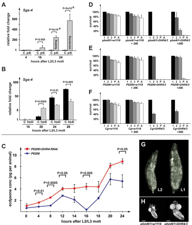
To test whether early wandering inP0206.DHR4-RNAi larvae (Figure 1C) is caused by precocious ecdysone signaling, we examined the expression levels of the glue gene Sgs-4 via quantitative real-time PCR (qPCR). Sgs-4 represents one of the first identified ecdysone-inducible genes in Drosophila [39,40]. Roughly 24 h after the molt to the L3 (,96 h after egg deposition), Sgs-4 is induced in salivary glands by a low titer ecdysone pulse [10,11], after which the gene maintains high expression levels until it is abruptly turned off at pupariation. At 24 h after the molt, we observed feeding and wandering larvae in
P0206.DHR4-RNAi populations, while controls show no sign of wandering behavior.Sgs-4expression was drastically higher in the 16 h and 24 h RNAi populations relative to controls, and,2-fold higher when one compares the wandering with the feeding larvae in the RNAi group at 24 h (Figure 2A). This indicates that the wandering cohort has received the 20E pulse that inducesSgs-4
earlier than the feeding cohort. Even at 16 h after the molt, when allP0206.DHR4-RNAi larvae are still feeding, we observe much higher expression levels ofSgs-4compared to controls, suggesting that the corresponding 20E pulse has occurred already before this time point, thereby preceding the wild type 20E peak (,20 h after the molt) by at least 4 h.
We also examined whether heat-inducedDHR4-RNAi in late L2 would trigger precociousSgs-4 induction, since this treatment triggers premature wandering behavior (Figure 1D). In contrast to PG-specific DHR4-RNAi, we did not observe induction at 16 h after the L2/L3 molt, however at the 20 h mark we found that RNAi larvae had,3-fold higherSgs-4mRNA levels than controls (Figure 2B). However, when we examinedSgs-4 levels at 24 and 28 h after the molt, we found higher expression of the gene in controls, suggesting that Sgs-4 is induced precociously, but
submaximally in hsDHR4-RNAi animals. Finally, when heat shocked in early L3, we do not observe differences in Sgs-4
expression betweenhsDHR4-RNAi and wild type larvae (Figure S2), consistent with our observation that only a heat treatment in late L2 triggers early wandering behavior.
To complement these findings, we analyzed the expression profiles of two isoforms of theE74gene, which are both ecdysone-regulated [41,42]. This approach has been used previously [43] and is based on the idea that the A and B isoforms of theE74gene
Figure 1. Disruption ofDHR4ring gland function affects developmental timing.(A) Pupal and prepupal phenotypes include size defects and malformations. From left to right:P427parental control line forDHR41mutants,DHR41,P0206-Gal4(x2),P0206.DHR4-RNAi (x2) prepupae of various sizes,Cg-Gal4(x2) pupa,Cg.DHR4-RNAi (x2). (B) Dwarf larvae.P427L3 and L2 are controls. A severe growth defect is observed in populations ofDHR41
mutants andphm22.RasV12
larvae. The two insets show the morphology of mouth hooks and anterior spiracles of aDHR41
L3 dwarf larva at high magnification. (C) Expression ofDHR4-RNAi in the RG causes premature pupariation. Percentages of embryos (staged within a 2-h interval) that reached pupariation, hours are after egg deposition. Unpaired Student’sttests betweenP0206.DHR4-RNAi (x2) (red,N= 386) andP0206-Gal4(x2) (blue,N= 143) for time points 104 to 118 are allp,0.0001 (not indicated in the graph).Cg-Gal4(x2) (black,N= 150) andCg.DHR4-RNAi (x2) (green,
N= 251) examine whether timing differences exist whenDHR4-RNAi is fat body-specific. (D, E) Time course shows percentage of clear gut larvae as a means to measure wandering behavior. Red:DHR4-RNAi (N= 122, 133 in D and E). Black:w1118controls (N= 157, 115 in D and E). Larvae were heat shocked in late L2 (D) or early L3 (E).pvalues (*p,0.05, **p,0.01) are based on Student’sttest and comparehsDHR4andw1118
at the same time point. (C–E) Error bars reflect standard deviation from three to six replicates.
Figure 2.DHR4-RNAi affects the timing of ecdysone-mediated responses.(A, B) qPCR ofSgs4transcript levels inP0206.DHR4-RNAi (p4i) (A) andhsDHR4-RNAi (hs4i) (B) animals, hours are relative to the L2/L3 molt, and fold changes are relative to the control of 4-h (A) or 16-h (B) time points. Controls are shown in black. Error bars represent 95% confidence intervals, andpvalues were calculated with the unpaired Student’sttest. (C) Ecdysteroid measurements during the first 24 h of the third instar. Larvae homozygous either forP0206.DHR4-RNAi (red) orP0206(blue) were compared. At least three samples were tested per time point, and each sample was tested in triplicate. Error bars represent standard error andp
respond inversely to ecdysone concentrations. In particular, if the ecdysone titer is high,E74Ais induced andE74Bis repressed, but when ecdysone titers drop to intermediate concentrations,E74Ais turned off, whileE74BmRNA levels rise. Therefore, by measuring both isoforms using qPCR, we can infer whether ecdysone concentrations have fallen or risen. WhenhsDHR4-RNAianimals are heat shocked as late L2, we find thatE74Alevels start to rise at 24 h after the molt and remain roughly 2-fold higher than controls at the 24, 28, 32, and 36 h time points, whileE74Blevels show a corresponding drop at 28–36 h (Figure S3, left panels). These findings suggest a preceding rise in ecdysone concentrations, consistent with the precocious induction ofSgs-4discussed above. When animals received a heat shock 4 h after the L2/L3 molt, we observed no significant differences inE74A and E74Btranscript levels between hsDHR4-RNAi animals and controls (Figure S3, right panels), agreeing with our finding that a later induction of RNAi fails to trigger precocious wandering behavior (Figure 1E).
DHR4Is Required for Repressing Ecdysone Pulses
To test whether the precocious ecdysone signaling in the mid-L3 larvae could be linked to the inappropriate timing and/or duration of a preceding ecdysone pulse, we measured ecdysone titers during the first 24 h of the L3 using a 20E EIA immunoassay (Cayman Chemical). The antibody appears to have similar affinities for 20E and its immediate precursor, ecdysone (E) (Naoki Yamanaka, personal communication), and therefore titers likely reflect a combination of both ecdysteroids. We find that knocking down DHR4 in the RG overall results in significantly higher ecdysteroid levels at all time points we examined (Figure 2C). Importantly, while we can resolve two minor ecdysteroid peaks in the control, we observe no recession of the first L3 pulse in
P0206.DHR4-RNAi larvae (Figure 2C, 16 h time point). Rather, the first and the second L3 pulse appear to be fused in RNAi animals, demonstrating that the first pulse was not properly repressed. It is likely that the combination of higher hormone levels and the inability to repress the first pulse causes the premature effects observed forSgs-4andE74transcripts, as well as the acceleration of wandering behavior and pupariation.
Overexpression of DHR4in the PG Blocks Molting by Suppressing Ecdysone Pulses
The derepression of ecdysone titers and the premature ecdysone signaling inDHR4-RNAi larvae suggest that the wild type function of this nuclear receptor is to inhibit ecdysone production and/or release. To test this idea, we reasoned that increasing DHR4
expression specifically in the PG using thephmN1- andP0206-Gal4
drivers should maintain low systemic ecdysone levels and prevent ecdysone pulses from occurring. Indeed, we found that DHR4
blocks molting when overexpressed in the PG, however the penetrance of this phenotype is dependent on the driver/ responder combination being used, as well as the chromosomal location of the transgene, suggesting that this effect is dose-sensitive. For instance, when UAS-DHR4(inserted on 2ndor 3nd chromosome) is used in combination with phmN1-Gal4, animals cannot progress beyond the L1 stage (Figure 2D). Similarly, using
P0206.DHR4/3 (inserted on the 3rd chromosome) results in all larvae being trapped in the L2 stage (Figure 2E), while
P0206.DHR4/2 (inserted on the 2ndchromosome) allows 5%–
10% escapers to reach the pupal stage. To test whether theDHR4 -mediated block in molting could be rescued by ecdysone, we added 20E to the medium and scored the percentage of animals that progress to later stages. Regardless of the driver used, we observed significant rescue when 20E was added to the diet. In the presence of the hormone,,80% of thephmN1.DHR4/3animals progress to the L2 stage, with another 20% reaching the L3 (Figure 2D). Similarly, most of theP0206.DHR4/3animals molt to the L3 in the presence of 20E, and,4% of the population pupariate (Figure 2E). Next, we tested whether the ability ofDHR4
to block molting was specific to the PG. Since the phmN1-Gal4
driver shows some expression in the fat body, we expressedDHR4
specifically in the fat body using theCg-Gal4driver. Similar to the results obtained with the PG drivers, we observe a developmental arrest in the L1 and L2 stages. In contrast to overexpressingDHR4
in the PG, however, the developmental arrest caused by fat body-driven expression of DHR4 cannot be rescued with 20E (Figure 2F). Taken together, these results demonstrate that expressing DHR4 in the PG blocks molting and interferes with systemic 20E levels in aDHR4-dose-dependent manner, where a strongerGal4driver leads to animals trapped in earlier stages.
We further characterized the ability of phmN1.DHR4 expres-sion to block larval development. Closer examination revealed that these animals survive and continue to grow for up to 10 d as first instar (L1) larvae. These L1 larvae grow very large, accumulate lipids in their fat bodies, and have larger organs than controls due to continued proliferation (Figure 2G,H). This demonstrates that expression ofDHR4in the PG specifically blocks molting and does not trigger lethality. Rather, it appears that these animals simply lack the ecdysone pulse to molt to the next stage and that all other aspects of larval life function normally.
The Subcellular Localization of DHR4 Oscillates between Cytoplasm and Nucleus
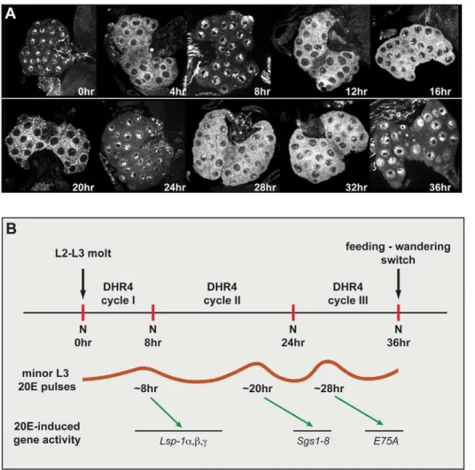
We previously reported that DHR4 is highly enriched in the cytoplasm of PG cells, even though the protein is nuclear in fat body cells in late L3 larvae [35]. Therefore, it appears that the subcellular localization of DHR4 is differentially controlled in these two tissues, raising the question as to whether DHR4 can enter the PG nucleus at all and, if so, how this translocation is regulated. To test whether DHR4 could enter the nucleus of PG cells at certain times during larval development, we stained ring glands isolated from carefully staged L3 larvae ranging from 0 to 36 h after the molt with affinity-purified DHR4 antibodies. Using this approach, we found that the subcellular localization of DHR4 changes periodically in PG cells during the L3. DHR4 appears to be entirely nuclear at 0, 8, 24, and 36 h, completely cytoplasmic at 4, 12, and 20 h, and present in both compartments at 16, 28, and 32 h after the L2/L3 molt (Figure 3A). During the first 36 h of the L3, DHR4 completes at least three cycles: It shifts from the nucleus to the cytoplasm and back during the first 8 h after the molt, while the next two cycles last 16 and 12 h, respectively (Figure 3B). These three oscillations show an intriguing correlation with the occurrence of the three low titer pulses during the L3. In particular, based on direct measurements of 20E titers [8] and indirect assessments based on ecdysone-regulated gene profiling in L3 larvae [11], the three minor 20E pulses have been mapped to 8, 20, and 28 h after the L2/L3 molt. It should be noted that 20E constitutes the final and active form of the molting hormone, and that a biosynthetic profile in the PG of its immediate
phmN1.DHR4/3cDNA in the PG gives rise to very large L1 larvae (right) when compared to newly molted L2phmN1.w1118
control larva (left). (H) Central nervous systems (CNS) were isolated from larvae equivalent to those pictured in ‘‘G’’ and stained with DAPI. The scale bars represent 25mm.
precursor, ecdysone, would therefore have to precede the depicted 20E curve. Taking this into account, it appears that DHR4 is cytoplasmic during a minor pulse, but nuclear between these peaks, consistent with the idea that DHR4 regulates the timing of these peaks.
In this context it is also important to note thatPTTHmRNA was shown to cycle with an 8-h periodicity in staged L3 larvae [30], raising the possibility that a causal link exists between the cyclic behaviors ofPTTHexpression and DHR4 localization. PTTH acts through Ras signaling, and larvae that express constitutively active
Rasin their prothoracic glands (phm22.RasV12) display shortened larval stages and small pupae [33], which are strikingly similar to
DHR4 loss-of-function phenotypes. We therefore investigated whether DHR4 acts in the PTTH pathway.
DHR4 Oscillation Depends on the PTTH Pathway
To examine the impact of altered PTTH activity on the subcellular distribution of DHR4, we analyzed the location of the DHR4 protein in PGs isolated from 0- to 8-h-old L3 larvae, which were carefully staged at the L2/L3 molt. We reasoned that this stage would not only allow us to follow DHR4 protein through an entire cycle, but also ensure that these animals are as precisely timed as possible. Manipulating components of the PTTH
pathway affects developmental timing, which makes later time points difficult to compare between the different genotypes. To test the effects of genetically removing PTTH function, we ablated PTTH-producing neurons in ptth.grim transgenic animals. In PTTH-abolished animals, DHR4 accumulated in the nucleus, with some residual protein residing in the cytoplasm (Figure 4). Ring glands from later L3 time points look comparable (unpublished data), strongly suggesting that nuclear export and/ or degradation of DHR4 is abrogated when PTTH signaling is disrupted. To validate these findings, we examined the effects of
torsoRNAi, which targets the PTTH receptor. Very similar to the PTTH ablation line, we observed nuclear enrichment of DHR4 and loss of oscillation inphm22.torso-RNAi animals (Figure 4). We surmised that hyperactivating the PTTH pathway via constitu-tively activeRasV12[34] should result in cytoplasmic rather than nuclear accumulation of DHR4. To test this, we carried out DHR4 antibody stains on ring glands isolated fromphm22.RasV12
larvae. In contrast to the nuclear accumulation observed in larvae without intact PTTH signaling, we found strong cytoplasmic enrichment of DHR4 in PG cells when RasV12 was expressed (Figure 4), indicating that Ras activity dictates whether DHR4 can accumulate in the nucleus or not. Note that constitutively active
RasV12results in overproliferating PGs, explaining the large and malformed glands we observe.
Figure 3. DHR4 oscillates between cytoplasm and nucleus in PG cells of L3 larvae.(A) Confocal images of ring glands isolated from carefully stagedw1118L3 larvae at different times relative to the L2/L3 molt. Ring glands were stained with affinity-purified DHR4 antibody. 15–20 ring glands were tested per time point. (B) Schematic representation of DHR4 oscillations. The three cycles observed in (A) correlate with the appearance of the three minor 20E pulses that are documented for the L3 [8]. These pulses likely induce theLsp1,Sgs, andE75Agenes [11]. N, nucleus. doi:10.1371/journal.pbio.1001160.g003
Taken together, our data demonstrate that the PTTH pathway controls DHR4 and that nuclear accumulation of the protein is only permitted when the pathway is inactive. This suggests that PTTH regulates DHR4 activity by controlling its subcellular localization, thereby permitting or preventing access of DHR4 to its target genes.
RasV12Prevents the Nuclear Localization of DHR4 in Fat Body Cells
To further examine the ability ofRasV12to prevent DHR4 from entering the nucleus, we tested whether RasV12 could abolish
DHR4 nuclear localization in larval fat body cells, where DHR4 was shown to be restricted to the nucleus once translation has occurred, at least in L2 and L3 larvae (Figures 5A, S1) [35]. When we specifically drive expression ofUAS-RasV12in the fat body using
Cg-Gal4, we found DHR4 to be virtually absent from the nuclei and to be strongly enriched in the cytoplasm instead (Figure 5B), indicating that constitutively active Ras is sufficient to trigger
cytoplasmic retention of DHR4. These findings corroborate our results obtained from the PG, providing strong evidence that nucleocytoplasmic distribution of DHR4 is controlled by PTTH signaling.
DHR4Overexpression in the PG RescuesRasV12
Phenotypes
The dependence of DHR4 on Ras is intriguing becauseRasV12
is—to the best of our knowledge—the only other known genetic alteration besides theDHR41mutation that results in accelerated larval development and small pupae [33,43]. We also observe the occurrence of dwarf larvae inRasV12animals (Figure 1B). These findings are consistent with our observation that RasV12prevents DHR4 from accumulating in the nucleus, thus disrupting its nuclear functions, similar to DHR41
mutants or DHR4-RNAi animals. Based on this observation, one would predict that some of the effects of RasV12could be blocked if one increases the level of DHR4 protein in the same tissue. We therefore asked whether
DHR4was epistatic toRasor, more specifically, if we could rescue
RasV12-induced phenotypes by overexpressingDHR4specifically in the PG. First, we determined the average time to puparium formation whenRasV12orDHR4, or both together, are expressed in the ring gland using the P0206-Gal4 driver. As mentioned previously, ,5%-10% of P0206.DHR4/2 larvae reach the prepupal stage, allowing us to determine their timing profiles. As previously reported by others, P0206.RasV12 animals develop much faster than controls [33,43], preceding the appearance of control prepupae by ,20 h (Figure 5C). In contrast, P0206.DHR4/2 prepupae form with a,20-h delay compared to controls. However, when RasV12 and DHR4 are expressed together in the PG, we observe normal timing of pupariation, and a partial rescue of the DHR4-mediated lethality (Figure 5C).
Since RasV12 overexpression results in a hyper-proliferation phenotype (Figures 4, 5B), we wondered whether this aspect of Ras activity could also be rescued byDHR4overexpression. For this, we examined the morphology of ring glands isolated from staged L3 larvae that expressRasV12and/orDHR4.P0206.RasV12larvae have very large ring glands, whileDHR4expression using the same driver results in slightly smaller ring glands compared to controls (Figure 5D). Importantly, whenDHR4is co-expressed withRasV12, hyper-proliferation of the ring gland appears to be repressed, strongly suggesting that increasing levels of DHR4 in this tissue blocks Ras activity.
Nuclear Localization of ERK/MAPK and DHR4 Are Inversely Correlated
PTTH acts on the PG by ultimately activating ERK, a MAP kinase, via phosphorylation. Upon activation, ERK can enter the nucleus and phosphorylate nuclear target proteins such as transcription factors or other kinases [44]. Since a key role of PTTH is the induction of ecdysone biosynthesis, and DHR4 appears to have the opposite function, we would predict that PTTH activity would remove nuclear DHR4 via activated ERK entering the nucleus. We therefore stained ring glands from staged early L3 larvae to examine the subcellular localization of ERK at various time points. At 0 and 8 h after the L2/L3 molt, we found ERK evenly distributed between nucleus and cytoplasm, however at the 4-h time point, ERK is strongly enriched in the nucleus (Figure 5E). We conclude that ERK shows an inverse relationship to DHR4, at least at the examined time points: ERK accumulates in the nucleus when DHR4 is enriched in the cytoplasm, and when DHR4 is abundant in the nucleus, we found no particular increase of nuclear ERK over cytoplasmic ERK. These data are
Figure 4. Effects on DHR4 subcellular localization by manipu-lating PTTH pathway components. DHR4 antibody stains.
phm22.w1118 and ptth.w1118 ring glands serve as controls. The
ptth.grim and phm22.torso-RNAi lines disrupt PTTH signaling.
phm22.RasV12
constitutively activates the PTTH pathway. Hours indicate time after the L2/L3 molt. 10–15 ring glands were tested per condition.
consistent with the idea that ERK plays a role in the displacement of DHR4 from the PG nuclei in response to PTTH, in line with our findings that the subcellular localization of DHR4 is regulated by the PTTH/Ras/Raf/ERK signaling pathway.
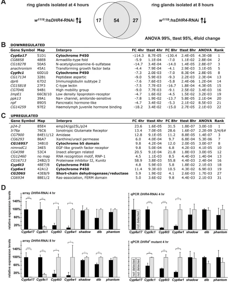
DHR4-RNAi Ring Gland Microarrays Reveal Misregulation of Cytochrome P450 genes
To identify possible target genes of DHR4, we triggered RNAi in late L2 using hsDHR4-RNAi and carried out microarray analysis of ring gland RNA isolated from larvae staged at 4 and 8 h after the molt. To reduce the number of false positives, we analyzed two adjacent time points, 4 and 8 h after the L2/L3 molt, allowing us to select for genes that exhibit significant expression changes at both time points. Using a stringent filtering approach we identified 54 genes whose transcript levels showed a greater than 4-fold difference between controls andDHR4-RNAi animals at both time points (Figure 6A). Selected genes from these 54 genes are shown in Figure 6B and C. Intriguingly, among these 54 genes are four cytochrome P450 genes, an enrichment that is highly unlikely to occur by chance (pvalue = 2.4E-11). Two of the P450 genes are downregulated (Cyp6a17 and Cyp9c1), while the
other two show increased expression (Cyp6t3andCyp6w1), and the effects are very similar between the two time points (Figure 6D, left panels). We also found a short-chain dehydrogenase/reductase (CG2065) among the affected genes, which belongs to the same enzyme family as the Halloween gene shroud. Finally, CG16957, which encodes a protein with a cytochrome b5 domain, is also affected by DHR4-RNAi. This protein family is functionally related to cytochrome P450 enzymes because both enzyme classes act as oxidoreductases and carry heme groups.
To validate some of these observations, we analyzed the expression of all four affected cytochrome P450 genes in brain-ring gland complexes isolated fromhsDHR4-RNAi animals as well as
DHR41mutants that were staged at 4 h after the L2/L3 molt. We also included the analysis of the Halloween genessad,dib, andphm
as additional controls, in case these genes were affected, but not identified by the microarray approach. As expected, all four cytochrome P450 genes identified by the array display very similar profiles in the qPCR validation experiments (Figure 6D, right panels). When we analyzed the samples derived from theDHR41
mutants, we confirmed thatCyp6t3 and Cyp6w1 are significantly higher whenDHR4function is impaired. However, we found that
Figure 5. DHR4 acts downstream of Ras and ERK.(A, B) DHR4 antibody stains (red) ofCg.w1118
(A) andCg.RasV12
(B) late L2 fat body cells. Blue: DAPI stain of nuclei. (C) Genetic epistasis analysis examining the timing of pupariation for transgenic lines expressingDHR4cDNA,RasV12, or both. Percentages indicate the fraction of embryos that developed into prepupae at a given time point. All populations were tested in triplicate, total
Nin brackets. Genotypes:P0206.RasV12(red, N= 151),P0206.w1118(blue,N= 223), P0206.DHR4/2; RasV12(black,N= 293), andP0206.DHR4/2
(green,N= 265). Error bars represent standard deviation. (D)DHR4overexpression inhibitsRasV12
-induced ring gland overgrowth. CNS-RG complexes isolated from early L3 larvae, pictures show same sample at 206and 406magnification. Blue: DAPI. Green:UAS-mCD8-GFPis recombined to the same chromosome asP0206-Gal4, and therefore reflects the expression pattern of theP0206driver. Genotypes are listed below the figure. The scale bars represent 25mm. (C–D)DHR4/2denotesUAS-DHR4cDNA inserted in the 2ndchromosome. (E) Upper panel: Anti-ERK antibody stains of L3 RGs isolated at 0, 4, and 8 h after L2/L3 molt. Lower panel: Nuclei stain with DAPI. (D–E) 10–15 ring glands were stained per condition.
doi:10.1371/journal.pbio.1001160.g005
Figure 6.DHR4-RNAi ring gland microarray reveals misregulated cytochrome P450 genes. (A) Comparison of microarray data sets representing 71 genes upregulated or downregulated more than 4-fold in 4-h L3 and 81 genes in 8-h L3hsDHR4-RNAi ring glands. Filtering criteria: $4-fold change, Student’sttest with apvalue of,0.05 for both time points, and an ANOVA pvalue of,0.01. (B, C) Selected genes either downregulated (B) or upregulated (C), sorted by the 4-hpvalue. Genes with possible roles in ecdysone biosynthesis are in bold.#indicates three different probe sets were detected forIr76a. (D) Selected microarray results and qPCR validation inhsDHR4-RNAi animals andDHR41
Cyp9c1 displayed higher rather than lower levels compared to controls. In addition, we were unable to detectCyp6a17inDHR41
mutants or in the corresponding parental line, P427, suggesting that both Cyp9c1 and Cyp6a17 expression varies substantially between different genetic backgrounds. Future experiments will address whether Cyp9c1 or Cyp6a17 are dependent on DHR4
function. Our qPCR analysis revealed no substantial effects on the tested Halloween genes (Figure 6D, all panels), confirming the microarray results. Taken together, our microarray data identified two cytochrome P450 genes, Cyp6t3 and Cyp6w1, which display significantly higher expression levels in the ring glands ofhsDHR4 -RNAi animals as well as in brain-ring gland complexes isolated from DHR41mutants, indicating that DHR4 normally represses these genes.
Cyp6t3Has a Novel Role in the Biosynthesis of Ecdysone
Of the two cytochrome P450 genes reproducibly affected by loss-of-DHR4function, Cyp6t3and Cyp6w1, we chose to analyze
Cyp6t3 in more detail for two reasons. First, Cyp6t3, but not
Cyp6w1 (unpublished data), is specifically expressed in the ring gland based on qPCR analysis, which shows,20-fold enrichment of Cyp6t3 transcripts in the ring gland compared to whole body (Figure S4). We confirmed this by in situ hybridization, which demonstrates that Cyp6t3 is specifically expressed in the protho-racic glands and thecorpus allatum(Figure 7E). Bleed-through of the tyramide-amplified signal did not allow us to determine whether
Cyp6t3 is also expressed in thecorpora cardiaca. Second, when we examined the changes in gene expression at 0, 4, 8, and 12 h after the L2/L3 molt, we found that Cyp6t3 levels, but not Cyp6w1
levels, oscillate during this time window, where lower concentra-tions ofCyp6t3correlate with nuclear DHR4, consistent with the idea that DHR4 represses this gene (compare Figures 7G and S5 with Figure 3A). For these reasons we examined whether interfering with Cyp6t3 function in the PG via RNAi results in any developmental defects. Specifically, we expressed Cyp6t3
RNAi (VDRC #109703) [45] in the PG by generating
phm22.Cyp6t3-RNAi animals. DisruptingCyp6t3 in this manner generates phenotypes typically observed in mutants that have defects in the ecdysone synthesis pathway [30,38,46,47]. For instance, we observe very large pupae (similar in size to phantom
(phm) anddisembodied(dib) RNAi pupae, Figure 7A), double larval mouthhooks (a common molting defect, Figure 7A inset), and L2 prepupae (Figure 7A). The latter phenotype occurs when larvae forego the molt to an L3 and directly molt from an L2 to a prepupa. This phenotype is relatively rare and has been only associated with mutations inE75[46],dre4[48], anditpr[49], all of which have dramatically lowered ecdysone levels. We also tested a second independently generated Cyp6t3 RNAi line (VDRC #30896, Figure S6), which is based on a smaller dsRNA construct. In this RNAi line, we also observe very large pupae, consistent with a longer feeding period, but failed to identify any L2 prepupae. Feeding 20E to these animals completely rescues the large body size (Figure S6). Since the VDRC line#109703 was stronger than the#30896 line, we continued our studies with the former line. No phenotypes are observed when Cyp6t3-RNAi is expressed in the fat body (unpublished data), indicating that the phenotypes induced by phm22.Cyp6t3-RNAi are specific to the PG.
Next, we examined whether Cyp6t3-RNAi animals have lower ecdysone titers. To test this, we compared ecdysteroid concentra-tions at multiple time points between L3 control larvae and delayed L2 larvae of the same absolute age. The latter ultimately develop into L2 prepupae. As expected,Cyp6t3-RNAi larvae have severely reduced ecdysteroid titers compared to controls, but generate a small pulse before they form L2 prepupae (Figure 7B, 100 h L2 time point). In addition, we also measured ecdysteroid concentrations in earlier stages and found that Cyp6t3-RNAi animals have lower hormone levels at all larval stages, but not as embryos (Figure S7). One would predict that feeding ecdysone to
phm22.Cyp6t3-RNAi animals should rescue at least some of the phenotypes, and indeed we observed that the occurrence of L2 prepupae is completely rescued when 20E is added to standard medium (Figure 7C), corroborating our finding that
phm22.Cyp6t3 RNAi affects ecdysone production. To further characterize at which step in the ecdysone biosynthetic pathway Cyp6t3 might act, we examined which 20E precursors might also result in a rescue. For this, we took advantage of the fact that the
Cyp6t3-RNAi phenotype was more pronounced on an instant medium (4–24, Carolina Biological Supply Company), hereafter referred to a ‘‘C424.’’ This medium is naturally low in cholesterol and other sterols, and has been used by us for sterol rescue studies before [50]. On this medium,phm22.Cyp6t3-RNAi animals very rarely progress beyond the L2/L3 molt (,0.5%), either dying as L2 larvae or L2 prepupae. When we supplemented C424 with carrier only (ethanol), cholesterol, or 7-dehydrocholesterol (7dC), we failed to see any rescue, defined by larvae developing to L3 larvae or later stages. In contrast, adding E or 20E to C424 medium resulted in.60% rescue, while supplementation with 5b -ketodiol rescued,15% of the Cyp6t3-RNAi population past the L2/L3 molt (Figure 7D), with some animals reaching the pupal stage (unpublished data). The lower percentage of rescued animals with 5b-ketodiol likely reflects the fact that this compound has to enter the PG, while E and 20E can act directly on the target tissues. This strongly suggests that Cyp6t3 plays a role in the black box, since mutations affecting enzymes acting downstream cannot be rescued with 5b-ketodiol [21,24].
Cyp6t3Is Epistatic toDHR4
While our data demonstrate thatCyp6t3is specifically expressed in the ring gland, it appears to be expressed at a fairly low level. Based on our microarray and qPCR experiments we estimate that in the ring gland, Cyp6t3 transcript levels are two orders of magnitude lower than those of phm, dib, and sad. A possible explanation for this is thatCyp6t3forms part of a ‘‘bottleneck’’ for ecdysone production and that a low abundance of transcripts rendersCyp6t3more susceptible to transcriptional regulation than its Halloween counterparts. We therefore wondered whether (a) overexpression of Cyp6t3 would be sufficient to alleviate this bottleneck and accelerate ecdysone synthesis and development and (b) if loss of Cyp6t3 function in aDHR41 mutant background is necessary for accelerated development of these animals. In the first experiment, we generated aphm22.Cyp6t3line that expresses a
Cyp6t3cDNA at high levels in the PG [51]. This resulted in no obvious phenotypes with respect to timing or overall morphology (unpublished data), suggesting that Cyp6t3 is not sufficient to accelerate developmental timing via an increased rate of ecdysone bars). Controls are shown in black,w1118
forhsDHR4-RNAi andP427forDHR41
mutant.Shadow,dib, andphantomfailed ANOVA testing at the 95% level, but were included for validation purposes. RNA from brain-ring gland complexes of animals staged at 4-h L3 was used for qPCR validation. Error bars for the array data represent standard deviation, and error bars for qPCR data show 95% confidence intervals. Asterisks indicate significant differences between groups (*p,0.05, **p,0.005, ***p,0.0005, ****p,0.00005 by Student’sttest).
doi:10.1371/journal.pbio.1001160.g006
production. In the second experiment we found that DHR41; phm22.Cyp6t3-RNAi larvae display delayed instead of accelerated development relative tow1118controls (Figure 7F). This indicates that upregulation of Cyp6t3 in DHR41 mutants (Figure 6D) is necessary for the accelerated development in these animals, and that lowering the levels ofCyp6t3 via RNAi effectively abolishes this effect. We conclude thatCyp6t3is necessary but not sufficient for accelerating development, suggesting that if Cyp6t3is indeed part of a bottleneck, it is not acting alone in this rate-limiting step.
Discussion
DHR4 Oscillation and Its Dependence on PTTH Signaling
A series of reports have provided ample evidence that PTTH utilizes the Ras/Raf/ERK pathway to regulate ecdysone biosyn-thesis inBombyx,Manduca, andDrosophila[33,52–56]. In the present study, we demonstrate that a critical readout of this pathway is the nuclear receptor DHR4. We suggest a simple model where PTTH represses DHR4 activity via its removal from the nucleus, while DHR4 in turn represses the occurrence of low-titer ecdysone peaks when nuclear (Figure 8). In laboratory fly cultures, PTTH was shown to be a non-essential gene, however when PTTH-producing neurons are ablated, development is substantially delayed and animals have a concomitant increase in body size [33]. We show here that disruptingDHR4specifically in the PG results in opposite phenotypes to loss-of-PTTHfunction, where animals are smaller and develop faster than controls. Like PTTH ablation lines, animals homozygous forP0206.DHR4-RNAi can be kept as a viable stock, consistent with the idea that DHR4 functions in the PG as a PTTH-dependent, non-essential developmental clock to generate appro-priately timed ecdysone pulses. It is of interest to note that the expression of constitutively active Ras in the PG ofP0206.RasV12
animals results in accelerated larval development and small pupae, very similar toDHR41mutants andP0206.DHR4-RNAi animals.
P0206.RasV12 animals are also viable, and we showed that L3 larvae of this genotype accumulate DHR4 in the cytoplasm of PG cells (Figure 4). This strongly suggests that P0206.RasV12larvae display these phenotypes precisely because DHR4 protein is prevented from entering PG nuclei, thereby mimicking the loss-of-function phenotypes observed inDHR4RNAi or mutant larvae. We have demonstrated that the PTTH pathway controls the subcellular location of DHR4. Loss of PTTH signaling results in nuclear presence of DHR4, while constitutively activating this pathway leads to cytoplasmic localization of the protein. It is unclear at this point whether the DHR4 oscillations represent shuttling or involve cycles of degradation and synthesis. Shuttling
would require a stable DHR4 protein that moves in and out of the nucleus, however this would be difficult to reconcile with the fact thatDHR4 RNAi works well in our hands, since a continuously shuttling protein would be impervious to RNAi. It is evident that sufficient turnover of the DHR4 protein must occur, at least around the L2/L3 molt, when we conducted our heat-induced
DHR4-RNAi experiments (Figures 1D–E, 2B, and S3). This raises the question of whetherDHR4mRNA levels are oscillating, given that the degraded protein must be replaced periodically. When we conducted a time course microarray of wild type ring glands, we observed very low and constant levels ofDHR4 mRNA, which does not support the idea that DHR4 transcripts levels are oscillating (Ou et al., manuscript in preparation). Based on these data, we suspect thatDHR4mRNA is highly stable in PG cells and translated when needed in L3 larvae. Alternatively, our current approach might be too insensitive to detect periodic changes in
DHR4mRNA levels.
It was shown in mammalian cell cultures that the ERK pathway controls the subcellular localization of the nuclear receptor PPARc. Specifically, mitogenic stimulation of resting cells causes the binding of nuclear MEK1 to PPARc, which is followed by rapid export of the protein complex from the nucleus [57,58]. Our study does not provide direct evidence that DHR4 is phosphor-ylated. However, our data are suggestive of the idea that ERK plays a role in removing DHR4 from the nucleus, since we found that ERK changes its nucleo-cytoplasmic distribution in early L3 larvae, in an apparent inverse relationship to DHR4, consistent with the notion that it acts upstream of this nuclear receptor (Figure 5E). Future experiments will have to examine whether DHR4 is a direct target of ERK and whether phosphorylation plays a role in the nucleo-cytoplasmic oscillations of DHR4.
An intriguing finding is that Drosophila PTTH mRNA levels oscillate with an apparent 8-h cycle time throughout the 48-h duration of the L3 stage [30]. DHR4, on the other hand, displays an 8-h, 16-h, and 12-h cycle time for the first 36 h of the L3 stage, raising the question as to how these ultradian periods are established. What could account for the difference in these cycle times? A simple possibility is that we were unable to detect all DHR4 cycles and that the DHR4 oscillations are well aligned with the PTTH cycles. In this study, we chose to conduct a time course based on a 4-h step size, because we consider 4 h a robust time interval that should compensate for the inherent asynchrony that exists in developingDrosophila larvae. We examined 15–20 ring glands per time point, and only found some discrepancies among ring glands from later time points, likely due to the asynchronous development of the population. However, it appears that during
Figure 7. Functional characterization ofCyp6t3.(A) PG-specificCyp6t3-RNAi phenotypes VDRC#109703), compared tophm22.w1118 control (left). Insets show the morphology of anterior spiracles and double mouth hooks ofphm22.Cyp6t3-RNAi compared to controls. (B) Whole-body ecdysteroid titer measurements comparing equivalent L2 and L3 stages betweenphm22.Cyp6t3-RNAi animals (orange) andphm22.w1118
controls (black). Time points indicate hours after the L2/L3 molt (control) or after the L1/L2 molt (phm22.Cyp6t3-RNAi). For every genotype/time point, 3–4 samples (N= 30–45 larvae) were each tested in triplicate. Error bars indicate standard error. (C) Feeding ecdysone toCyp6t3-RNAi larvae rescues L2 pupae phenotype. Percentages of L2 pupae (striped) and L3 pupae (black) ofphm22.w1118
andphm22.Cyp6t3-RNAi in populations fed a standard medium with or without 20E. Error bars indicate standard deviation,N= 150–200 for each condition. (D) Feeding 5b-ketodiol toCyp6t3-RNAi larvae rescues larvae beyond the L2 stage. C424 instant fly medium (Carolina) was supplemented with different ecdysteroid precursors or the carrier alone (ethanol). Percentages show fraction of embryos reaching the L3 stage. Grey: phm22.w1118. Orange:phm22.Cyp6t3-RNAi. Error bars indicate standard deviation,N= 150–200 for each condition. ETOH, ethanol; C, cholesterol; 7dC, 7-dehydrocholesterol; 5bKD, 5b-ketodiol; E, ecdysone; 20E, 20-hydoxyecdysone. (E) In situ hybridization ofCyp6t3antisense and sense probes. Early L3 larval CNS-RG complexes with eye-antenna imaginal discs were examined at 206magnification. A DAPI stain of the nuclei is included. (F) Genetic epistasis analysis examining the timing of pupariation in animals carrying aDHR41mutation,phm22-Gal4//Cyp6t3-RNAi transgenes, or both. Percentages were normalized to the final number of pupae for each genotype and represent the fraction of larvae that formed pupae at a given time point.DHR41mutants (red,N= 54),phm22
.w1118(black,
N= 180),phm22.Cyp6t3-RNAi (orange,N= 600), andDHR41; phm22
.Cyp6t3-RNAi (blue,N= 107). (G) Transcriptional profile ofCyp6t3in early L3. Brain-ring gland complexes were dissected from carefully stagedw1118larvae of indicated time points. Fold changes are relative to 0 h after the L2 to L3 molt. Error bars in (G) represent 95% confidence intervals.pvalues (Student’sttest) are relative to the previous time point. (B,C,D,F)Cyp6t3i: short forCyp6t3-RNAi.
doi:10.1371/journal.pbio.1001160.g007
some time points, such as 16 h after the L2/L3, DHR4 is detected in the nucleus and the cytoplasm, and it is possible that this reflects a transition phase of a cycle we might have missed. Future studies could attempt a time course with a 2-h step size, ideally in combination with aSgs3-GFPreporter line to re-stage animals in the mid third instar [8].
Alternatively, the differences in cycle duration between DHR4 and PTTH could reflect the possibility of another cyclic process that may contribute to the timing of DHR4 periodicity. An attractive possibility is that circadian rhythms are superimposed on the PTTH oscillations to determine DHR4 cycle times. Anatomical evidence indicates that the central circadian pacemaker cells found in the
Drosophilabrain, the Lateral Neurons, indirectly innervate the PG [59]. A critical effector of these neurons is the neuropeptide PDF (Pigment Dispersing Factor), and a mutation in pdf alters the periodicity of PTTH mRNA oscillations [30]. Future experiments will address whether PDF and other components of the circadian clock impinge on the oscillatory behavior of DHR4 in PG cells.
PTTH and the Transcriptional Control of Ecdysone Biosynthetic Genes
The mechanism by which PTTH regulates ecdysone biosyn-thesis has been the subject of intense research for the last 35 years. PTTH triggers a complex array of signaling events in the PG that precede the synthesis of ecdysone. These include second messengers like Ca2+
influx and the synthesis of cAMP, followed by the stimulation of Protein kinase A, the activation of p70S6K and the concomitant phosphorylation of ribosomal protein S6, a myriad of tyrosine phosphorylations, as well as the activation of the ERK pathway discussed in this report [56]. Clearly, PTTH triggers a range of events, which, among others, results in the transcriptional upregulation of genes required for ecdysteroid biosynthesis. The first evidence that PTTH is sufficient to increase transcript levels of an ecdysteroidogenic gene is based on the observation that theBombyx disembodiedgene (dib-Bm) is upregulated by administering PTTH to cultured prothoracic glands [32]. The upregulation ofphmandspoappears to be more moderate [31,32],
Figure 8. Models for DHR4 function.DHR4 represses ecdysone pulses dependent on whether PTTH signaling is active or inactive (middle panel). In the presence of PTTH signaling (upper panel, left), DHR4 is removed from the nucleus either by shuttling to the cytoplasm or by protein degradation, which allows for ecdysone biosynthesis to occur. In the absence of PTTH (lower panel), DHR4 remains in the nucleus and represses
Cyp6t3and possibly other genes with roles in ecdysone production, thereby lowering ecdysone titers. During molts, DHR4 acts as a component of the EcR-controlled gene hierarchy in some target tissues of the hormone (upper panel, right). See text for details.
while sad transcription appears not to be induced under these conditions [32]. Attempts to generate functional recombinant
Drosophila PTTH have so far been unsuccessful [33], but loss-of-function studies have confirmed a role for PTTH in the transcriptional regulation of ecdysteroidogenic genes. Specifically, ablation of PTTH-producing neurons resulted in a strong reduction of Drosophila dib(,10-fold down) and had a moderate effect onphm,sad, andspok(2–3-fold down). While similar studies with a torso RNAi line have not been published, indirect results come from a recent paper where the authors knocked down
dSmad2 function in the PG, which results in a strong downregu-lation of torso [60]. Concomitantly, spok and dib are strongly reduced in phm.dSmad2-RNAi larvae, but similar to the above findings, no effect was seen forphmand sad. Taken together, the findings from Bombyx and Drosophila seem to suggest that the transcriptional effect of PTTH is most clearly established fordib, whilephmandsadappear to be less dependent on PTTH signaling. One aspect of PTTH-mediated transcriptional regulation that has not been satisfactorily addressed is whether some of the ecdysteroidogenic genes require PTTH to reach high expression levels in the first place or whether the hormone provides a ‘‘boost’’ to elevate transcript levels even further. According to our RG microarray and qPCR data presented here, we conclude that expression levels of the Halloween genes are very high in L3 larvae, comparable to that of ribosomal genes. We also conducted a microarray time course (Ou et al., manuscript in preparation), which suggests that the Halloween genes are expressed at very high levels during the first 36 h of the L3, without much fluctuation in their expression levels. Therefore, it would seem unlikely, at least according to our data, that transcriptional downregulation of the highly expressed ecdysteroidogenic genes likediborphmcan be the mechanism by which the three minor ecdysone peaks are generated, which is in line with our finding that knocking down DHR4 in the RG has no effect onphm,sad, and dib (Figure 6D). Rather, it appears that DHR4 negatively regulates Cyp6t3 and possibly other uncharacterized genes that play critical roles in the production of ecdysone (Figure 8).
DHR4 appears to mainly act as a repressor [35], similar to what has been reported for its vertebrate ortholog GCNF [61]. It remains to be seen whether the genes that are downregulated in ourhsDHR4-RNAi microarrays are indirect targets of DHR4 or whether this nuclear receptor can act as an activator on some promoters. We have previously shown that DHR4 acts down-stream of the 20E receptor EcR as a key component of the ecdysone hierarchy during puparium formation [35]. It therefore appears that DHR4 acts upstream of ecdysone in the PG, but downstream of the hormone in target tissues (Figure 8), which nicely reflects the duality of DHR41 mutant phenotypes: the mutation affects developmental timing (due to its role in the PG) and puparium formation (due to its role in the fat body).
While some transcription factors have been identified for their roles in the ecdysone production pathway, none have been reported to repress ecdysteroidogenesis, and none have been directly linked to PTTH. A mutation in the woc(without children) gene, which encodes a putative zinc finger transcription factor, causes low ecdysone levels. This phenotype can be rescued with feeding 7-dehydrocholesterol (7dC), suggesting that Woc might control the conversion of cholesterol to 7dC by regulating the genes required for this step [62]. The nuclear receptorbFTZ-F1 is expressed in the PG of late L3 larvae and is required for normal levels of Phm and Dib protein [27]. Another nuclear receptor, E75A, acts in a feed-forward loop to maintain normal ecdysone levels, possibly by acting upstream ofbFTZ-F1 in the PG [46].
Cyp6t3Is a Downstream Target of DHR4
Our ring gland microarray and qPCR data revealed thatCyp6t3
transcript levels are significantly elevated in hsDHR4 RNAi animals andDHR41mutants. In addition, we showed thatCyp6t3
expression levels oscillate during the first 12 h after the L2/L3 molt, with lower levels of Cyp6t3 when DHR4 is nuclear (Figure 7G). It therefore appears plausible thatCyp6t3is a direct transcriptional target of DHR4, however direct evidence for this is lacking. No DNA recognition sites have been identified for DHR4, nor is it known whether this nuclear receptor acts as a homodimer, heterodimer, or monomer. According to our microarray data, as well as judging by the cycle numbers required to detectCyp6t3in qPCR experiments, we estimate that transcript levels of this gene are relatively low, probably by two orders of magnitude lower than the Halloween mRNAs forphm,dib,spookier, andshadowin PG cells (unpublished data). This is consistent with the finding thatCyp6t3
was previously neither detected by in situ hybridization in any larval tissue, nor amplifiable from larval cDNA, while the aforementioned 4 Halloween genes showed strong expression under the same conditions in the PG of Drosophila larvae [63]. However, using tyramide amplification coupled to in situ hybridization, we were able to validate the ring gland-specific expression of Cyp6t3 (Figure 7E). According to our wild type microarray study, Cyp6t3 is one out of nine cytochrome P450 transcripts that have a higher than 10-fold enrichment in the ring gland compared to the whole body signal (Ou et al., manuscript in preparation), supporting the idea that this gene has an important role in ecdysteroid biosynthesis.
The fact that Cyp6t3is expressed at very low levels raises the possibility that the Cyp6t3 enzyme is scarce and therefore rate-limiting with respect to the production of ecdysone. An attractive model is that DHR4-mediated repression of Cyp6t3 suffices to reduce ecdysone production to basal levels. If true, one would predict that Cyp6t3 turnover is controlled so that transcriptional control of theCyp6t3gene becomes a relevant factor in controlling ecdysone synthesis. Conversely, derepression ofCyp6t3due to loss-of-DHR4function could result in faster accumulation of ecdysone, which would account for the timing defects we observe. However, when we overexpressed aCyp6t3cDNA specifically in the PG, we did not observe any obvious phenotypes or effects on development timing, suggesting that changing levels of Cyp6t3 alone is not sufficient for this response (unpublished data). In contrast, we could show thatCyp6t3 function is necessary for the accelerated developmental phenotype ofDHR41mutants, strongly supporting the notion that Cyp6t3 is a key target of DHR4-mediated repression of ecdysone pulses (Figure 7F).
The degree by which Cyp6t3 is repressed might directly correspond to the amount of DHR4 protein allowed to enter the nucleus. In support of this idea we find that overexpression of
DHR4cDNA in the PG results in varying degrees of larval arrest, depending on the strength of the Gal4 driver being used (Figure 2D–F), which in turn suggests that the strength of the phenotype depends on how much DHR4 can enter the nucleus. Therefore, it is conceivable that the nuclear functions of DHR4 are dose-sensitive, giving rise to the idea that the oscillations of this nuclear receptor do not necessarily represent an all or nothing response, but may in fact fine-tune the expression levels of target genes instead.
search (Figure S8) and analyzed their phylogenetic relationship using the program Seaview (Figure S9) [65]. Due to the large size of the Cyp450 family 6 [66] and the sequence similarities within this family, we were not able to identify a definitive Cyp6t3
ortholog outside theDrosophilagenus. We reached this conclusion based on a reverse BLAST search strategy, which revealed that the top hits from theD. melanogasterquery did not retrieve Cyp6t3 as the best hit when used as queries themselves. Despite this, Cyp6t3 is highly similar to sequences found in other insect species, and it is likely that very similar paralogs are masking the true ‘‘functional’’ ortholog. Often, reverse BLAST searches fail to reveal definitive orthologs. For instance, the best characterized nuclear receptor in
Drosophila, EcR, is most similar to both FXR and LXR, depending on whether one uses the DNA-binding domain or the ligand-binding domain as a query [67]. Future studies in other insect species will have to address the question of whether Cyp6t3
orthologs are also important for ecdysone production.
Materials and Methods
Drosophila Stocks
w1118(#3605) andUAS-RasV12(#4847) were ordered from the Bloomington stock center. Gal4 drivers were obtained from labs indicated by the references. Ring gland:P0206-Gal4, UAS-mCD8-GFP [68],phm22-Gal4[33];phmN1-Gal4[69]. Fat body:Cg-Gal4
[70]. PTTH-Gal4 driver and PTTH ablation line: UAS-Grim/CyO-act-GFP & ptth-Gal4/Ser-UAS-Grim/CyO-act-GFP[30];DHR41/FM7h,& hsDHR4-RNAi[35]; RNAi lines were obtained from ViennaDrosophilaRNAi Center [45]. UAS-Torso-RNAi (VDRC #4300 & #1016); UAS-Cyp6t3-RNAi (VDRC #109703 & #30896); UAS-phm-RNAi: (VDRC#100811),UAS-dib-RNAi (VDRC#101117).
Developmental Timing Analysis
Before embryos were collected on grape juice agar plates, flies were allowed to lay eggs twice for 2 h in order to reduce egg retention. After 2-h egg collection intervals at 25uC, eggs were transferred to petri dishes containing fresh yeast paste and reared at 25uC. Pupariation was scored in 2-h intervals. For the hsRNAi experiments, larvae were reared on yeast until the late L2 stage, at which w1118 controls and hsDHR4-RNAi L2 larvae were heat shocked for 35 min at 37.5uC. After a 4-h recovery, newly molted L3 larvae were transferred to yeast paste supplemented with 0.05% bromophenol blue to monitor their gut clearing status [71,72].
qPCR
All qPCR data shown here are based on 3–4 biological samples each tested in triplicate. Whole larvae were collected in distilled water and snap-frozen in liquid nitrogen, while dissected tissue samples were prepared in ice-cold PBS, rinsed twice with fresh PBS, transferred to TRIzol (Invitrogen), and snap-frozen in liquid nitrogen. Total RNA of whole larvae was isolated following a modified TRIzol protocol, where we substituted sodium acetate with lithium chloride for RNA precipitation. Total RNA from tissue samples was extracted using the RNAqueous-Micro Kit (Ambion) following the manufacturer’s instruction. RNA samples (0.5–2mg/reaction) were reverse transcribed using ABI High Capacity cDNA Synthesis kit, and the synthesized cDNA was used for qPCR (StepOnePlus, Applied Biosystems) using PowerSYBR Green PCR master mix (Applied Biosystems) with 5 ng of cDNA template with a primer concentration of 200 nM. Samples were normalized to rp49 based on the DDCT method. All primer sequences can be found in Table S1.
Transgenic Constructs
To generatepUAST-DHR4cDNA, a 6.3 kb fragment contain-ing the full-length synthetic cDNA ofDHR4[35] was cut withEco
RI andXbaI from Litmus 28 and cloned intopUASTdigested with the same enzymes. For the pUAST-DHR4-RNAi construct, the same inverted repeat used in thehsDHR4-RNAi [35] was used to clone the fragment intopUASTusingXbaI for all restriction cuts. Transgenic flies were generated by injecting DNA at a concentration of 0.5mg/ml along with 0.1mg/ml helper plasmid pD2–3 into embryos following standard procedures [73,74].
Immunostaining
Tissues were dissected from larvae in PBS, fixed in 4% paraformaldehyde (EMS #15710) in PBST (PBS containing 0.3% Triton-X 100) for 20 min at room temperature (RT), and washed in PBST. Tissues were then blocked for 2 h at RT or overnight at 4uC in PBST/5% NGS. Primary antibodies were incubated at 4uC overnight, while the secondary antibody was either incubated overnight at 4uC or 4 h at RT. Nuclei were stained with DAPI (1:5000). After several wash steps, tissues were mounted in SlowFade Gold Antifade Reagent (Invitrogen). Images were captured on a Nikon C1 plus confocal microscope. Anti-DHR4 antibody was used at a dilution of 1:500, and anti-ERK antibody was used at a dilution of 1:100 (Cell Signaling#4695). Secondary antibodies (anti-rabbit Cy3) were used at a dilution of 1:200 (Rockland#611-104-122).
Ring Gland Microarrays
DHR4-RNAi andw1118populations were heat shocked as late L2 larvae for 35 min at 37.5uC. To carefully stage larvae at the L2/L3 molt, L3 larvae were discarded 4 h after the heat treatment, and L3 larvae that molted in the following hour were allowed to feed for either 4 h or 8 h before their ring glands were dissected in ice-cold PBS. Ten ring glands were dissected and washed twice in PBS before being transferred to ice-cold TRIzol reagent (Invitrogen). The lysates were then vortexed for 5 s at RT, flash frozen, and stored at280uC. Total RNA was isolated by Ambion RNAqueous-Micro Kit. Isolated RNA was quantified by RiboGreen Quanti Kit (Invitrogen) and RNA integrity was analyzed by Agilent Bioanalyzer Pico Chips. RNA linear amplification was based on the MessageII RNA Amplification kit (Ambion): First-strand cDNA synthesis was carried out by a T7-(dT) primer and ArrayScript reverse transcriptase using 50 ng RNA of each ring gland sample. Second-strand cDNA synthesis was performed according to the provided protocol. Purified cDNA was then fed into the IVT reactions. The amplified RNA (aRNA) was column-purified and analyzed by Agilent Bioanalyzer Nano chips. 1mg of aRNA was used for double-stranded cDNA synthesis (Invitrogen SuperScript One-Cycle cDNA Kit) and 1mg of the purified cDNA was Cy3-labeled by Roche NimbleGen
one-color cDNA labeling kit. From this, 4mg of Cy3-labeled
cDNA was hybridized on aDrosophila126135K Array
(Nimble-Gen). Each condition was analyzed by three independent biological samples. Chip hybridization and scanning was per-formed by the Alberta Transplant Applied Genomics Center. Raw data were normalized with the NimbleScan software (NimbleGen) using the RMA algorithm [75], and data were analyzed with Arraystar 4.0 (DNAstar) as well as Access (Microsoft).
Ecdysteroids Measurements