Signaling Regulates the Differentiation of Germline Cells
in Testes of
Drosophila melanogaster
Alicia G. Hudson1, Benjamin B. Parrott1,2, Yue Qian1, Cordula Schulz1*
1Department of Cellular Biology, University of Georgia, Athens, Georgia, United States of America,2Department of Obstetrics and Gynecology, Medical University of South Carolina, Charleston, South Carolina, United States of America
Abstract
Tissue replenishment from stem cells follows a precise cascade of events, during which stem cell daughters first proliferate by mitotic transit amplifying divisions and then enter terminal differentiation. Here we address how stem cell daughters are guided through the early steps of development. In Drosophila testes, somatic cyst cells enclose the proliferating and differentiating germline cells and the units of germline and surrounding cyst cells are commonly referred to as cysts. By characterizing flies with reduced or increased Epidermal Growth Factor (EGF) signaling we show that EGF triggers different responses in the cysts dependent on its dose. In addition to the previously reported requirement for EGF signaling in cyst formation, a low dose of EGF signaling is required for the progression of the germline cells through transit amplifying divisions, and a high dose of EGF signaling promotes terminal differentiation. Terminal differentiation was promoted in testes expressing a constitutively active EGF Receptor (EGFR) and in testes expressing both a secreted EGF and the EGFR in the cyst cells, but not in testes expressing either only EGF or only EGFR. We propose that as the cysts develop, a temporal signature of EGF signaling is created by the coordinated increase of both the production of active ligands by the germline cells and the amount of available receptor molecules on the cyst cells.
Citation:Hudson AG, Parrott BB, Qian Y, Schulz C (2013) A Temporal Signature of Epidermal Growth Factor Signaling Regulates the Differentiation of Germline Cells in Testes ofDrosophila melanogaster. PLoS ONE 8(8): e70678. doi:10.1371/journal.pone.0070678
Editor:Andreas Bergmann, University of Massachusetts Medical School, United States of America
ReceivedMarch 4, 2013;AcceptedJune 21, 2013;PublishedAugust 5, 2013
Copyright:ß2013 Hudson et al. This is an open-access article distributed under the terms of the Creative Commons Attribution License, which permits unrestricted use, distribution, and reproduction in any medium, provided the original author and source are credited.
Funding:The work was supported by a grant from the American Foundation for Aging Research given to B.B.P. (no grant#), and start-up funds from University of Georgia, Athens, and NSF grant#0841419 given to C.S. The funders have no role in study design, data collection and analysis, decision to publish, or preparation of the manuscript.
Competing Interests:The authors have declared that no competing interests exist.
* E-mail: cschulz@uga.edu
Introduction
Tissue homeostasis depends on adult stem cells that constantly self-renew and produce differentiated cells [1,2]. Self-renewal of stem cells and differentiation of stem cell daughters are regulated by interactions with other cell types. For example, in the hair follicle of the skin, melanocyte stem cells are closely associated with epithelial stem cells and signaling between the two lineages is an important mechanism in coordinating the differentiation of the two stem cell lineages to make pigmented hair [3,4]. Also in the skin, follicular stem cell activation is regulated by signals from underlying intradermal adipocytes, and in the bone marrow, hematopoietic stem cell fate and proliferation depend on mesenchymal stem cells [5–7]. One of the best described examples of the dependence of a stem cell lineage on another cell type is the development of germline cells in the male gonad of Drosophila melanogaster[8].
Within theDrosophilatestis, the germline cells and their somatic support cells are arranged in a spatio-temporal order along the apical to basal axis. The germline stem cells (GSCs) are attached to a single group of post-mitotic, apical hub cells and enclosed by cytoplasmic extensions from two somatic stem cells, the cyst stem cells (CySCs, Figure 1A) [9,10]. Both stem cell populations undergo asymmetric mitotic cell divisions, producing gonialblasts and cyst cells respectively [11,12]. Once produced, cyst cells
interconnected spermatogonia enter terminal differentiation. The germline cells are now referred to as spermatocytes. Spermatocytes first grow in size and produce the majority of mRNAs and proteins required for the subsequent steps in differentiation. The spermatocytes are significantly larger cells than the spermatogonia and located further away from the apical tip than the spermato-gonia (Figure 1B). After growth, the spermatocytes undergo the two divisions of meiosis and differentiate into elongated spermatids (Figure 1A) [9,10]. Germline and cyst cells dissociate from each other only at the end of spermatogenesis for sperm individuali-zation and release [8,24,25].
The cell fate decision of the GSC daughters to either self-renew or to initiate development depends on signals from the hub and the CySCs. The hub cells signal via the Signal Transducer and Activator of Transcription (JAK/STAT) and the Hedgehog signaling pathways to induce and maintain stem cell fate in the CySCs [26–30]. CySCs then signal for stem cell fate and maintenance to the enclosed GSCs via the Transforming Growth Factor b (TGFb) signaling pathway [28,31,32]. Several lines of evidence suggest that the exit of the spermatogonia from TA-divisions is also regulated by TGFbsignaling [33–35]. However, in this case, the TGFbpathway is activated in the surrounding cyst cells [36]. Thus, the literature provides some understanding of how communication between germline and soma regulates critical steps in tissue homeostasis, but the concepts governing how the cysts proceed from a precursor state (GSCs surrounded by CySCs), through the proliferative phase (spermatogonia surround-ed by early-stage cyst cells) to differentiatsurround-ed cysts (spermatocytes surrounded by late-stage cyst cells) have not been fully addressed. In both genders of Drosophila, germline differentiation is dependent on EGF signaling, a highly conserved pathway that plays multiple roles in development and has been associated with many forms of human cancers [14,37–43]. The major ligand for the pathway, Spitz (Spi), and the EGFR are ubiquitously expressed in many tissues while pathway activation depends on the activity of cell-type specific ligand processing proteases [41,44,45]. In testes, Spi is activated by the germline-specific protease, Stet, and stimulates the EGFR on CySCs and cyst cells [14,39,40,46,47]. We previously showed that EGF signaling regulates cyst forma-tion. Inspiorstetmutant animals, germline cells and cyst cells were present but the cyst cells did not enclose the germline cells [14,48]. It appears essential that exactly two cyst cells enclose the germline cells. The two cyst cells express different molecular markers and eventually become morphologically distinct, as one of them will develop into a relatively small head cyst cell and the other will develop into a much larger tail cyst cell [8,17]. Temperature-sensitive mutations in theegfrand inspiled to defects in germline enclosure, as multiple cyst cells were associated with one cluster of germline cells. In both situations, the germline cells accumulated at early stages and failed to differentiate [39,48]. A different effect on cyst cells was observed in animals mutant forraf, which encodes a Mitogen Activated Protein Kinase (MAPK) that can act downstream of the EGFR [49]. Testes from animals mutant forrafcontained somatic cells in the CySC position that expressed late stage cyst cell markers. Though it was not addressed whether the cyst cells inrafmutant testes enclosed the germline cells, the testes contained over-proliferating germline cells similar to testes fromegfr and spi mutant animals [40]. Together, these observations suggested that signals from the cyst cells to the germline cells are essential for restricting germline proliferation. However, the literature does not reveal if overproliferation of the germline cells in the spi, egfr, or rafmutant animals was due to defects in germline-soma association or due to a direct
require-ment for EGF signaling in the cyst cells for the production of these signals.
To separate the role of EGF signaling during germline enclosure from the potential role of EGF past the enclosure event, we specifically addressed the behavior of the germline cells when
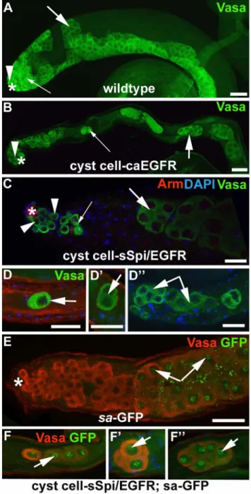
Figure 1. Reduction in EGF signaling disrupts germline differentiation. (A) Cartoon illustrating the cell types and their arrangement in a wildtype testis. Germline stem cells (GSCs) are located around the apical hub (white cells) and are enclosed by cyst stem cells (CySCs). Each gonialblast (GB) and its progeny are completely enclosed by two early-stage cyst cells (CC). The GB proliferates through exactly four rounds of TA-divisions and produces a cluster of 16 spermatogonia (SGs). After ceasing mitosis, the SGs enter terminal differentiation. They become spermatocytes (SCs), grow in size, undergo meiosis I and II, and finally differentiate into elongated spermatids. The surrounding cyst cells differentiate into late-stage cyst cells. (B) Apical region of a wildtype testis showing the germline cells in green (anti-Vasa) and the surrounding cyst cells in red (anti-GFP, UAS-GFP is driven by a cyst cell Gal4-transactivator,C784-Gal4). (C–F) Whole testes stained with DAPI. Genotypes and temperature regimens as indicated. Arrowheads: nuclei of early-stage germline cells, small arrows: spermatocyte nuclei, large arrows: spermatid heads, asterisks: apical tips of the testes. (C) A wildtype testis contains small, strongly DAPI-stained nuclei of early-stage germline cells exclusively in the apical region. (D) Astet/stet-testis is filled with small, strongly DAPI-stained nuclei. (E) A testis from astet/ stet; germline-sSpi-animal raised at 29uC appears wildtype. (F) A testis from astet/stet; germline-sSpi animal raised at 29uC and shifted to 18uC with several clusters of small, strongly DAPI-stained nuclei. Scale bars: 30mm.
properly enclosed by cytoplasmic extensions from exactly two early-stage cyst cells, as normally seen in wildtype testes. We discovered that EGF signaling has a dose-dependent effect on cyst development. When we reduced EGF signaling in testes, the germline cells appeared to be properly enclosed by cyst cells but were trapped in TA-divisions. When we increased EGF signaling in testes, the germline cells also appeared properly enclosed by cyst cells but the cysts entered terminal differentiation before the germline cells completed all four rounds of TA-divisions. These observations strongly suggest that EGF signaling from the germline to the cyst cells normally increases as the cysts develop and that this increase in EGF signaling leads to the production of different return signals with different effects on the germline. We further observed that simultaneous over-expression of EGF and EGFR promoted germline differentiation, while high levels of either EGF or EGFR alone had no effect on germline development. We conclude that the doses of EGF signaling are controlled at the level of both the active ligand produced by the germline cells and the available receptor molecules on the cyst cells as these two cell lineages develop. We propose that the increase in EGF signaling provides a temporal signature that guides the cysts through the early steps of development. This is a novel concept that may shed light on the principles how stem cell daughters differentiate into highly specialized cells.
Results
EGF Signaling from the Germline to the Surrounding Cyst Cells is Required for Spermatogonia to Proceed through TA-divisions
To investigate a dose-dependent effect of EGF signaling we used the UAS/Gal4- system that allows for temporal control of tissue-specific expression of target genes by exposing Drosophila to different temperatures. When Drosophila are exposed to a temperature of 18uC, the Gal4 transcription factor has low activity. In contrast, whenDrosophilaare exposed to a temperature of 29uC, Gal4 is highly active [50–52]. To address whether and how EGF signaling plays a role in cyst development past the enclosure event, we first generated testes in which we decreased EGF signaling and investigated the resulting effect on the cysts.
In wildtype testes, early-stage germline cells have small, bright nuclei (Figure 1C, arrowhead) when stained with the DNA-dye 4, 6-diamidino-2-phenylindole (DAPI). The more basally located spermatocytes have larger, less bright DAPI-stained nuclei (Figure 1C, small arrow), and the bundles of spermatids at the base have sickle-shaped DAPI-stained nuclei (Figure 1C, large arrow). We previously established that testes from stet1 mutant animals (stet/stet) are short and contain only cells with small, bright DAPI-stained nuclei (Figure 1D, arrowheads), while testes from
stet1 mutant animals expressing constructs for either the Stet protease or for secreted EGF ligand (stet/stet; germline-sSpi) are indistinguishable from wildtype testes when raised at 29uC (Figure 1E) [14]. We reasoned that Gal4 activity should decrease or cease upon shiftingstet/stet;germline-sSpi-animals from 29uC to 18uC. The germline cells within one cyst instet/stet;germline- sSpi-testes should continue to undergo TA-divisions but eventually have a lower dose of Spi compared to germline cells within a cyst in control testes. Hence a mutant phenotype should develop.
Testes from control siblings did not display any defects in spermatogenesis when shifted from 29uC to 18uC. However, after one day at 18uC, testes from stet/stet; germline-sSpi-animals frequently contained clusters of cells with small, bright DAPI-stained nuclei (Figure 1F, arrowheads, 80% of testes, n.100). We do not have the tools to measure the amount of Spi molecules.
However, the appearance of the clusters of over-proliferating germline cells instet/stet;germline-sSpi-testes shifted from 29uC to 18uC strongly suggests that the level of Spi in these testes is lower than the level of Spi molecules in a wildtype testes. Similar clusters of over-proliferating germline cells were observed in a variety of genetic backgrounds in which EGF signaling was reduced (Table 1), confirming that their appearance was not due to a genetic background mutation but characteristic for defects in EGF signaling.
In wildtype testes and testes fromstet/stet;germline-sSpi-animals raised at 29uC, we detected two cyst cells positive for the nuclear cyst cell marker Traffic jam (Tj, Figure 2A, arrowheads) associated with each cluster of transit amplifying spermatogonia (blue in Figure 2A) [53]. The two cyst cells enclosed the spermatogonia in cytoplasmic extensions, as visualized by the cell surface marker Armadillo (Arm, Figure 2A, arrows) [54]. Germline cells and cyst cells remained properly associated instet/stet;germline-sSpi-testes even after shifting the animals to 18uC for one or two days. In these testes, the clusters of over-proliferating early stage germline cells (blue in Figure 2B) were still enclosed by cytoplasmic extensions from two cyst cells, based on anti-Tj (Figure 2B, arrowheads) and anti-Arm staining (Figure 2B, arrows). We conclude that shiftingstet/stet;germline-sSpi-animals to 18uC does not disrupt germline enclosure, but does disrupt germline differentiation.
All cells within each enclosed cluster of over-proliferating germline cells in stet/stet; germline-sSpi-testes were small and expressed germline-specific markers, such as anti-Vasa (compare Figure 2D to 2C) [55]. The enclosed germline cells, however, did not undergo all four rounds of TA-divisions. In a wildtype testis, each GSC normally divides asynchronously to produce two distinct daughter cells, a new GSC and a gonialblast. The gonialblast and its daughters, in contrast, divide in a synchronous manner to produce equal precursor cells, the spermatogonia [10]. When control testes from wildtype animals were labeled with the M-phase marker anti-phosphorylated Histone H3 (pHH3), pHH3-positive spermatogonia were always seen in groups of two, four, or eight cells (Figure 2E, numbered 1–8, n.100). In testes fromstet/ stet;germline-sSpi-animals shifted from 29uC to 18uC for one or two days, only a fraction of the germline cells (Figure 2F–F99, blue)
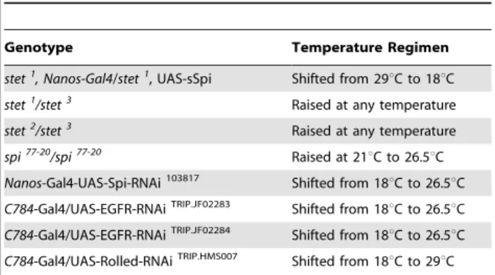
Table 1.Summary of genotypes examined.
Genotype Temperature Regimen
stet1
, Nanos-Gal4/stet1
, UAS-sSpi Shifted from 29uC to 18uC
stet1 /stet3
Raised at any temperature
stet2
/stet3 Raised at any temperature
spi77-20/spi77-20 Raised at 21uC to 26.5uC
Nanos-Gal4-UAS-Spi-RNAi103817 Shifted from 18uC to 26.5uC
C784-Gal4/UAS-EGFR-RNAiTRIP.JF02283 Shifted from 18
uC to 26.5uC
C784-Gal4/UAS-EGFR-RNAiTRIP.JF02284 Shifted from 18
uC to 26.5uC
C784-Gal4/UAS-Rolled-RNAiTRIP.HMS007 Shifted from 18uC to 29uC
within a single cyst were positive for pHH3 (Figure 2F–2F99, pHH3-positive cells are marked by asterisks, n.20). We only detected either single pHH3-positive cells within a cyst containing over-proliferating germline cells (Figure 2F), several single
pHH3-positive cells within a cyst containing over-proliferating germline cells (Figure 2F9), or small groups of pHH3-positive cells within a cyst containing over-proliferating germline cells (Figure 2F99shows an example of a cyst containing two pHH3-positive cells, next to a cyst with only one pHH3-positive cell). Confirming the role for EGF signaling in guiding TA-divisions, we observed the same phenotype in the testes of animals carrying a temperature sensitive allele ofspi, spi77-20(spi/spi), raised at semi-permissive temperature (data not shown). These findings strongly suggest that EGF signaling within the cysts is required for the production of a return signal that allows the germline cells to undergo all four rounds of TA-divisions. They further suggest that the dose of EGF signaling required for the production of this return signal is higher than the dose of EGF signaling required for germline enclosure.
A High Dose of EGF Signaling caused Spermatogonia to Bypass TA-divisions
To generate cysts with increased EGF signaling we also took advantage of the UAS-Gal4-system. Flies carrying constructs for the expression of constitutively active versions of the EGFR (caEGFR), UAS-EGFRltop and UAS-EGFRA887T, cause hyper-activity of the EGF pathway when expressed inDrosophilaovaries or eyes [56,57]. We expressed these two well-established tools, as well as constructs expressing secreted Spi (UAS-sSpi) and a wildtype version of the EGFR (UAS-EGFR) in testes of otherwise wildtype animals. The constructs were specifically expressed in the cyst cells using two different cyst cell Gal4-transactivators,C784 -Gal4 and tj-Gal4 (cyst cell-caEGFR-testes and cyst cell-sSpi/ EGFR-testes). To circumvent expression of the constructs during development, animals were raised at 18uC and also carried a ubiquitously expressed, temperature sensitive Gal80 (tub-Gal80ts), a known negative regulator of Gal4, that has high activity at 18uC and low activity at 29uC [52,58].
Testes from adult wildtype and sibling controls (non-shifted cyst cell-caEGFR-animals, non-shifted cyst cell-sSpi/EGFR-animals, animals carrying only the Gal4-transactivators or only the UAS-constructs shifted to 29uC for seven days) did not display observable defects in spermatogenesis. They contained Vasa-positive GSCs (Figure 3A, arrowhead), gonialblasts, and sper-matogonia (Figure 3A, small arrow) in the apical region, followed by clusters of spermatocytes (Figure 3A, large arrow) in more basal regions of the testes. Testes from cyst cell-caEGFR-animals and from cyst cell-sSpi/EGFR-animals shifted to 29uC for seven days were much thinner than wildtype testes and contained fewer germline cells than control testes (Figures 3B, 3C). We detected single germline cells in the apical region of cyst cell-caEGFR-testes and cyst cell-sSpi/EGFR-testes (Figures 3B, 3C, arrowheads), followed by very few clusters of spermatogonia (Figures 3B, 3C, small arrows), and, more basally, clusters of spermatocytes (Figures 3B, 3C, large arrows, Table 2).
Many of the spermatocytes in cyst cell-sSpi/EGFR-testes were single (Figure 3D, arrow), or in clusters of only two (Figure 3D9, arrow), four (Figure 3D99, arrows), or eight cells instead of the normal 16, 32, or 64-cell clusters and expressed spermatocyte-specific molecular markers, such as a spermatocyte arrest-Green Fluorescent Protein (sa-GFP, compare Figures 3F–F99to Figure 3E, arrows point to GFP in the germline nucleoli) [59]. Our data suggest that a high dose of EGF signaling caused the germline cells to enter the spermatocyte differentiation program prior to completing all four rounds of TA-divisions. Single spermatocytes and small clusters of spermatocytes were detected in 95% of the cyst cell-sSpi/EGFR-testes (n = 76) but were not found in testes fromw1118-animals (n = 80), in testes from cyst cell- sSpi/EGFR-animals prior to the temperature shift (n = 37), or in control testes
Figure 2. The germline cells within cysts in testes with decreased EGF signaling divided out of synchrony. (A–F99) Germline and cyst cells in testes from wildtype animals and stet, germline-sSpi-animals shifted from 29uC to 18uC for two days. Genotypes and stainings as indicated. (A, B) In (A) the apical region of a wildtype testis and B) astet/stet, germline-sSpi-testis, germline cells (blue) are enclosed by Arm-positive (red) cytoplasmic extensions (arrows) from two Tj-positive (green) cyst cells (arrowheads). Note that the cyst in the stet/stet, germline-sSpi-testis contains more than 16 early-stage germline cells. (C, D) The Arm-enclosed (red) germline cells (arrowheads) express the germline-specific marker Vasa (green) in testes from (C) wildtype and (D)stet/stet, germline-sSpi-animals. (E–F99) In a (E) wildtype testis, all spermatogonia (blue) within a single cyst are in division (red, and numbered 1–8), while in (F-F99)stet/stet, germline-sSpi-testes, only a fraction of the germline cells within one cyst divide (red and marked by asterisks). Note that the germline cells are enclosed in Arm-positive cyst cell cytoplasmic extensions (green). Large asterisks: apical tips of the testes, small arrows: cytoplasmic extensions of cyst cells, scale bars: 30mm.
from animals carrying only the Gal4-transactivators (n = 42) or only the UAS-constructs (n = 58) shifted to 29uC. Thus, their appearance in the testes of cyst cell-sSpi/EGFR-animals shifted to 29uC was specifically due to the increase in EGF signaling. Conversely, over-expression of sSpi and EGFR in the germline via
the nanos-Gal4 transactivator did not result in any observable
defects in the testes (Table 2). This confirms that the mutant phenotype was due to increased EGF signaling in cyst cells.
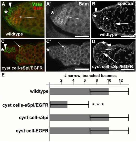
Testes from cyst cell-sSpi/EGFR-animals already contained fewer germline cells in the apical region after only two days at 29uC, based on expression of the molecular germline markers Vasa,bag-of-marbles(bam), anda-spectrin. Wildtype testes contain a wide area of cells that co-express Vasa and the spermatogonial marker Bam in the apical region (Figures 4A, 4A9, arrow) [60,61]. In addition, the spermatogonia containa-spectrin-positive fusomes of characteristic shape and size [62]. In wildtype testes, the GSCs at the apical tip contained round a-spectrin-positive fusomes (Figure 4B, arrowhead), the spermatogonia contained narrow, branched a-spectrin-positive fusomes (Figure 4B, small arrows), and the spermatocytes contained wide, branched a -spectrin-positive fusomes (Figure 4B, large arrow). After only two days at 29uC, testes from cyst cell-sSpi/EGFR-animals contained very few cells in the apical region that co-expressed Vasa and Bam (Figures 4C, 4C9, arrow). Furthermore, these testes contained hardly any narrowa-spectrin-positive fusomes, normally found in spermatogonia (Figure 4D, small arrow). The cyst cell-sSpi/ EGFR-testes did contain rounda-spectrin-positive fusomes at the apical tip, indicating that GSCs were present (Figure 4D, arrowhead) and wide, branched a-spectrin-positive fusomes, as normally found in spermatocytes (Figure 4D, large arrow). To demonstrate the difference in the number of spermatogonia found in control and cyst cell-sSpi/EGFR-testes, we counted and compared the narrow fusomes detected in a single focal plane per testis. Within one focal plane, wildtype testes contained an average of eight (+/2three) narrow, branched fusomes (n.50,
Figure 4E). Cyst cell-sSpi/EGFR-testes either lacked, or contained less than five fusomes (average of three (+/2three) within one focal plane (n = 36, Figure 4E). The difference in the number of narrow fusomes is statistically relevant, with a P-value,0.0001 (according
Table 2.Summary of over-expression of EGF signaling components and the observed effects on the germline cells.
Genotype Few SGs ,16SCs/cyst
C784-Gal4/tub-Gal80ts; UAS-EGFRA887T Yes Yes
Tj-Gal4/tub-Gal80ts; UAS-EGFRA887T Yes Yes
C784-Gal4/tub-Gal80ts; UAS-EGFRltop Yes Yes
Tj-Gal4/tub-Gal80ts; UAS-EGFRltop Yes Yes
C784-Gal4/tub-Gal80ts; UAS-sSpi/UAS-EGFR Yes Yes
Tj-Gal4/tub-Gal80ts; UAS-sSpi/UAS-EGFR Yes Yes
Nanos-Gal4-UAS-sSpi/UAS-EGFR No No
C784-Gal4/tub-Gal80ts; UAS-EGFR No No
Tj-Gal4/tub-Gal80ts;UAS-EGFR No No
C784-Gal4/tub-Gal80ts; UAS-EGFR/UAS-GalH No No
C784-Gal4/tub-Gal80ts;UAS-sSpi No No
Tj-Gal4/tub-Gal80ts;UAS-sSpi No No
C784-Gal4/tub-Gal80ts;UAS-sSpi/UAS-sSpi No No
Animals from various genotypes were shifted from 18uC to 29uC within one to seven days after hatching and their testes investigated at seven days after the shift. Only expression of caEGFR or co-expression of Spi and EGFR in the cyst cells produced mutant phenotypes (n.30). The testes contained fewer spermatogonia (SGs) compared to controls and contained clusters of less than 16 spermatocytes (SCs) per cyst.
doi:10.1371/journal.pone.0070678.t002
Figure 3. A high dose of EGF signaling promoted differentia-tion of germline cells into the spermatocyte stage.(A–C) Apical regions of testes showing Vasa-positive germline cells (green). Note that (B) cyst cell-caEGFR-testes and (C) cyst cell-sSpi/EGFR-testes raised at 18uC and shifted to 29uC for seven days contain fewer spermatogonia and spermatocytes compared to a (A) wildtype testis. (D–D99) Germline cells along the testis coil are the size of spermatocytes and found in groups of less than 16 cells. (E) Spermatocytes but not the early germline cells in the apical region of a control testis express GFP (arrows) from asa-GFP construct. (F–F9) The large germline cells along the coil of cyst cell-sSpi/EGFR-testes express GFP from the sa-GFP construct (arrows). Genotypes as indicated. Arrowheads: GSCs, small arrows: spermatogonia, large arrows: spermatocytes. Asterisks: apical tips of the testes, scale bars: 30mm.
to the student’s t-test, and marked by asterisks). Together, our data suggest that by two days after the temperature shift, many of the spermatogonia had already differentiated into spermatocytes. Consistent with the idea that the spermatogonia differentiated into spermatocytes, no apparent hallmarks of apoptotic cell death were observed in testes from cyst cell-sSpi/EGFR-animals (n = 50) compared to control-animals (n = 50) at one, two, or seven days after the shift to 29uC on the basis of the cell death assay, TUNEL (not shown).
A High Dose of EGF Signaling Promoted Cyst Cell Differentiation
Communication between germline cells and cyst cells did not appear to be disrupted in cyst cell-sSpi/EGFR-testes as the germline cells and the cyst cells remained properly associated. The early-stage germline cells at the apical tip (Figures 5A, 5A9) and the small clusters of spermatocytes (Figure 5B) along the testes coil were both enclosed by cyst cell cytoplasmic extensions (arrows in
Figures 5A9, B), even after seven days at 29uC. The cyst cells in cyst cell-sSpi/EGFR-testes appeared to differentiate along with the enclosed germline cells upon the temperature shift. After the germline cells cease TA-divisions and become spermatocytes, both the nuclei of the germline cells and the nuclei of the surrounding cyst cells grow in size (in the following referred to as the growth phase) [10]. Cyst differentiation is characterized by changes in the expression of the transcription factors, Tj and Eya. Tj is considered an early-stage cyst cell marker due to its expression in CySCs, cyst cells prior to the growth phase, and cyst cells during the growth phase [53]. Eya is considered a late-stage cyst cell marker as it is expressed at high levels in cyst cells after the growth phase [63]. We detected a low level of Eya in the small size cyst cell nuclei shortly before growth phase begins. During the growth phase, the level of Eya expression increased as the cyst cell nuclei became larger. After the growth phase, the large cyst cell nuclei continued to express Eya at high levels (Figure 5C, 5C9, compare the size and staining intensity of the cyst cell nuclei within the
Figure 4. Testes with increased EGF signaling contained few spermatogonia.(A) A wildtype testis showing co-expression of Vasa (green) and Bam (red) in many germline cells in the apical region. (A9) Same testis as in (A) showing Bam expression only. B) A wildtype testis labeled with antibodies againsta-spectrin. Arrowhead: GSC containing a round fusome, small arrows: narrow fusomes interconnecting spermatogonia, large arrow: wide fusomes interconnecting spermatocytes. (C) A cyst cell-sSpi/EGFR-testis showing co-expression of Vasa (green) and Bam (red) in only a few germline cells. (C9) Same testis as in (C) showing Bam expression only. (D) A cyst cell-sSpi/EGFR-testis showing the presence of only few narrow fusomes (arrows), and the presence of GSCs with a round fusome (arrowhead), and spermatocytes with wide fusomes (large arrow). (E) Bar graph showing the differences in numbers of narrow branched fusomes between testes from control and experimental animals. Three asterisks mark a statistically significant difference compared to the control. Genotypes and stainings as indicated, asterisks in (A-D): apical tips of the testes, scale bars: 30mm.
white circles). When control testes from cyst cell-sSpi/EGFR-animals raised at 18uC (n = 20) were double-labeled with Anti-Tj and anti-Eya, a small fraction of the cyst cell nuclei that are associated with germline cells expressed both markers (yellow in Figure 5). By analyzing each testis in all focal planes, we detected an average of 14 (+/2eight) cyst cells that were both Tj-positive and Eya-positive (Tj+
, Eya+
-cyst cells, note that the testis in Figure 5C9 shows four Tj+
, Eya+
-cyst cells in one focal plane). Testes from cyst cell-sSpi/EGFR-animals shifted to 29uC also contained Tj+
, Eya+
-cyst cells yet their numbers changed dependent on how long the animals were at 29uC. At 36 hours after the temperature shift, the cyst cell-sSpi/EGFR-testes (n = 20) contained more Tj+
, Eya+
-cyst cells than the control testes (compare Figures 5D, 5D9 to 5C, 5C9). Cell counts revealed an average of 22 (+/211) Tj+
, Eya+
-cyst cells (statistical difference in the number of Tj+
, Eya+
-cyst cells compared to non-shifted cyst cell-sSpi/EGFR-testes: P,0.0001, according to the student’s t-test). This shows that over-expression of sSpi and EGFR increased the number of cysts that have entered the growth phase. By 48 hours after the temperature shift cyst cell-sSpi/EGFR-testes (n = 20) contained fewer Tj+
, Eya+
-cyst cells (Figure 5E, E9) with an average of only eight (+/2eight). These are statistically fewer Tj+
, Eya+
-cyst cells compared to Tj+
, Eya+
-cyst cells in non-shifted testes (P,0.001). This suggests that after 48 hours at 29uC most of the cyst cells that were associated with germline cells have differentiated into Tj-negative, Eya-positive late-stage cyst cells. These observations suggest that a high dose of EGF either directly or indirectly promotes cyst cell differentiation.
The Reduction in Spermatogonia was Dependent on Co-expression of Both EGF and EGFR
Interestingly, while the expression of caEGFR or the co-expression of sSpi and EGFR in cyst cells caused a phenotype shortly after the shift to 29uC, expression of sSpi (cyst cell- sSpi-testes) or the EGFR (cyst cell-EGFR-sSpi-testes) alone using either of the two cyst cell Gal4-transactivators did not have an effect on the cysts (n.30, Table 2). For example, over-expression of only the sSpi-ligand or the EGFR in cyst cells did not cause a reduction in the number of narrow fusomes that interconnect spermatogonia. Testes from either genotype contained an average of eight (+/
2three) thin, branched fusomes (n.30) in a single focal plane (Figure 4E) compared to controls with an average of eight (+/
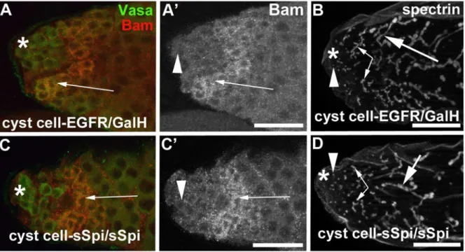
2three, Figure 4E). This is a surprising observation, as we should have the same number of Gal4 molecules in all experiments and the same or even higher levels of sSpi or EGFR expression as we used the same UAS-sSpi and UAS-EGFR insertions for these experiments. We next increased the amount of EGFR molecules produced in the cyst cells by co-expressing EGFR with GalH, a potentiator of Gal4 (cyst cell-EGFR/GalH) [64]. We did not observe an effect on the germline cells in testes from cyst cell-EGFR/GalH-animals (Table 2). Similarly, increasing the number of sSpi molecules by expressing two copies of secreted Spi in the cyst cells (cyst cell-sSpi/sSpi) also did not have an effect on the germline cells (Table 2). As in the controls, cyst cell-EGFR/GalH-testes (Figures 6A, 6A9, 6B) and cyst cell-sSpi/sSpi-testes (Figures 6C, 6C9, 6D) contained a wide area of spermatogonia co-expressing Vasa and Bam (arrows in Figures 6A, 6A9, 6C, 6C9) and contained many narrow fusomes as normally seen in interconnected spermatogonia (Figures 6B, 6D, small arrows). We conclude that the reduction in the number of early-stage cysts containing spermatogonia and the presence of late-stage cysts containing less than 16 spermatocytes was dependent on the over-expression of both the ligand and the receptor.
Discussion
Here we propose that a temporal increase in EGF signaling between germline cells and surrounding cyst cells regulates cyst development. We present genetic evidence that a low dose of EGF signaling is required for the early stages of germline development, the progression of the germline cells through TA-divisions, and that a high dose of EGF signaling promotes the entry of germline and cyst cells into terminal differentiation. Reduction in EGF signaling led to the accumulation of cyst cell-enclosed, tumor-like aggregates of early-stage germline cells. The germline proliferation defects in EGF mutant testes were different from the germline proliferation defects reported for other mutants displaying germ-line tumors. For example, germgerm-line cells mutant forbamorbenign gonial cell neoplasm (bgcn)over-proliferated beyond the 16-cell stage, producing large clusters of interconnected spermatogonia [60,65]. The same phenotype was observed when germline cells were associated with cyst cells mutant for TGFbsignaling or when the germline cells overexpressed the TGFbligand decapentaplegic(dpp) [33,36,66]. Notably, the germline cells in testes with dpp over-expression divided in synchrony, as large cysts were detected in which all of the spermatogonia expressed the mitosis marker pHH3 [34]. In testes fromstet/stet;germline-sSpi-animals shifted from 29uC to 18uC and in testes from spi77-20- animals raised at semi-permissive temperature, large cysts were detected as well. However, the germline cells within a single cyst divided out of synchrony as we observed that only a few of them were positive for pHH3. It has not been revealed yet how the decision of the gonialblast to divide synchronously is regulated. Synchronous versus asynchronous cell division could depend on morphology changes of the fusome. When a GSC or a gonialblast divides, the fusome morphology changes from spherical to dumbbell-shaped. During the division of a GSC, the fusome breaks and one spherical fusome is formed in each of the resulting daughter cells. When a gonialblast divides, the dumbbell structure of the fusome is maintained in the resulting 2-cell stage spermatogonia [62,67]. We did not find indications that the asynchronous divisions observed in testes with reduced EGF signaling is due to a direct role of EGF in preventing fusome breakage in the germline cells. More likely than having a direct effect on fusome morphology, we hypothesize that EGF signaling regulates a return signal from the cyst cells to the germline cells that promotes synchronous cell divisions. We propose that EGF signaling normally regulates a return mecha-nism from the cyst cells that reduces stem cell characteristics in the spermatogonia which, in turn, promotes the progression of the spermatogonia through TA-divisions.
and cyst cell-sSpi/EGFR-animals suggests that a mechanism exists that prevents the entry of spermatogonia into the spermatocyte-stage prior to the completion of exactly four rounds of TA-divisions and that the EGF pathway plays a role in this mechanism. However, how EGF signaling from the germline to the cyst cells ties into germline-intrinsic activities of Bam and Nup98-96 is yet to be explored.
Together, our phenotypic analyses suggest that EGF signaling plays a rather complex role in the developing cysts. Based on the mutant phenotypes caused by the loss, reduction, and increase of EGF signaling, we propose that EGF signaling between germline and cyst cells increases as the cysts develop and that different doses of EGF signaling, in turn, induce distinct responses in the cyst cells: germline enclosure, the production of return signals that allow for the germline cells to progress through the early steps of
development, the spermatogonia TA-divisions, and the production of return signals that promote terminal differentiation of the cysts (Figure 7). It is possible that the three different responses depend on three threshold levels of EGF signaling (black lines in Figure 7). Alternatively, a continuous increase in EGF signaling (blue line in Figure 7) may promote a continuous progression of the cysts towards terminal differentiation, thereby gradually reducing stem cell characteristics and increasing differentiation competence. The view that spermatogonia may retain stem cell characteristics is consistent with the observation that they can revert into GSCs when exposed to Unpaired, the ligand activating the JAK/STAT pathway [70–72].
Different doses of EGF signaling also guide the development of other tissues, for example photoreceptor development in the developing eye [57,73]. Our findings suggest a different
mecha-Figure 5. A high dose of EGF signaling promoted cyst cell differentiation.(A) Apical region of a testis from a cyst cell-sSpi/EGFR-animal showing that the germline cells (green) are enclosed by Arm-positive cytoplasmic extensions (red) of cyst cells. (A9) Same testis as in (A) showing the Arm-positive cytoplasmic extensions of cyst cells (arrows) only. (Note that not all cytoplasmic extensions are in focal plane) (B) A cluster of two spermatocytes (green) in a cyst cell-sSpi/EGFR-testes is enclosed in Arm-positive cyst cell cytoplasmic extensions (red, arrows). (C–E9) Apical regions of testes labeled with (left panel) only Eya and (right panel) co-labeled with Eya (red) and Tj (green). Cyst cells appear to accumulate Eya as they differentiate. The left-most circles in each testis show the most apical cyst cell nuclei that express low levels of Eya. These early-stage cyst cells also express high levels of Tj. The middle circles show cyst cells in the growth phase that express high levels of both, Eya and Tj. Small numbers indicate the numbers of the Eya+
, Tj+
-cells in one focal plane. The right-most circles show cyst cells after the growth phase expressing high levels of Eya, but low levels of Tj. Genotypes as indicated, scale bars: 30mm.
nism by which the different doses of EGF signaling are generated. In otherDrosophilatissues, a spatial EGF gradient is generated by the diffusion of the ligand away from the source [73–75]. In testes, over-expression of either the ligand or the receptor alone was not sufficient to promote germline differentiation, arguing against a diffusion model. This is not surprising as the germline cells are completely enclosed by cyst cells and EGF thus cannot diffuse
outside the cysts. Only when both the ligand and the receptor were overexpressed was a strong phenotype observed. We propose that different doses of EGF signaling are created by the coordinated increase of EGF signaling components in the germline cells and the cyst cells as they mature. This, in turn, could create a temporal signature that determines the behavior of the individual cysts. It remains to be ascertained if expression data, once reliable tools for
Figure 6. Increased EGF signaling depends on both the dose of EGF and the dose of EGFR.(A–D) In testes expressing either (A, A9, B) EGFR and GalH or (C, C9, D) two copies of sSpi, many Vasa-positive germline cells (green in A and C) also expressed Bam (A, A9, C, C9arrows) and contained many narrow, brancheda-spectrin-positive fusomes (B, D, small arrows). Arrowheads: round fusomes in GSCs, small arrows: narrow fusomes interconnecting spermatogonia, large arrows: wide fusomes interconnecting spermatocytes, asterisks: apical tips of testes, scale bars: 30mm.
doi:10.1371/journal.pone.0070678.g006
Figure 7. A temporal signature of EGF regulates early stages of cyst development.Model demonstrating the dose-dependent effects of EGF signaling on the germline and cyst cells as they develop through the early stages of spermatogenesis (bottom). Blue line indicates a gradual increase in EGF and EGFR, and black lines indicate increasing threshold doses of EGF and EGFR. Examples of genotypes in which the observed defects were studied are listed on top.
imaging EGF expression levels or receptor stimulation become available, can confirm our hypothesis.
How could such an increase in ligand and receptor be regulated? A simple explanation for an increase in EGF would be that its amount depends on the number of germline cells. Within one cyst, the number of spermatogonia that can produce the active ligand increases during TA-divisions. At the 16-cell stage, the amount of ligand may be sufficient to induce the production of a signal in the cyst cells that promotes spermatocyte differentiation. However, such a simple mechanism is not likely as cysts in testes with reduced EGF signaling contained a large number of germline cells that eventually should have produced a high enough dose of EGF for differentiation, yet these germline cells still over-proliferated. More likely, the amount of EGF may depend either on transcriptional up-regulation of signaling molecules within the developing germline cells or on an increase of ligand secretion from the developing germline cells. Ligand secretion is a critical step during development of otherDrosophila
tissues, such as the eye and the nervous system. In the eye, for example, the cleaved Spi ligand is retained in the endoplasmatic reticulum to regulate the range of EGF signaling [76,77]. In a similar manner, transcriptional up-regulation of the EGFR or a reduction in the amount of negative regulators of the EGFR may explain the increase in EGFR availability as the cysts develop. Along with this, the increase in the size of the cyst cells as they develop may serve as a vehicle for a more efficient display of EGFR molecules.
In summary, our data strongly support the idea that EGF signaling between germline cells and cyst cells increases as they transition from early to later stages of development, and that this increase guides early steps of gametogenesis. It will be interesting to learn if threshold doses of signaling molecules guide differen-tiation of stem cell lineages in mammals as well.
Materials and Methods
Fly Strains
Flies were raised on standard cornmeal molasses agar medium.
white1118,tub-Gal80ts, UAS-Rolled-RNAiTRIP.HMS007, and balanc-er chromosomes are as described in [78] and wbalanc-ere obtained from the Bloomington stock center. Likewise, EGFR [79], UAS-GalH [64], C784-Gal4 [80], and nanos-Gal4 [81] were obtained from the Bloomington stock center. UAS-EGFR-RNAiTRIP.JF02283 and UAS-EGFR-RNAiTRIP.JF02284 were obtained from the HarvardDrosophilaRNAi Screening Center, UAS-Spi-RNAi103817 from the Vienna Drosophila Research Center, tj-Gal4 from the Kyoto Stock Center, UAS-EGFRA887T[57] from Nick Baker, and UAS-EGFRltop [56] from Trudi Schupbach. stet-alleles are
described in [14] and the temperature sensitive spi77-20-allele is described in [48]. The UAS-sSpi-construct [82] was obtained
from Ben Shilo and injected into flies by the company The Best Gene.
UAS/Gal4 Expression Studies
Crosses were set up either at 18uC or at 29uC and the animals were shifted to different temperatures as outlined in the results.
Immunofluorescence
Immunofluorescence experiments were performed as previously described [14]. Staining was observed with a Zeiss Axiophot microscope and images were taken with a CCD camera using an Apotome and Axiovision Rel Software. Single slices and z-stack-composites of up to ten slices (for DAPI and anti-a-Spectrin images) are shown. Mouse anti-a-Spectrin 3A9 (1:10) developed by D. Branton and R. Dubreuil, mouse-anti-Bam (1:5) developed by D. McKearin, and mouse anti-Armadillo (1:10) developed by E. Wieschaus were obtained from the Developmental Studies Hybridoma Bank developed under the auspices of the NICHD and maintained by The University of Iowa, Department of Biological Sciences, Iowa City, IA 52242. Rabbit anti-phosphor-ylated Histone-H3 (Ser-10, 1:500) was obtained from Millipore and goat anti-Vasa (1:100) was obtained from Santa Cruz Biotechnology (dc-13). Guinea pig-anti-Tj (1:5000) was a gift from Dorothea Godt. Fluorophore-coupled secondary antibodies (Mo-lecular Probes) were used at 1:1000.
TUNEL-assay
Testes were dissected in Tissue Isolation Buffer [14], incubated with the TUNEL solutions following the manufacturer’s (Roche-Applied Scientific) instructions, and then used for immunofluo-rescence as described above.
Acknowledgments
The authors are indebted to Ricky Zoller and Sampreet Reddy for technical assistance. We thank Xin Chen for the sa-GFP flies, Scott Dougan, Robert Beckstead, Steve DiNardo, Helen McNeill, and the UGA Alliance for Integrated Research for helpful discussions, and Ricky Zoller, Robert Ng, Wolfgang Lukowitz, and unknown reviewers for comments on the manuscript.
Author Contributions
Conceived and designed the experiments: CS AGH. Performed the experiments: AGH YQ BBP CS. Analyzed the data: AGH YQ BBP CS. Contributed reagents/materials/analysis tools: AGH YQ BBP CS. Wrote the paper: AGH CS.
References
1. Weissman IL (2000) Translating stem and progenitor cell biology to the clinic: barriers and opportunities. Science 287: 1442–1446.
2. Pellettieri J, Sanchez Alvarado A (2007) Cell turnover and adult tissue homeostasis: from humans to planarians. Annu Rev Genet 41: 83–105. 3. Rabbani P, Takeo M, Chou W, Myung P, Bosenberg M, et al. (2011)
Coordinated activation of Wnt in epithelial and melanocyte stem cells initiates pigmented hair regeneration. Cell 145: 941–955.
4. Nishimura EK (2011) Melanocyte stem cells: a melanocyte reservoir in hair follicles for hair and skin pigmentation. Pigment Cell Melanoma Res 24: 401– 410.
5. Devine SM, Hoffman R (2000) Role of mesenchymal stem cells in hematopoietic stem cell transplantation. Curr Opin Hematol 7: 358–363.
6. Festa E, Fretz J, Berry R, Schmidt B, Rodeheffer M, et al. (2011) Adipocyte lineage cells contribute to the skin stem cell niche to drive hair cycling. Cell 146: 761–771.
7. Li T, Wu Y (2011) Paracrine molecules of mesenchymal stem cells for hematopoietic stem cell niche. Bone Marrow Res 2011: 353878.
8. Zoller R, Schulz C (2012) The Drosophila cyst stem cell lineage: Partners behind the scenes? Spermatogenesis 2: 145–157.
9. Hardy RW, Tokuyasu KT, Lindsley DL, Garavito M (1979) The germinal proliferation center in the testis of Drosophila melanogaster. J Ultrastruct Res 69: 180–190.
10. Fuller MT (1993) Spermatogenesis inDrosophila. In: Bate M, Martinez Arias, A., editor. The development ofDrosophila melanogaster. Cold Spring Harbor, New York, USA: Cold Spring Harbor Laboratory Press. 71–148.
11. Yamashita YM, Jones DL, Fuller MT (2003) Orientation of asymmetric stem cell division by the APC tumor suppressor and centrosome. Science 301: 1547– 1550.
12. Cheng J, Tiyaboonchai A, Yamashita YM, Hunt AJ (2011) Asymmetric division of cyst stem cells in Drosophila testis is ensured by anaphase spindle repositioning. Development 138: 831–837.
14. Schulz C, Wood CG, Jones DL, Tazuke SI, Fuller MT (2002) Signaling from germ cells mediated by the rhomboid homolog stet organizes encapsulation by somatic support cells. Development 129: 4523–4534.
15. Riparbelli MG, Colozza G, Callaini G (2009) Procentriole elongation and recruitment of pericentriolar material are downregulated in cyst cells as they enter quiescence. J Cell Sci 122: 3613–3618.
16. Gonczy P, Viswanathan S, DiNardo S (1992) Probing spermatogenesis in Drosophila with P-element enhancer detectors. Development 114: 89–98. 17. Papagiannouli F, Mechler BM (2009) discs large regulates somatic cyst cell
survival and expansion in Drosophila testis. Cell Res 19: 1139–1149. 18. Potten CS, Loeffler M (1990) Stem cells: attributes, cycles, spirals, pitfalls and
uncertainties. Lessons for and from the crypt. Development 110: 1001–1020. 19. Huckins C (1971) The spermatogonial stem cell population in adult rats. I. Their
morphology, proliferation and maturation. Anat Rec 169: 533–557. 20. Blanpain C, Fuchs E (2006) Epidermal stem cells of the skin. Annu Rev Cell Dev
Biol 22: 339–373.
21. Metcalf D (2007) Concise review: hematopoietic stem cells and tissue stem cells: current concepts and unanswered questions. Stem Cells 25: 2390–2395. 22. Hanahan D, Weinberg RA (2000) The hallmarks of cancer. Cell 100: 57–70. 23. Reya T, Morrison SJ, Clarke MF, Weissman IL (2001) Stem cells, cancer, and
cancer stem cells. Nature 414: 105–111.
24. Desai BS, Shirolikar S, Ray K (2009) F-actin-based extensions of the head cyst cell adhere to the maturing spermatids to maintain them in a tight bundle and prevent their premature release in Drosophila testis. BMC Biol 7: 19. 25. Rotkopf S, Hamberg Y, Aigaki T, Snapper SB, Shilo BZ, et al. (2011) The
WASp-based actin polymerization machinery is required in somatic support cells for spermatid maturation and release. Development 138: 2729–2739. 26. Kiger AA, Jones DL, Schulz C, Rogers MB, Fuller MT (2001) Stem cell
self-renewal specified by JAK-STAT activation in response to a support cell cue. Science 294: 2542–2545.
27. Tulina N, Matunis E (2001) Control of stem cell self-renewal in Drosophila spermatogenesis by JAK-STAT signaling. Science 294: 2546–2549. 28. Leatherman JL, Dinardo S (2010) Germline self-renewal requires cyst stem cells
and stat regulates niche adhesion in Drosophila testes. Nat Cell Biol 12: 806– 811.
29. Michel M, Kupinski AP, Raabe I, Bokel C (2012) Hh signalling is essential for somatic stem cell maintenance in the Drosophila testis niche. Development 139: 2663–2669.
30. Amoyel M, Sanny J, Burel M, Bach EA (2013) Hedgehog is required for CySC self-renewal but does not contribute to the GSC niche in the Drosophila testis. Development 140: 56–65.
31. Shivdasani AA, Ingham PW (2003) Regulation of stem cell maintenance and transit amplifying cell proliferation by tgf-beta signaling in Drosophila spermatogenesis. Curr Biol 13: 2065–2072.
32. Kawase E, Wong MD, Ding BC, Xie T (2004) Gbb/Bmp signaling is essential for maintaining germline stem cells and for repressing bam transcription in the Drosophila testis. Development 131: 1365–1375.
33. Bunt SM, Hime GR (2004) Ectopic activation of Dpp signalling in the male Drosophila germline inhibits germ cell differentiation. Genesis 39: 84–93. 34. Schulz C, Kiger AA, Tazuke SI, Yamashita YM, Pantalena-Filho LC, et al.
(2004) A misexpression screen reveals effects of bag-of-marbles and TGF beta class signaling on the Drosophila male germ-line stem cell lineage. Genetics 167: 707–723.
35. Li CY, Guo Z, Wang Z (2007) TGFbeta receptor saxophone non-autonomously regulates germline proliferation in a Smox/dSmad2-dependent manner in Drosophila testis. Developmental Biology 309: 70–77.
36. Matunis E, Tran J, Gonczy P, Caldwell K, DiNardo S (1997) punt and schnurri regulate a somatically derived signal that restricts proliferation of committed progenitors in the germline. Development 124: 4383–4391.
37. Wiley LM, Adamson ED, Tsark EC (1995) Epidermal growth factor receptor function in early mammalian development. Bioessays 17: 839–846.
38. Nilson LA, Schupbach T (1999) EGF receptor signaling in Drosophila oogenesis. Curr Top Dev Biol 44: 203–243.
39. Kiger AA, White-Cooper H, Fuller MT (2000) Somatic support cells restrict germline stem cell self-renewal and promote differentiation. Nature 407: 750– 754.
40. Tran J, Brenner TJ, DiNardo S (2000) Somatic control over the germline stem cell lineage during Drosophila spermatogenesis. Nature 407: 754–757. 41. Shilo BZ (2003) Signaling by the Drosophila epidermal growth factor receptor
pathway during development. Exp Cell Res 284: 140–149.
42. Moghal N, Sternberg PW (2003) The epidermal growth factor system in Caenorhabditis elegans. Exp Cell Res 284: 150–159.
43. Normanno N, De Luca A, Bianco C, Strizzi L, Mancino M, et al. (2006) Epidermal growth factor receptor (EGFR) signaling in cancer. Gene 366: 2–16. 44. Bier E, Jan LY, Jan YN (1990) rhomboid, a gene required for dorsoventral axis establishment and peripheral nervous system development in Drosophila melanogaster. Genes Dev 4: 190–203.
45. Urban S (2006) Rhomboid proteins: conserved membrane proteases with divergent biological functions. Genes Dev 20: 3054–3068.
46. Urban S, Lee JR, Freeman M (2002) A family of Rhomboid intramembrane proteases activates all Drosophila membrane-tethered EGF ligands. EMBO J 21: 4277–4286.
47. Parrott BB, Hudson A, Brady R, Schulz C (2012) Control of germline stem cell division frequency - a novel, developmentally regulated role for epidermal growth factor signaling. PLoS One 7: e36460.
48. Sarkar A, Parikh N, Hearn SA, Fuller MT, Tazuke SI, et al. (2007) Antagonistic roles of Rac and Rho in organizing the germ cell microenvironment. Curr Biol 17: 1253–1258.
49. Schlessinger J (2004) Common and distinct elements in cellular signaling via EGF and FGF receptors. Science 306: 1506–1507.
50. Brand AH, Perrimon N (1993) Targeted gene expression as a means of altering cell fates and generating dominant phenotypes. Development 118: 401–415. 51. Phelps CB, Brand AH (1998) Ectopic gene expression in Drosophila using GAL4
system. Methods 14: 367–379.
52. Duffy JB (2002) GAL4 system in Drosophila: a fly geneticist’s Swiss army knife. Genesis 34: 1–15.
53. Li MA, Alls JD, Avancini RM, Koo K, Godt D (2003) The large Maf factor Traffic Jam controls gonad morphogenesis in Drosophila. Nat Cell Biol 5: 994– 1000.
54. Peifer M, Orsulic S, Sweeton D, Wieschaus E (1993) A role for the Drosophila segment polarity gene armadillo in cell adhesion and cytoskeletal integrity during oogenesis. Development 118: 1191–1207.
55. Raz E (2000) The function and regulation of vasa-like genes in germ-cell development. Genome Biol 1: REVIEWS1017.
56. Queenan AM, Ghabrial A, Schupbach T (1997) Ectopic activation of torpedo/ Egfr, a Drosophila receptor tyrosine kinase, dorsalizes both the eggshell and the embryo. Development 124: 3871–3880.
57. Lesokhin AM, Yu SY, Katz J, Baker NE (1999) Several levels of EGF receptor signaling during photoreceptor specification in wild-type, Ellipse, and null mutant Drosophila. Dev Biol 205: 129–144.
58. Lee T, Luo L (1999) Mosaic analysis with a repressible cell marker for studies of gene function in neuronal morphogenesis. Neuron 22: 451–461.
59. Chen X, Hiller M, Sancak Y, Fuller MT (2005) Tissue-specific TAFs counteract Polycomb to turn on terminal differentiation. Science 310: 869–872. 60. Gonczy P, Matunis E, DiNardo S (1997) bag-of-marbles and benign gonial cell
neoplasm act in the germline to restrict proliferation during Drosophila spermatogenesis. Development 124: 4361–4371.
61. McKearin D, Ohlstein B (1995) A role for the Drosophila bag-of-marbles protein in the differentiation of cystoblasts from germline stem cells. Development 121: 2937–2947.
62. Lin H, Yue L, Spradling AC (1994) The Drosophila fusome, a germline-specific organelle, contains membrane skeletal proteins and functions in cyst formation. Development 120: 947–956.
63. Fabrizio JJ, Boyle M, DiNardo S (2003) A somatic role for eyes absent (eya) and sine oculis (so) in Drosophila spermatocyte development. Dev Biol 258: 117–128. 64. Liu Y, Lehmann M (2008) A genomic response to the yeast transcription factor
GAL4 in Drosophila. Fly (Austin) 2: 92–98.
65. McKearin DM, Spradling AC (1990) bag-of-marbles: a Drosophila gene required to initiate both male and female gametogenesis. Genes Dev 4: 2242– 2251.
66. Li CY, Guo Z, Wang Z (2007) TGFbeta receptor saxophone non-autonomously regulates germline proliferation in a Smox/dSmad2-dependent manner in Drosophila testis. Dev Biol 309: 70–77.
67. de Cuevas M, Spradling AC (1998) Morphogenesis of the Drosophila fusome and its implications for oocyte specification. Development 125: 2781–2789. 68. Insco ML, Leon A, Tam CH, McKearin DM, Fuller MT (2009) Accumulation
of a differentiation regulator specifies transit amplifying division number in an adult stem cell lineage. Proc Natl Acad Sci U S A 106: 22311–22316. 69. Parrott BB, Chiang Y, Hudson A, Sarkar A, Guichet A, et al. (2011)
Nucleoporin98-96 function is required for transit amplification divisions in the germ line of Drosophila melanogaster. PLoS One 6: e25087.
70. Brawley C, Matunis E (2004) Regeneration of male germline stem cells by spermatogonial dedifferentiation in vivo. Science 304: 1331–1334.
71. Sheng XR, Brawley CM, Matunis EL (2009) Dedifferentiating spermatogonia outcompete somatic stem cells for niche occupancy in the Drosophila testis. Cell Stem Cell 5: 191–203.
72. Wong C, Jones DL (2012) Efficiency of spermatogonial dedifferentiation during aging. PLoS One 7: e33635.
73. Shilo BZ, Barkai N (2007) EGF receptor signaling - a quantitative view. Curr Biol 17: R1038–1041.
74. Gabay L, Seger R, Shilo BZ (1997) In situ activation pattern of Drosophila EGF receptor pathway during development. Science 277: 1103–1106.
75. Cheung LS, Schupbach T, Shvartsman SY (2011) Pattern formation by receptor tyrosine kinases: analysis of the Gurken gradient in Drosophila oogenesis. Curr Opin Genet Dev 21: 719–725.
76. Schlesinger A, Kiger A, Perrimon N, Shilo BZ (2004) Small wing PLCgamma is required for ER retention of cleaved Spitz during eye development in Drosophila. Dev Cell 7: 535–545.
77. Li-Kroeger D, Witt LM, Grimes HL, Cook TA, Gebelein B (2008) Hox and senseless antagonism functions as a molecular switch to regulate EGF secretion in the Drosophila PNS. Dev Cell 15: 298–308.
78. Consortium TF (2003) The FlyBase database of the Drosophila genome projects and community literature. Nucleic Acids Res 31: 172–175.
80. Hrdlicka L, Gibson M, Kiger A, Micchelli C, Schober M, et al. (2002) Analysis of twenty-four Gal4 lines in Drosophila melanogaster. Genesis 34: 51–57. 81. Van Doren M, Williamson AL, Lehmann R (1998) Regulation of zygotic gene
expression in Drosophila primordial germ cells. Curr Biol 8: 243–246.