Development and validation of HPLC
method with Fluorometric detection for
quantification
of
bisnaphthalimidopropyldiaminooctane
in
animal tissues following administration in
polymeric nanoparticles
Marcela A. Segundo
a,∗, Vera L.R.G. Abreu
a, Marcelo V.
Osório
a, Sonia Nogueira
b, Paul Kong Thoo Lin
c, Anabela
Cordeiro-da-Silva
b,d, Sofia A.C. Lima
a,ba
UCIBIO, REQUIMTE, Department of Chemical Sciences, Faculty of
Pharmacy, University of Porto, Rua de Jorge Viterbo Ferreira, 228,
4050-313 Porto, Portugal;
b
Instituto de Investigac¸ ão e Inovac¸ ão em Saúde, Universidade do Porto,
Portugal and IBMC, Rua Campo Alegre, 824, 4150-180 Porto, Portugal;
c
School of Pharmacy and Life Sciences, Robert Gordon University,
Riverside East, Garthdee Road, Aberdeen AB10 1GJ Scotland, UK;
d
Department of Biological Sciences, Faculty of Pharmacy, University of
Porto, Rua de Jorge Viterbo Ferreira, 228, 4050-313 Porto, Portugal;
Send correspondence to
msegundo@ff.up.pt
.
Keywords:
Poly(d,l-lactide-co-glycolic
acid),
Bisnaphthalimido-propyldiamino-octane,
Anti-leishmanial
compound,
Monolithic
column, Fluorometry.
Originally published J. Pharm. Biomed. Anal., 2015, 120:290-6, doi: 10.1016/j.jpba.2015.12.033,
ABSTRACT
A simple, sensitive and specific high-performance liquid chromatography method for the quantification of bisnaphthalimidopropyldiaminooctane (BNIPDaoct), a potent anti-Leishmania compound, incorporated into poly(d,l-lactide-co-glycolic acid) (PLGA) nanoparticles was developed and validated toward bioanalysis application. Biological tissue extracts were injected into a reversed-phase monolithic column coupled to a fluorimetric detector (_exc = 234 nm, _em = 394 nm), using isocratic elution with aqueous buffer (acetic acid/acetate 0.10 M, pH 4.5, 0.010 M octanesulfonic acid) and acetonitrile, 60:40 (v/v) at a flow rate of 1.5 mL min−1. The run time was 6 min, with a BNIPDaoct retention time of 3.3 min.
Calibration curves were linear for BNIPDaoct concentrations ranging from 0.002 to 0.100 _M. Matrix effects were observed and calibration curves were performed using the different organ (spleen, liver, kidney, heart and lung) extracts. The method was found to be specific, accurate (97.3–106.8% of nominal values) and precise for intra-day (RSD < 1.9%) and inter-day assays (RSD < 7.2%) in all matrices. Stability studies showed that BNIPDaoct was stable in all matrices after standing for 24 h at room temperature 20 ◦C) or in the autosampler, and after three freeze–thaw cycles. Mean recoveries of BNIPDaoct spiked in mice organs were >88.4%. The LOD and LOQ for biological matrices were ≤0.8 and ≤1.8 nM, respectively, corresponding to values ≤4 and ≤9 nmol g−1 in mice organs. The method developed was successfully applied to biodistribution assessment.
INTRODUCTION
Naphthalimides and bisnaphthalimides are cytotoxic DNAintercalating compounds, with well-established activity against several cancers [1]. Bisnaphthalimidopropyl derivatives (BNIPs) linked to natural polyamines were designed and synthesized to exhibit good cytotoxicity against cancer cells and parasites [2–7]. In particular, bisnaphthalimidopropyldiaminooctane (BNIPDaoct, Fig. 1) was shown to exert promising activity against certain cancer cells (pancreas, breast and leukaemia) and Leishmania infantum protozoa, eliciting cell death by apoptosis with DNA damage [4, 8, 9]. However the lack of aqueous solubility and some toxic effects to normal cell at higher doses has made BNIPDaoct in vivo testing difficult and highly limited [4]. To overcome these problems, BNIPDaoct was incorporated into polymeric nanoparticles of poly(lactic-coglycolic) acid (PLGA), a biodegradable and biocompatible polymer approved by Food Drug Administration for therapeutic applications. By applying such a drug delivery system one can reduce compound cytotoxic activity side effects, increase their aqueous solubility properties and alter compound pharmacokinetics profile [10]. In a previous report, it was demonstrated that PLGA nanoparticles provided controlled and effective delivery of BNIPDaoct for treatment of visceral leishmaniasis caused by Leishmania infantum protozoa [11]. In this respect it has emerged a need for rapid, sensitive and reliable analytical method that can be used to accurately quantify BNIPDaoct in biological samples.
Concerning the analytical determination of bisnaphthalimides, reports are scarce. A validated method for determination of bisnafide in human plasma by HPLC with UV detection has been described, requiring extensive sample treatment, involving removal of sample proteins, pH adjustment, extraction to ethyl ether followed by back-extraction to phosphoric acid aqueous solution [12]. In fact, fluorescent properties of bisnaphthalimides
[13, 14] have been applied for monitoring their binding to biomolecules [15], but their application in validated analytical methods have not been described yet.
In this context, HPLC coupled to fluorometric detection is a suitable tool to bioanalysis of nanoparticles loaded with bioactive compounds. In fact, C18 monolithic columns, consisting of microand mesopores [16,17], have been shown as suitable alternatives for bioanalysis [18,19], fostering minimal sample treatment. Hence, the objective of the present work was the development and validation of HPLC method based on monolithic column for determination of BNIPDaoct in biological samples, targeting the evaluation of its biodistribution as free compound or loaded in nanoparticles.
EXPERIMENTAL
Chemicals. Sodium acetate and octanesulfonic acid were purchased from Sigma–Aldrich
(St. Louis, MO, USA). Acetonitrile (LiChrosolv HPLC grade), dimethyl sulfoxide (DMSO) and acetic acid were obtained from Merck (Darmstadt, Germany). Water from Arium water purification system (resistivity > 18 M_ cm, Sartorius, Göttingen, Germany) was used for the preparation of solutions. PLGA (lactide:glycolide [65:35], molecular weight: 40,000–75,000 Da) and poly(vinyl alcohol) (PVA; 87–89% hydrolyzed, molecular weight: 13,000–23,000 Da) were acquired from Sigma–Aldrich. BNIPDaoct (containing 2HBr, molecular weight of 780.5 Da), represented in Fig. S1, was synthesized as described previously [4].
Preparation and characterization of PLGA nanoparticles containing BNIPDaoct. The biodegradable and biocompatible PLGA was used for the production of the nanoparticles by a nanoprecipitation method described in detail elsewhere [11]. Briefly, the polymer was dissolved in acetone at ∼10 mg mL−1 to form the diffusing phase. BNIPDaoct in DMSO was then added to reach 10% drug loading (w/w). This phase (c.a. 1 mL) was added to the PVA 1% (w/v) dispersing phase (10–15 mL) and the organic solvent was evaporated overnight, at room temperature. The formed nanoparticles were then recovered and washed by centrifugation, resuspended in phosphate buffered saline (PBS) at pH 7.4. Unloaded PLGA nanoparticles were also prepared, using the same procedure without the addition of BNIPDaoct. Particle size and distribution (polydispersity index, PI) were determined by dynamic light scattering (DLS), using a Zetasizer Nano ZS laser scattering device (Malvern Instruments Ltd., Malvern, UK) as described elsewhere [11].
Chromatographic analysis: Equipment and analytical conditions. Samples were injected (20 _L) into a reversed-phase monolithic column (Chromolith RP-18e, 100 mm °ø 4.6 mm i.d., Merck), connected to a Jasco (Easton, USA) HPLC system (pump PU-2089, autosampler AS-2057 and LC-Net II/ADC controller) coupled to a fluorimetric detector (Jasco FP-2020, _exc = 234 nm, _em = 394 nm). The chromatographic separation was achieved by isocratic mode using a mobile phase consisting of aqueous buffer (acetic acid/acetate 0.10 M, pH 4.5, 0.010 M octanesulfonic acid)- acetonitrile (60:40, v/v) at a flow rate of 1.5 mL min−1. Mobile phase was filtered through a 0.22 _m Millipore GVWP filter. Prior to use, the mobile phase was degassed in an ultrasonic bath for 15 min. Determinations were performed at room temperature (20 °æ 2 ◦C).
Chromatographic analysis: Preparation of stock and standard solutions. Stock solutions of BNIPDaoct were prepared daily in mobile phase at 20 _M, followed by intermediate dilution to 1 _M. Working standards were prepared from 1 _M intermediate stock solution, ranging from 2 to 100 nM. QC samples at three diferente levels (low, middle
and high) were prepared (6, 20 and 100 nM) in mobile phase from the intermediate stock solution.
Chromatographic analysis: Preparation of biological matrices samples. Firstly, methanol was added to each organ at 1:2 (w/v, g/mL) for kidney, liver, and lung or at 1:5 (w/v, g/mL) for heart and spleen, following homogenization using a high-intensity IKA ultra-turrax. Next, the homogenates were dried under N2 and then they were reconstituted using mobile phase (1:5, w/v, g/mL). This mixture was vigorously vortexed for 30 s, followed by sonication during 1 min, and then vortexed again for 30 s. The extract obtained was filtered (PVDF, 0.22 _M) and analyzed for BNIPDaoct content by HPLC. QC samples were also prepared using the extract, as described in Section 2.3.2.
Six-week-old male BALB/c mice were used (Charles River, Barcelona, Spain). Animals were housed five per cage for acclimatization one week before the experiments at the animal resource facilities of the IBMC (Porto, Portugal). All experiments were approved by and conducted in accordance with the IBMC/INEB Animal Ethics Committee and the Portuguese Veterinary Director General guidelines. Formulations containing BNIPDaoct in solution or nanoencapsulated (165 _M) were administered intravenous via the lateral tail vein (1.0 mg kg−1) to each group of four healthy male BALB/c mice. The animals were sacrificed 1 h after BNIPDaoct injection with lethal dose of anaesthesia. The organs were collected and preserved in ice during all the procedure. The concentration of BNIPDaoct in the biological matrices (spleen, heart, liver, lungs, and kidneys) was determined by processing the samples as described above, with further assessment of BNIPDaoct content by the developed and validated HPLC method.
Method validation. The chromatographic method was validated for specificity, linearity, accuracy, precision, range and stability in accordance with EMA guidelines [20].
Selectivity. Selectivity of the method was determined by analyzing six blanks of each matrix, including mobile phase, heart, kidney, liver, lung or spleen obtained from healthy mice.
Linearity and calibration range. To evaluate linearity, calibration curves were prepared and analyzed in triplicate, comprising three independent experiments. Data were fitted to least squares linear regression concerning peak area vs. concentration for ten standards prepared in mobile phase (2, 4, 6, 8, 10, 20, 40, 60, 80 and 100 nM) or six standards prepared in each biological matrix (2, 6, 10, 20, 60 and 100 nM), and by analysis of the respective response factors (i.e., peak area divided by concentration of each standard sample). Back calculated concentrations were also obtained [20].
Accuracy and precision. The accuracy and precision were determined by analyses of QC samples at three concentration levels (6, 20 and 100 n). The accuracy was expressed by percent of the nominal concentration value ((mean measured concentration)/(nominal concentration) °ø 100% and the precision by the coefficient of variation (standard deviation/ mean) °ø 100%). Intra-day (within-run) values were obtained by replicate analyses (n = 6) followed by interpolation in calibration curves prepared on the same day. Inter-day (between-run) values were obtained from 3 independent experiments (n = 3).
Limit of detection and quantification. The limits of detection (LOD) and quantification (LOQ) for each matrix were calculated as f °ø _/S, where f is 3 (LOD) or 10 (LOQ), _ is the statistics sy/x, and S is the slope of the calibration curve [21].
Stability. The short-term stability was assessed by maintaining the QC samples at RT for 24 h. Freeze–thaw stability of the samples was obtained over three freeze–thaw cycles, by thawing at RT for 2 h and refreezing for 24 h. Autosampler stability of BNIPDaoct was tested by analysis of QC samples, which were stored in the autosampler tray of the HPLC instrument for 24 h. For each concentration and each storage condition, six replicates were analyzed in one analytical batch. The concentration of BNIPDaoct after each storage period was compared to the initial concentration determined for the fresh samples, processed immediately after preparation.
Recovery of BNIPDaoct from biological tissue. Extractions for the recoveries of BNIPDaoct from biological matrices at three QC levels were performed by spiking the organ extract (Section 2.4.2) in mobile phase or by spiking the organ tissue before extraction. Recovery and precision were determined as described in Section 2.4.3.
EXPERIMENTAL
Development of chromatographic method. Considering the method application to studies of biodistribution and pharmacokinetics, the main goal to be achieved in the development of this chromatographic method was low detection and quantification limits, using low sample volumes. Furthermore, as a high sample throughput was aimed, a monolithic column coupled to fluorescence detection was chosen to meet these needs.
Moreover, the development of a simplified HPLC method with isocratic elution was aimed. Acetonitrile was chosen as organic component and the aqueous component was composed by 0.10 M acetic acid/acetate buffer, pH 4.5 and 0.010 M octanesulfonic acid in order to guarantee pH control (buffer) and to avoid peak tailing (sulfonate as ionic pairing agent). The effect of aqueous to organic ratio was investigated using 0.05 _M BNIPDaoct standard. As expected, when the percentage of the organic phase increases, the retention time of BNIPDaoct decreases. Hence, in order to attain a fast separation while maintaining good separation of BNIPDaoct from the void volume signal (non-retained matrix components), the proportion of aqueous to organic components in the mobile phase was fixed at 60:40 (v/v). Considering that monolithic columns can provide analyte separations using high flow rates with an increase of pressure that can be withstand by any HPLC system, the flow rate applied was studied, in order to maximize sample throughput and minimize mobile phase consumption. The retention time decreased to 25.7, 33.7 and 50.2% when applying 2.0, 1.5, and 1.0 mL min−1, compared to the value obtained for 0.5 mL min−1. Hence, a flow rate of 1.5 mL min−1 was selected, considering that it afforded low acetonitrile consumption and a pressure of 2.5 MPa within the HPLC system. The run time was 6 min, with a retention time of 3.3 min for BNIPDaoct compound.
Method validation and application to biological samples. The validation of the analytical HPLC method was carried out by evaluating its selectivity, linearity, intra-day and inter-day precision and accuracy, stability, and extraction recovery.
For selectivity, analyses of blank samples of all the matrices were performed as shown in Fig. 2, where a chromatogram for 0.10 _M BNIPDaoct standard is also presented. No interference for BNIPDaoct detection from endogenous compounds present in tissue extracts was observed as no peaks at the BNIPDaoct retention time were detected (Fig. 2). Any potential interfering compound was washed from the column together with the solvent front, particularly
for liver and kidney samples.
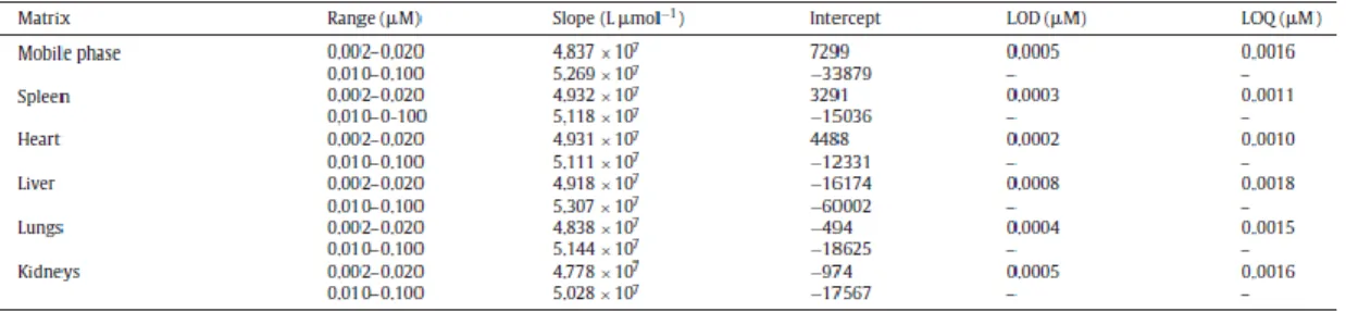
Regression analysis data from calibration curves in mobile phase and biological tissue extracts are given in Table 1. Two calibration ranges were established for BNIPDaoct: a low range for concentrations between 0.002 and 0.020 _M and a high range between 0.010 and 0.100 _M. Sensitivity, assessed as the slope of the calibration curve, was 5 to 10% lower for the low range. Calibration curves for BNIPDaoct in mobile phase were linear and reproducible, with correlation coefficient >0.9996. The back calculated concentrations presented deviations <5.9% from the nominal value, meeting the requirements of EMA guideline [20]. The calculated LOD was 0.5 nM and the LOQ was 1.6 nM.
Calibration curves for BNIPDaoct in the biological matrices tested were also linear for the two ranges correlation coefficients ≥0.9997 (Table 1). The LOD and LOQ for biological matrices were ≤0.8 and ≤1.8 nM, respectively (Table 1), corresponding to values ≤4 and ≤9 nmol g−1 in mice organs.
Accuracy and precision (inter- and intra-day) were also estimated. The intra-day precision and accuracy were calculated by analyzing the QC of all matrices at concentrations of 0.006, 0.020 and 0.100 _M and the 0.020 _M standard was interpolated in both calibration curves (Table 2). The intra-day precision was ≤1.9% and accuracy ranged between 97.3 and 106.8%. The inter-day precision was ≤7.2% and accuracy ranged between 96.6 and 105.6% (Table 2). Both intra- and inter-day precision and accuracy found have acceptable values, because precision for each concentration level, represented as CV, did not exceed 15% and the accuracy range was between 85 and 115% [20].
The stability of BNIPDaoct at laboratory temperature was assessed analyzing fresh samples and analyzing the same samples after 24 h at RT (20 °æ 2 ◦C). BNIPDaoct showed stability in all matrices after this period as can be seen from data in Table 3, where recoveries ranged from 92.6 to 105.9%, with a mean value of 100.0 °æ 2.7%. The autosampler stability was assessed analyzing fresh samples and analyzing the same samples after 24 h after the first injection. BNIPDaoct was stable in the autosampler for at least 24 h (Table 3), providing recovery values in all matrices between 94.1 and 103.2%, with a mean value of 99.1 °æ 2.4. The freeze–thaw stability of BNIPDaoct over three freeze–thaw cycles was also assessed, providing values of 89.1–102.5% for analyte recovery (Table 3) and a mean value of 97.1 °æ 2.8, showing the stability during sample storage and handling, according to EMA guidelines, which recommend that mean concentration at each level should be within 20% of the nominal concentration.
Finally, extraction efficiency was assessed, by spiking organs harvested from healthy animals and processing as described in Section 2.3.3 (Table 4). Recoveries were similar to all tissues and for both BNIPDaoct in solution (mean recovery 93.2 °æ 2.1%) and nanoencapsulated BNIPDaoct (mean recovery 92.3 °æ 2.4%). Therefore, recovery values are acceptable for all organs and suitable for reliable bioanalysis.
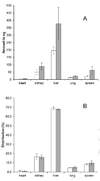
Application to biodistribution assessment. The validated HPLC method was applied to assess the biodistribution of free and nanoencapsulated BNIPDaoct. PLGA nanoparticles containing BNIPDaoct presented a mean size of 156 °æ 3 nm, with a polydispersity index of 0.08 °æ 0.03, and Zeta potential of −5.1 °æ 0.7 mV. The amount found in each organ and the relative distribution is given in Fig. 3. A larger amount of BNIPDaoct (3.1% of administered mass as free drug) was found compared to BNIPDaoct administered in nanoencapsulated form (1.5% of administered mass). The distribution per harvested organs was similar for both
formulations, with a large amount found in liver (68%), and followed by kidney (16%), spleen (10%), lung (5%) and heart (1%). Considering the organs were harvested 1 h after drug administration, a similar distribution profile was observed, with a larger amount of free drug recovered. Nevertheless, we would expect an increased amount of encapsulated BNIPDaoct if quantification was performed following a longer time (e.g., 24 h) after drug administration because the bio-accumulation of encapsulated BNIPDaoct requires more time due to their interactions with complement system and inespecific cellular interactions.
CONCLUSIONS
A simple, sensitive, accurate and precise HPLC method was developed and fully validated for determination of BNIPDaoct in biological samples. This method was convenient for the quantification of BNIPDaoct in mice organ samples, allowing future application to bioavailability assessment for longer harvest times (e.g., 12 and 24 h). Further application will also target pharmacokinetic studies and application of other formulations for BNIPDaoct delivery. Furthermore, this validated method can potentially be applied in the biodistribution of other mono and bisnaphthalimido molecules.
ACKNOWLEDGEMENTS
Authors acknowledge financial support from the Q3 European Union (FEDER funds through COMPETE) and National Funds (FCT) through project UID/Multi/04378/2013, from U.Porto/Santander Totta—Project 155/2010 and from FEDER funds under the framework of QREN through Project NORTE-07-0162-FEDER-000124.
REFERENCES
[1] M.F. Brana, A. Ramos, Naphthalimides as anti-cancer agents: synthesis and biological activity, Curr. Med. Chem. 1 (2001) 237–255.
[2] T. P. Kong, V.A. Pavlov Lin, The synthesis and in vitro cytotoxic studies of novel bis-naphthalimidopropyl polyamine derivatives, Bioorg. Med. Chem. Lett. 10 (2000) 1609–1612. [3] A.M. Dance, L. Ralton, Z. Fuller, L. Milne, S. Duthie, C.S. Bestwick, P.K.T. Lin, Synthesis and biological activities of bisnaphthalimido polyamines derivatives: cytotoxicity, DNA, binding, DNA damage and drug localization in breast cancer MCF 7 cells, Biochem. Pharmacol. 69 (2005) 19–27.
[4] J. Oliveira, L. Ralton, J. Tavares, A. Cordeiro-da-Silva, C.S. Bestwick, A. McPherson, P.K.T. Lin, The synthesis and the in vitro cytotoxicity studies of bisnaphthalimidopropyl polyamine derivatives against colon cancer cells and parasite Leishmania infantum, Bioorg. Med. Chem. 15 (2007) 541–545.
[5] L.D. Ralton, C.S. Bestwick, L. Milne, S. Duthie, P.K.T. Lin, Bisnaphthalimidopropyl spermidine induces apoptosis within colon carcinoma cells, Chem. Biol. Interact. 177 (2009) 1–6.
Bisnaphthalimidopropyl derivatives as inhibitors of Leishmania SIR2 related protein 1, ChemMedChem 5 (2010) 140–147.
[7] J. Tavares, A. Ouaissi, A.M. Silva, P.K.T. Lin, N. Roy, A. Cordeiro-da-Silva, Anti-leishmanial activity of the bisnaphthalimidopropyl derivatives, Parasitol. Int. 61 (2012) 360– 363.
[8] C. Hoskins, M. Ouaissi, S.C. Lima, W.P. Cheng, I. Loureirio, E. Mas, D. Lombardo, A. Cordeiro-da-Silva, A. Ouaissi, P.K.T. Lin, In Vitro and In Vivo anticancer activity of a novel nano-sized formulation based on self-assembling polymers against pancreatic cancer, Pharm. Res. 27 (2010) 2694–2703.
[9] J. Tavares, A. Ouaissi, P.K.T. Lin, A. Tomas, A. Cordeiro-da-Silva, Differential effects of polyamine derivative compounds against Leishmania infantum promastigotes and axenic amastigotes, Int. J. Parasitol. 35 (2005) 637–646.
[10] P. Couvreur, C. Vauthier, Nanotechnology: intelligent design to treat complex disease, Pharm. Res. 23 (2006) 1417–1450.
[11] S.A. Costa Lima, M. Resende, R. Silvestre, J. Tavares, A. Ouaissi, P.K.T. Lin, A. Cordeiro-da-Silva, Characterization and evaluation of BNIPDaoct-loaded PLGA nanoparticles for visceral leishmaniasis: in vitro and in vivo studies, Nanomedicine 7 (2012) 1839–1849. [12] C.M. Lai, D.M. Garner, J.E. Gray, B.L. Brogdon, V.C. Peterman, H.J. Pieniaszek, Determination of bisnafide, a novel bis-naphthalimide anticancer agent, in human plasma by high-performance liquid chromatography with UV detection, J. Pharm. Biomed. Anal. 17 (1998) 427–434.
[13] S. Banerjee, E.B. Veale, C.M. Phelan, S.A. Murphy, G.M. Tocci, L.J. Gillespie, D.O. Frimannsson, J.M. Kelly, T. Gunnlaugsson, Recent advances in the development of 1,8-naphthalimide based DNA targeting binders, anticancer and fluorescent cellular imaging agents, Chem. Soc. Rev. 42 (2013) 1601–1618.
[14] R. Ferreira, C. Baleizao, J.M. Munoz-Molina, M.N. Berberan-Santos, U. Pischel, Photophysical study of bis(naphthalimide)-amine conjugates: toward molecular design of excimer emission switching, J. Phys. Chem. A 115 (2011) 1092–1099.
[15] M. Lv, H. Xu, Overview of naphthalimide analogs as anticancer agents, Curr. Med. Chem. 16 (2009) 4797–4813.
[16] G. Guiochon, Monolithic columns in high-performance liquid chromatography, J. Chromatogr. A 1168 (2007) 101–168.
[17] K.K. Unger, R. Skudas, M.M. Schulte, Particle packed columns and monolithic columns in high-performance liquid chromatography-comparison and critical appraisal, J. Chromatogr. A 1184 (2008) 393–415.
[18] K.C. Saunders, A. Ghanem, W.B. Hon, E.F. Hilder, P.R. Haddad, Separation and sample pre-treatment in bioanalysis using monolithic phases: a review, Anal. Chim. Acta 652 (2009) 22–31.
[19] A.R. Neves, S. Reis, M.A. Segundo, Development and validation of a HPLC method using a monolithic column for quantification of trans-resveratrol in lipid nanoparticles for intestinal permeability studies, J. Agric. Food Chem. 63 (2015) 3114–3120.
[20] European Medicines Agency, Guideline on bioanalytical method validation, EMEA/CHMP/EWP/192217/2009 (2011).
[21] J.N. Miller, J.C. Miller, Statistics and Chemometrics for Analytical Chemistry, Pearson Education, Harlow, 2005.
TABLES & FIGURES
Table 1. Calibration curve parametersa, limit of detection (LOD) and quantification (LOQ) for BNIPDaoct in
mobile phase and tissue extracts.
Version: Postprint (identical content as published paper) This is a self-archived document from i3S – Instituto de Investigação e Inovação em Saúde in the University of Porto Open Repository For Open Access to more of our
A
0
1
/0
Table 3. Stability of BNIPDaoct at different experimental conditions for all matrices tested.
Figure 3. Amount (A) and relative distribution (B) of BNIPDaoct recovered from mice organs (n = 4) after administration of nanoencapsulated (white bars) or free drug (grey bars).