Development of a Simulator for Tomographic Images
Generated by Radiation Transmission
Gevaldo L. de Almeida, Maria Ines Silvani, Rosanne C. A. A. Furieri,
Instituto de Engenharia Nuclear – CNEN, C.P. 68550, Ilha do Fund ˜ao, 21945-970 Rio de Janeiro, RJ, Brasil
Marcelo J. Gonc¸alves, and Ricardo T. Lopes
Universidade Federal do Rio de Janeiro, COPPE, Centro de Tecnologia, Bloco 6, 21945-970, Rio de Janeiro, RJ, Brazil
Received on 9 September, 2003
A computer program to simulate tomographic images generated by transmitted radiation was developed. The algorithm uses a deterministic approach to generate the projections, which supply an existing image recon-struction software. A dispersion associated with the counting statistics is also incorporated into the algorithm, in order to simulate the influence of the detector efficiency and counting time on the final image quality. The detector resolution is also included in the algorithm by assuming a gaussian shape for its line spread function -LSF. The program deals with cylindrical objects containing any desired number of cylindrical rods inside and requires their positions, dimensions and attenuation properties as input data. Images of such objects, acquired with a thermal neutron tomograph equipped with a position sensitive detector, have been compared with those simulated by the developed program in order to evaluate its ability to reproduce those images.
1
Introduction
Computer Assisted Tomography - CAT is a growing branch of non-destructive assay applied in technology, engineering and medical examinations. A good review of the histori-cal development of this methodology is presented by Brooks and Di Chiro [1] who also addressed a comprehensive dis-cussion of the mathematical basis for the techniques used for image reconstruction, its constraints and limitations. The recent development and incorporation [2] of a Position Sen-sitive Detector - PSD to a thermal neutron tomographic sys-tem has stimulated several related works [3,4,5,6] devoted to explore the advantages and potential capabilities of an image acquiring system of this kind, classified as a 2ndgeneration tomograph, for it no longer requires a translation of the test objects as in the 1stgeneration ones.
Disregarding their generation, one cannot forecast the quality of the final image unless the Modulation Transfer Function - MTF for the system is known. However, to ob-tain this function, it is necessary to carry out measurements using a special test object constituted by an opaque mask, containing alternating slits and spaces that increase in spa-tial frequency. Since these objects are very expensive, an alternative way is to perform a Fourier transform of the Line Spread Function, henceforth referred as LSF [7]. This func-tion is difficult and somewhat cumbersome to be directly measured, but it can be approximated by a numerical dif-ferentiation of the Edge Response Function – ERF, which is easier to be obtained, for it deals with higher count rates due to the lower collimation constraints. [7,8].
Anyhow, none of the outlined options would avoid a cer-tain amount of experimental work to get the MTF. Even so,
one could only estimate the expected degradation of the ac-quired image.
Therefore, a program to simulate a tomographic system, forecasting what image quality should be expected, has been developed for use as a guide to plan experiments. It fur-nishes projections of an user specified test object, which are then unfolded by a reconstruction program Recpar2000 [9]. This approach would cut down experimental efforts, reduc-ing costs and eventual radiological burden associated with them.
2
Tomographic principles
translated to generate the aimed projections.
According to the CAT approach, an infinite number of projections would reproduce perfectly the attenuating struc-ture of the object. In practice however, such a perfection would never be reached because the detector finite resolu-tion spoils the image, making a line source to appear like a more or less blurred image, as dictated by the LSF - a curve reminding a Gaussian function, with its FWHM represent-ing the detector resolution.
The non-zero detector resolution affects the quality of the image, degrading its contrast and resolution due to the overlapping of the LSF tails. In practice, these parameters are further worsened due to beam divergence and its non-monochromatic character. Moreover, neutron scattering (or scattering via Compton effect when gamma or X-rays are used as incident radiation) take place contributing to an ad-ditional degradation of the final image.
3
Algorithm
The algorithm used by the simulator treated in this work, assumes an ideally parallel and monochromatic radiation beam, suffering no scattering as it interacts with the object and the detector.
The average count-rate per position-channel produced by an undisturbed beam is the result of an infinite number of infinitely narrow beams reaching the whole extension of each channel. Under such ideal conditions only those beams hitting directly each channel would contribute to the count rate. In practice however, even if a very thin slab beam is used to simulate a line source, the finite resolution of the detector would spoil that ideal conception. Since the LSF can be fairly represented by a Gaussian function, any beam hitting the detector would be seen by the detector as a Gaussian-shaped source, with its tail invading all the po-sition spectrum, and thus, contributing to the count-rate at any channel. The degree of this contribution would depend on how far the Gaussian epicenter is from each particular channel and from their widths.
Taking the case of a position sensitive detector, having a total active length L coupled to a multi-channel analyzer and submerged into an homogeneous perfectly parallel radi-ation beam, then the average count-rateT¯jproduced at each channel j would be expressed by,
¯ Tj=L.δA0
L
0
xj+δ
xj e
−[
x−c w ]
2
dxdc (1)
with,
A0= constant expressing the beam intensity. δ= channel width.
w= width of the Gaussian expressing the LSF.
C= epicenter of the above Gaussian.
Xj= left side abcissa of the jthchannel.
X= position along the position spectrum.
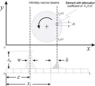
When a rotating object with its axis perpendicular to the detector intercepts a beam, its intensity is reduced by the in-ner attenuating structure of the object, gein-nerating a different projection for each angle that the object assumes. Applying
a coordinate system to the region covering the cross-section of the object, one can define a functionµk(x, y)expressing the attenuation coefficient as a function of the coordinates. Due to the rotation, this function will be different for each angle, since a new element, having a different attenuation coefficient from the previous one, will occupy a given point
(x, y).This angle-dependent character is expressed by the indexk. The average count-rateT¯kj produced at the jth channel by the radiation beam disturbed by the object varies for each projectionk, and is expressed as
¯ Tkj= L.δA0
L
0
xj+δ
xj e
−
y2(x)
y1(x)
µk(x,y)dy e−[xw−c]
2
dxdc (2)
an expression which differs from (1) only due to the attenua-tion term containing the variables and parameters as follows,
µk(x, y)= attenuation coefficient for the point (x,y) at the
kthprojection. y
1(x), y2(x) = lower and upper integration
limits respectively as shown in Fig. 1
The count-rates produced by equations (1) and (2) de-pend, among other parameters, on the integration limit L, for a Gaussian never vanishes but decreases asymptotically. Those count-rates are integrated over the acquisition time in-terval to yield the average counts per position channel stored in the multi-channel analyzer as ordinates of the position spectrum.
Figure 1. Diagram showing the relevant parameters and variables affecting the projection produced by a homogeneous parallel radi-ation beam.
4
Effects of detector resolution and
total counts on simulated images
In order to evaluate the effects of detector resolution and to-tal counts on the images reconstructed from projections pro-duced by the simulator, a cylinder with sets of thin inserts was used.
The cylinder and the rods are made of hypothetical ma-terials with attenuation coefficients of 0.1mm−1 and 0.5 mm−1respectively for the incoming radiation, disregarding its nature and energy. The purpose of this first test was to verify the soundness of the simulator. Within this frame, the nature and energy of the incident radiation do not play any role as long as they produce similar attenuation maps.
The responses of the simulator - for a channel widthδgf 0.08 mm - to changes in the detector resolution and total counts are shown in Fig. 2. A degradation of the image res-olution (merging dots) and contrast (changes of color) are observed as the detector resolution decreases as it should be theoretically expected. The decrease of contrast is caused by the overlapping of the LSF tails which grows withw.
Figure 2. Response of the simulator to changes in the detector res-olutionw(top) and total counts per channel (bottom).
Although not promptly recognizable - since the recon-struction software uses a color scale that differs from the natural and intuitive rainbow sequence - a careful observa-tion shows that for w=1.5 the rods and the cylinder exhibit the colors yellow and green respectively. For w=0.25 these colors are respectively gray and pink, which, according to the color scale are more distant from each other than the pair yellow-green.
When the total count increases, the image flickering di-minishes and the merging of single features are better ob-served, but the image resolution remains the same, as in a real system.
5
Materials and methods
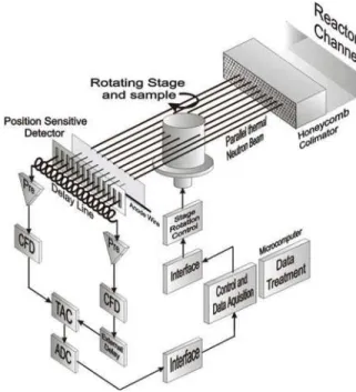
The tomographic system used to acquire the images and compare them with those simulated ones is schematized in Fig. 3. The Argonauta reactor at the Instituto de Engenharia Nuclear (IEN-CNEN-Brazil) has been utilized as the source of thermal neutrons, furnishing a flux of 4.5x10⁵ncm−2s−1 at the energy range 0.005-0.1eV. A 40mm-deep honeycomb-type collimator (hexagon side=2mm) made of a 40µthick gadolinium foil was used to improve the beam parallelism. The test object and the detector were placed at 18cm and 26cm respectively from the collimator front face. The PSD has an active length L of 80 mm and an active width of 5mm, as defined by its 2mm-thick aluminum window. The soft-ware Recpar2000 uses the filtered back-projection approach to unfold the projections and reconstruct the images. Further details can be found elsewhere [3].
Figure 3. Scheme of the thermal neutron tomographic system used to acquire the images.
6
Comparison with experimental
re-sults
to estimate by visual inspection the resolution of the real system through that comparison, i.e. associating thewof the simulator detector to thew of the real system. After this approach, three different test objects have been used to compare the images generated by the simulator with those actually experimentally acquired.
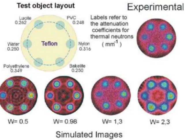
Figure 4 focuses the capability of the real system to de-tect plastic materials within a teflon matrix and to differenti-ate them. Comparing the synthetic images with the real one, one can estimate the resolution of the system used to acquire it at about 1.3 mm. As far as a visual inspection can infer, such a result is ratified by the similar contrasts achieved by the two kinds of images, both produced with about 4,000 counts per position channel. It can be furthermore recog-nized that the colors of the spots produced by the different materials are consistent with their attenuation coefficients and the color scale shown in Fig. 2.
Figure 4. Simulated images under several detector resolutions (w) compared with the experimental one. The test object is a 23mm-diameter teflon cylinder containing 4mm-23mm-diameter rods of different materials.
Images of the same teflon cylinder where the plastic rods have been replaced by metallic ones are shown in Fig. 5. One can conclude that the resolution of the system lies within the range of 1.3-1.7 mm. It is worthwhile to point out that the two kinds of images (simulated and acquired) exhibit additional matching results in terms of contrast, as a visual inspection demonstrates. Indeed, in both of them alu-minum is hardly detected, while lead is not detected at all, for its attenuation coefficient is very close to the teflon ma-trix, a behavior consistent with their attenuation coefficients for thermal neutrons.
The third test object used for comparison intended to as-sess the capability of the simulator to reproduce the perfor-mance of the real tomograph to detect and resolve small-size features.
A small-size feature requires a high attenuation coeffi-cient for its detection, and to attain this condition, 1mm-thick cadmium wires have been introduced in orifices cast in a 23mm-diameter aluminum cylinder, after the scheme shown in Fig. 6, where the results are also presented.
Figure 5. Same as Fig. 4, except for the rods materials.
Figure 6. Simulated and experimental images of a 23mm-diameter Al cylinder containing 1mm-diameter Cd wires. The gap between the closest wires is 1mm.
Although the tomographic system could furnish a fair image of the test object, the simulator was unable to per-form its task properly. The difficulty comes from the high incident-to-transmitted beam intensity ratio reaching 1017
for some projections generated by the simulator. The recon-struction software fails to handle such high numbers arising from the high attenuation coefficient of cadmium, for it was designed to deal with much lower values. Indeed, a tomo-graphic image of acceptable quality is obtained when this ra-tio remains approximately within the range of 3 – 150 [10], a feature which has been incorporated to the reconstruction software, making it incapable to process unexpected high ratios.
no physical meaning, it has been performed as a cross-check to assure that the difficulties really were caused by the high attenuation coefficient rather than by an eventual miscon-ception in the architecture of the simulator. A reduction fac-tor of about 20 was required to match the generated images with the real ones. Such a factor seems to be caused by beam divergence and neutron scattering. Indeed, both divergence and scattering would contribute to the average count-rate at a given channel, otherwise exposed only to those neutrons fol-lowing a straight path intercepting the cadmium wire. The increased count-rate of the transmitted beam would then re-duce the apparent attenuation coefficient for that particular path, reducing thus the incident-to-transmitted beam inten-sity ratio to a level acceptable by the reconstruction soft-ware.
Although the tomographic system could furnish a fair image of the test object, the simulator was unable to per-form its task properly. The difficulty comes from the high incident-to-transmitted beam intensity ratio reaching 1017 for some projections generated by the simulator. The recon-struction software fails to handle such high numbers arising from the high attenuation coefficient of cadmium, for it was designed to deal with much lower values. Indeed, a tomo-graphic image of acceptable quality is obtained when this ra-tio remains approximately within the range of 3 – 150 [10], a feature which has been incorporated to the reconstruction software, making it incapable to process unexpected high ratios.
Therefore, to supply acceptable projections to the recon-struction software, the attenuation coefficient for the cad-mium - given as input data to the simulator - had to be strongly reduced. Although such a reduction has obviously no physical meaning, it has been performed as a cross-check to assure that the difficulties really were caused by the high attenuation coefficient rather than by an eventual miscon-ception in the architecture of the simulator. A reduction fac-tor of about 20 was required to match the generated images with the real ones. Such a factor seems to be caused by beam divergence and neutron scattering. Indeed, both divergence and scattering would contribute to the average count-rate at a given channel, otherwise exposed only to those neutrons fol-lowing a straight path intercepting the cadmium wire. The increased count-rate of the transmitted beam would then re-duce the apparent attenuation coefficient for that particular path, reducing thus the incident-to-transmitted beam inten-sity ratio to a level acceptable by the reconstruction soft-ware.
7
Conclusions
The images reconstructed from simulator generated projec-tions exhibit the expected features and behavior according to the changes in the detector resolution and counting statis-tics, demonstrating thus the soundness of the developed al-gorithm. For this simulator, the system resolution coincides with that of its detector, since this virtual device is the only component in the chain contributing to the degradation of the final image.
In a real tomograph however, the system resolution is
poorer than its detector resolution due to beam divergence, alignment, scattering and other processes. Therefore, chang-ing the resolution w of the simulator until the synthetic image matches visually with that experimentally acquired would furnish an estimation of the tomograph resolution, namely w. Once this wis known, the simulator could be used to plan experiments and to forecast the expected im-age quality prior to its acquisition, sparing thus experimental work and the associated radiological burden. The simulator didn’t succeed in reproducing images of objects containing small-size features with extremely high attenuation proper-ties, as the real tomograph did. This shortcoming has been imputed to its inability to take into account beam divergence and neutron scattering. The contribution of these processes to the reduction of the apparent attenuation coefficient of the test object is unknown. However, since the first one is simpler to be treated, and taking into account the fair results obtained, making the program capable of handling divergent beams is a further improvement and an worthwhile task.
In a real tomograph however, the system resolution is poorer than its detector resolution due to beam divergence, alignment, scattering and other processes. Therefore, chang-ing the resolution w of the simulator until the synthetic image matches visually with that experimentally acquired would furnish an estimation of the tomograph resolution, namely w. Once this wis known, the simulator could be used to plan experiments and to forecast the expected im-age quality prior to its acquisition, sparing thus experimental work and the associated radiological burden. The simulator didn’t succeed in reproducing images of objects containing small-size features with extremely high attenuation proper-ties, as the real tomograph did. This shortcoming has been imputed to its inability to take into account beam divergence and neutron scattering. The contribution of these processes to the reduction of the apparent attenuation coefficient of the test object is unknown. However, since the first one is simpler to be treated, and taking into account the fair results obtained, making the program capable of handling divergent beams is a further improvement and an worthwhile task.
References
[1] R. A. Brooks and G. Di Chiro, Phys. Med. Biol. 21, 5 689 (1976).
[2] M. I. Silvani, Computed Tomography with thermal
neu-trons and a Position Sensitive Detector, DSc. Thesis,
COPPE/UFRJ-Brazil (2001). In Portuguese.
[3] M. I. Silvani, R. T. Lopes, E. F. Jesus, G. L. de Almeida, and A. F. Barbosa, Nucl. Instr. Meth. in Phys. Research A 505, 568 (2003).
[4] M. I. Silvani, R. T. Lopes, E. F. Jesus, G. L. de Almeida, A. F. Barbosa, and D. Braz, Conversion of a X-ray Position
Sensitive Detector for use in Thermal Neutron Tomographic Systems, V International Topical Meeting on Industrial
Radi-ation and Radioisotope Measurement ApplicRadi-ations, Bologna, Italy (2002).
[6] M. J. Gonc¸alves, Optimization of a gaseous position
sensi-tive detector for use in a thermal neutron tomographic sys-tem. M.Sc. Thesis, COPPE/UFRJ, Rio de Janeiro, RJ, Brazil
(2003) In Portuguese.
[7] ASTM E 1441-95 and 1570-95a, Non-destructive Testing,
Radiation Methods, computed Tomography, Guide for Imag-ing and Practice Examination, ISO/TC 135/SC5, N118
(1996).
[8] G. L. de Almeida, M. I. Silvani, R. T. Lopes, A Hybrid
Technique to Evaluate the Line Spread Function, VI ENAN,
E10 142, Rio de Janeiro, Brazil (2002).
[9] Recpar2000, LIN-COPPE/UFRJ. Unpublished.