Web-based evaluation of experts’ opinions on impacted
maxillary canines forced eruption using CBCT
Amirfarhang Miresmaeili1, Nasrin Farhadian1, Vahid Mollabashi2, Faezeh Yousefi3
How to cite this article: Miresmaeili A, Farhadian N, Mollabashi V, Yousefi F. Web-based evaluation of experts’ opinions on impacted maxillary canines forced eruption using CBCT. Dental Press J Orthod. 2015 Mar-Apr;20(2):90-9. DOI: http://dx.doi.org/10.1590/2176-9451.20.2.090-009.oar
» Patients displayed in this article previously approved the use of their facial and intraoral photographs.
Contact address: Vahid Mollabashi E-mail: [email protected] » The authors report no commercial, proprietary or financial interest in the
prod-ucts or companies described in this article.
Submitted: April 23, 2014 - Revised and accepted: October 20, 2014
1 Associate professor, Hamadan University of Medical Science, School of
Dentistry, Department of Orthodontics, Hamadan, Iran.
2 Assistant professor, Hamadan University of Medical Science, School of
Dentistry, Department of Orthodontics, Hamadan, Iran.
3 Associate professor, Hamadan University of Medical Science, School of
Dentistry, Department of Dento-maxillofacial radiology, Hamadan, Iran.
Aim: This study aims at examining the difficulty in performing forced eruption of impacted maxillary canines, using CBCT information and according to experienced orthodontist’s opinion. The second aim was to find the most impor-tant factors related to this decision. Methods: Based on a careful literature review on impacted maxillary canines, ten main factors were selected to assess difficulties associated with impacted teeth. Thirty six consecutive patients with 50 impacted maxillary canines were examined and variables were measured for each impacted tooth using Dolphin 3D software. Ten orthodontists assessed the radiographs of teeth and provided their opinion on the difficulty in bringing impacted teeth into occlusion named subjective degree of difficulty (SDD). The correlation established between mean SDD of each tooth and measured variables were analyzed by means of linear regression. Results: Mean SDD was 6.45 ± 1.22 for all 50 teeth. Linear regression showed a high coefficient of correlation between mean SDD and age, dilacerations, vertical height, angulation and horizontal overlap (P < 0.05). Conclusion: To predict the difficulty of impacted maxillary canines forced eruption, according to the opinion of experienced orthodontists, the factors age, dilaceration, angulation, overlap and vertical distance from the occlusal plane are the most important variables.
Keywords:Cuspid. Cone-beam computed tomography. Tooth eruption. Ectopic. Internet. DOI: http://dx.doi.org/10.1590/2176-9451.20.2.090-099.oar
Objetivo: o presente estudo tem o objetivo de avaliar, de acordo com a opinião de ortodontistas experientes, a dificulda-de dificulda-de se realizar a erupção forçada dificulda-de caninos superiores impactados, com base em informações fornecidas por tomografia computadorizada de feixe cônico. O objetivo específico foi identificar os fatores mais importantes relacionados a essa opção de tratamento. Métodos: com base em uma revisão meticulosa da literatura sobre caninos superiores impactados, selecionamos os dez principais fatores utilizados para avaliar a dificuldade em tratar dentes impactados. Trinta e seis pa-cientes, com um total de 50 caninos superiores impactados, foram avaliados. As variáveis para cada dente impactado foram obtidas utilizando o programa Dolphin 3D. Dez ortodontistas avaliaram as radiografias referentes aos dentes selecionados e opinaram sobre a dificuldade em alinhar dentes impactados. O grau de dificuldade foi denominado Grau de Dificuldade Subjetivo (GDS). A relação entre o valor médio de GDS de cada dente e as variáveis obtidas foi analisada por regressão linear. Resultados: o valor médio de GDS foi de 6,45 ± 1,22, para todos os dentes. A regressão linear gerou um alto coe-ficiente de relação entre o valor médio de GDS e as variáveis idade, dilaceração, altura vertical, angulação e sobreposição horizontal (p < 0,05). Conclusão: de acordo com a opinião de ortodontistas experientes, para prever a dificuldade em se realizar a erupção forçada de caninos superiores impactados, as variáveis idade, dilaceração, angulação, sobreposição e distância vertical do plano oclusal são as mais importantes.
original article
Miresmaeili A, Farhadian N, Mollabashi V, Yousefi F
INTRODUCTION
With the exception of third molars, maxillary ca-nines are the most frequently impacted teeth, with
prevalence ranging from 0.8 to 3.0%.1,2,3 Maxillary
ca-nines are considered to be important esthetically and functionally, and patients with impacted maxillary ca-nines are found to be more diicult and
time-consum-ing to treat than the average orthodontic patient.4
Localization of impacted canines can be chal-lenging with conventional radiographic methods due to image distortion, superimposition of three-dimensional structures, image artifacts, projection
errors and sometimes poor image quality.1,5,6,7 More
recently, three-dimensional volumetric imaging sys-tems (CBCT) have allowed more precise localization of impacted canines, using spatial relationships, with
excellent tissue contrast.5 The costs, efficiency, and
benefits of CBCT imaging are very favorable, as one single imaging session can provide many important
views to locate the position of the impacted tooth.3
Several variables have been proposed to predict the diiculty of treating impacted maxillary canines and
the likelihood of complications or failure.8 In a study
by Fleming et al, angulation of the canine, vertical po-sition from the occlusal plane, anterior-posterior posi-tion of the root apex and the degree of overlap of the adjacent incisor correlate with the prognosis of ectopic
canines.2 Zucatti et al reported a strong association
be-tween the number of visits and increasing age, vertical
height, and mesial displacement of the cusp tip.8
Canines that are angulated towards the horizontal
plane, according to Pitt et al,9 have a poorer
align-ment prognosis. A buccopalatal position of the canine crown also influences treatment decisions, with pala-tally impacted canines being more likely to be surgi-cally exposed, whereas those in the line with the den-tal arch or buccally positioned are more likely to be
removed.9 It has been reported that the higher above
the occlusal plane the canine is positioned, the poorer
the prognosis for alignment.9 McSherry described
this as “the vertical rule of thirds”.10
Maxillary lateral incisor root resorption is the most common adverse effect associated with an impacted
maxillary canine.11 Previous studies have shown that
root resorption less than 0.60 mm in diameter and 0.30 mm in depth cannot be detected with 2D
ra-diography.12,13 Alqerban14 found that CBCT imaging
was significantly better than panoramic radiography in determining the degree of root resorption in the categories of slight and severe resorption.
Impacted teeth are notoriously more difficult to
treat in adults.15 A study found that the success rate
among patients over 30 years old was 41%, where-as the success rate for those aged between 20 and
30 years old was 100%.8
Presently, the prediction of impacted canines treat-ment success has been largely based on personal clini-cal experience and anecdotal evidence; therefore, a system that ofers an improved assessment technique of the degree of diiculty in bringing a displaced ca-nine into alignment will be beneicial for both patient
and clinician.9 The related studies are mainly based on
conventional radiographs, and CBCT have not been used widely for estimation of diiculty.
In the present study, the primary objective was to find the opinion of experienced orthodontists about difficulties in treating a sample of impacted canines, using CBCT information. The secondary objective was to find the main factors related to this decision.
MATERIAL AND METHODS
There is no general agreement on the criteria used to distinguish between impacted maxillary ca-nines that could be treated orthodontically or not. After a careful literature review on ISI website for “impacted maxillary canine”, “ectopic maxillary canine”, “treatment difficulty”, “orthodontic treat-ment”, “CT” and “CBCT”, 237 articles were found. Among these, 11 articles were selected according to
their citation and relevance.2,4,5,6,8,9,15-19 One expert
orthodontist evaluated the articles. Age, horizontal position, vertical position, apex position, angulation, buccopalatal position and rotation factors were used
in the articles with 2D radiograph.2,4,8,9,15,16 3D view
provides more information about impacted canines, but the studies using 3D views only evaluated
inci-sor reinci-sorption, canine crown or root position.5,6,17,18,19
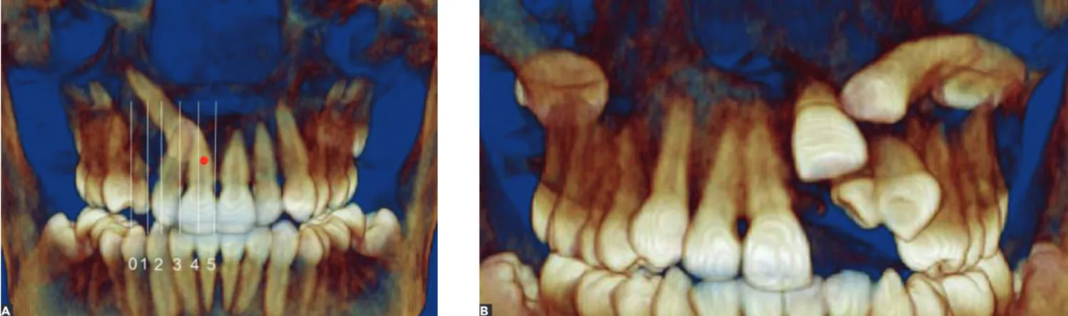
Figure 1 - Four-step evaluation. Step one: Reconstructed 3D image in frontal view used to examine impacted canine. A) Overlap and B) Transposition. we decided to combine these data with expert
ortho-dontists’ opinions.
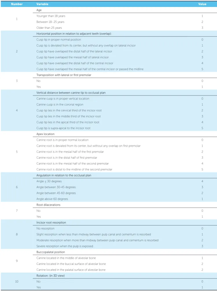
These ten factors all have different scales of mea-surements and a different range of ratings depending on the nature and importance of each factor which were generally based on the reviewed literature. With the exception of age, all other factors were measured on the CBCT scan. Because CBCT provided a great amount of information for each patient, we decided to use a more structured format so that we could ana-lyze tooth location and surrounding structures in a smooth and convenient way. Each factor was deter-mined using the following sequence in CBCT:
» Step one: frontal view
A) The horizontal position of the impacted tooth was evaluated in relation to adjacent incisors (Fig 1A).
B) Transposition was evaluated, if present (Fig 1B).
» Step two: lateral view
(Right and left side depending on the location of the impacted tooth).
A) The vertical position of canine tip was mea-sured in relation to adjacent teeth (Fig 2A). B) The apex position of the impacted tooth was
recorded in relation to adjacent teeth (Fig 2B). C) The angulation of the impacted tooth was
calcu-lated in relation to the occlusal plane (Fig 2C).
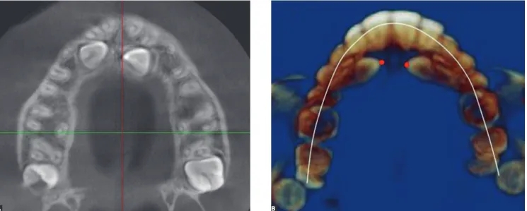
» Step three: axial view
A) The extent of possible incisor root
resorp-tion was measured at the locaresorp-tion where the
canine tip was closest to incisor root (according to the distance between pulp and cementum) (Fig 3A).
B) The buccopalatal position of the impacted
tooth was determined in relation to the center of the dental arch (Fig 3B).
» Step Four
A) Dilacerations and their location were recorded. B) Rotations were recorded (mesial and distal 3D
views of canine crown were determined and its angulation with the line of arch circumference was measured. If this angle was zero, the teeth had no rotation).
A sample of 36 patients with 50 impacted maxillary canines were collected and had CBCT scans taken by a Newtom 3G device (Quantitative Radiology, Verona, Italy), with minimum slice thickness of 0.4 mm, in which the maxilla and impacted canines could be seen completely. To measure these factors, we imported the DICOM iles into Dolphin 3D sotware designed for analysis of CBCT data. Subsequently, we adjusted the orientation and used the transparency tool to increase image clarity so that any impacted teeth could be easily seen. Then, each factor was measured on Dolphin 3D.
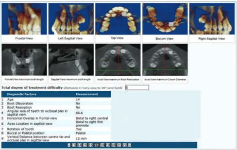
According to our second aim, we planned to as-sess the difficulty of impacted maxillary canine forced eruption to the occlusal level using the opinion of a group of experienced orthodontists. To facilitate our four-step examination, for this phase, we prepared four 2D and five 3D images for each impacted tooth to be uploaded easily in a website devoted to this study (www.canineimpaction.com shown in Fig 4).
original article
Miresmaeili A, Farhadian N, Mollabashi V, Yousefi F
Table 1 - Scales and grading proposed to establish the ten variables assessed.
Number Variable Value
1
Age
Younger than 18 years 1
Between 18- 25 years 2
Older than 25 years 3
2
Horizontal position in relation to adjacent teeth (overlap):
Cusp tip in proper normal position 0
Cusp tip is deviated from its center, but without any overlap on lateral incisor 1
Cusp tip have overlaped the distal half of the lateral incisor 2
Cusp tip have overlaped the mesial half of lateral incisor 3
Cusp tip have overlaped the distal half of the central incisor 4
Cusp tip have overlaped the mesial half of the central incisor or passed the midline 5
3
Transposition with lateral or first premolar
No 0
Yes 1
4
Vertical distance between canine tip to occlusal plan
Canine cusp is in proper vertical location 0
Canine cusp is in the coronal region 1
Cusp tip lies in the cervical third of the incisor root 2
Cusp tip lies in the middle third of the incisor root 3
Cusp tip lies in the apical third of the incisor root 4
Cusp tip is supra-apical to the incisor root 5
5
Apex location
Canine root is in proper normal location 0
Canine root is deviated from its center, but without any overlap on irst premolar 1
Canine root is in the mesial half of the irst premolar 2
Canine root is in the distal half of irst premolar 3
Canine root is in the mesial half of the second premolar 4
Canine root is distal to the midline of the second premolar 5
6
Angulation in relation to the occlusal plan
Angle ≤ 30 degrees 4
Angle between 30-45 degrees 3
Angle between 45-60 degrees 2
Angle above 60 degrees 1
7
Root dilacerations
No 0
Yes 1
8
Incisor root resorption
No resorption 0
Slight resorption when less than midway between pulp canal and cementum is resorbed 1
Moderate resorption when more than midway between pulp canal and cementum is resorbed 2
Severe resorption when the pulp is exposed 3
9
Buccopalatal position
Canine located in the middle of alveolar bone 1
Canine located in the buccal surface of alveolar bone 2
Canine located in the palatal surface of alveolar bone 2
10
Rotation: (in 3D view)
No 0
We sent a participation request to 20 well-known national and international orthodontists.
Ater examining the records of each patient, the evaluators were asked to suggest a grade for the dii-culty in aligning, or bringing into occlusion, impacted canines. This was done by allocating a score based on a scale from 1 to 10, with 1 being very easy and 10 ex-tremely diicult. The evaluators’ score for each tooth was termed subjective degree of diiculty (SDD).
Following data collection, correlation between mean SDD and the measured variables was estab-lished by means of linear regression in SPSS 16.
RESULTS
Thirty six consecutive patients (29 females and 7 males), with 50 impacted maxillary canines, referred
to the orthodontic clinic of the School of Dentist-ry for orthodontic treatment, were included in this study. Fourteen patients had bilateral impaction. Nine canine impactions were on the right, while 13 were on the left side. The subjects ranged in age from 12 to 34 years old (mean 19.08 ± 5.8 years).
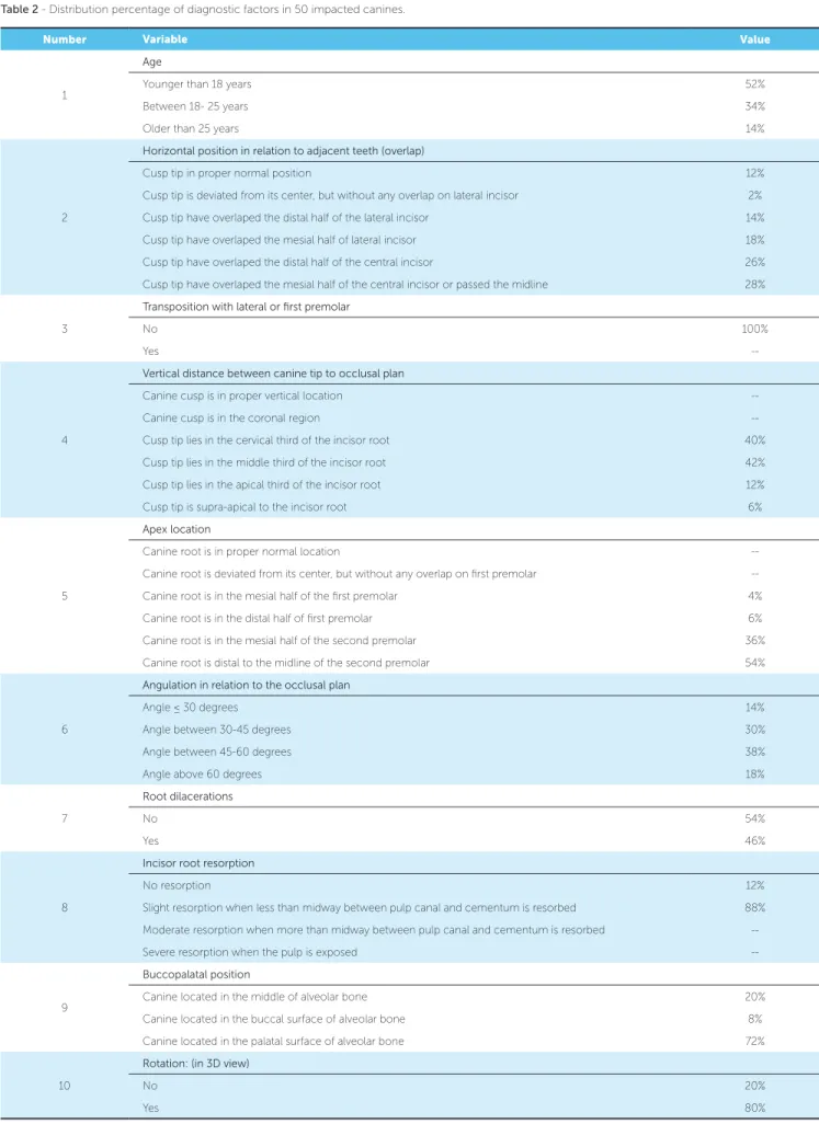
The main observer examined all impacted maxil-lary canines and measured each one of the ten fac-tors. Distribution of percentages for each factor can be seen in Table 2.
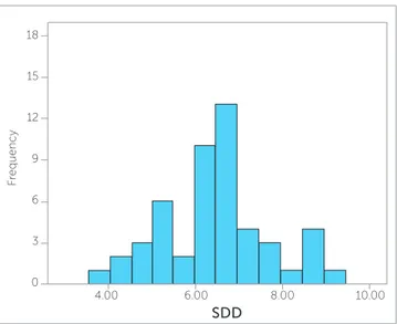
Ten orthodontists accepted to participate in the study and had an average number of years in practice of 22.7 ± 12.02 years. The frequencies of SDD by each evaluator are shown in Figure 5. Mean SDD was computed by ten evaluators for each tooth (Fig 6) and its total mean was 6.45 ± 1.22 for all impacted teeth. Figure 2 - Four step evaluation; Step two: Reconstructed 3D image from sagittal view to examine impacted canine A) Vertical position B) Apex location and C) Angulation in relation to occlusal plan.
Figure 3 - Four-step evaluation. Step three: Axial view to examine impacted canine. A) Induced root resorption at the closest contact with incisor root and B) Buccopalatal position at the crown level.
A
A B
original article
Miresmaeili A, Farhadian N, Mollabashi V, Yousefi F
Figure 4 - Web-base questionnaire for 50 im-pacted canines that each expert orthodontist evaluated for subjective degree of difficulty. Measurements of all variables for each tooth were recorded.
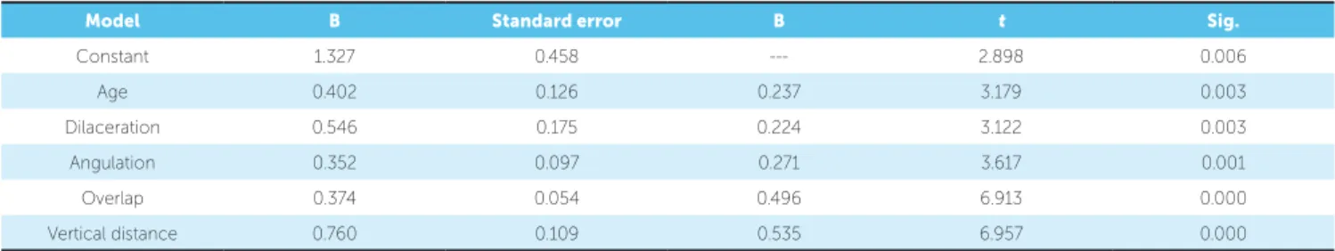
Simple linear regression was used to assess the corre-lation between mean SDD and nine factors. The ef ec-tive variables entered the model at P = 0.001 and were excluded at P = 0.05 in backward stepwise by SPSS 16.
Results show that age, dilacerations, angulation, overlap and vertical distance had significant correla-tion with mean of SDD (Table 3).
DISCUSSION
When orthodontists consider the possibility of treating an impacted canine, they are facing several problems. The first is the potential duration of treat-ment, if guided eruption is considered. Should that be the case, the tooth is brought into occlusion by a combination of surgical and orthodontic interven-tions. The second problem is the esthetic and func-tional results of treatment, which may not fulfill pa-tients’ best interests. The third problem is the risk of damage to neighboring teeth during treatment, espe-cially in cases in which surgical exposure is needed. It would be of great benefit to both the patient and the clinician if the orthodontist, before treatment, had a better idea of treatment difficulties involved in
treating an impacted tooth. Becker et al16 concluded
that the major reasons for failure of impacted canine force eruption were inadequate anchorage, mistaken location, directional traction and ankylosis.
It could be easier for the clinician to determine whether to extract an impacted tooth or attempt to force erupt it if a systematic approach were avail-able. A treatment difficulty index has been proposed
according to conventional radiographs,9 but rare
stud-ies6 are available using CBCT to assess treatment
dif-ficulty. Bjerklin and Ericson18 found that treatment
planning of 43.7% os patients were modified when new information gleaned from computed tomogra-phy were presented to the examiner.
CBCT provides detailed information about im-pacted teeth. It is important that this information be organized so as to prevent confusion and reduce the time needed for evaluation in the diagnosis and treatment planning process. The evaluation of each one of the ten factors was structured in a four-step approach, so that the clinician could view impacted teeth conveniently in a simple protocol, in addition to measuring each factor systematically.
Kau CH5 introduced a new measuring scale, in
cases of impacted canines, based on three different viewpoints of CBCT, in order to grade the difficulty of impaction and the potential efficacy of treatment. The author believed that the sum of scores for the cusp tip and root tip in the three views determined the anticipated difficulty of treatment. This grading may be useful in treatment planning, but its clinical usefulness and reliability have not yet been evaluated.
In our study, the most important factors listed in published articles about canine impaction and their
possible role with respect to treatment dii culty were
Figure 5 - Histograms of subjective degree of difficulty (SDD) of the whole sample derived from each one of the ten evaluators.
18 18 18
18 18 18 18
18 18
18
15 15 15
15 15 15 15
15 15
15
12 12 12
12 12 12 12
12 12
12
9 9 9
9 9 9 9
9 9
9
F
requency Frequency Frequency
F
requency
F
requency
F
requency
F
requency
F
requency
F
requency
F
requency
6 6 6
6 6 6 6
6 6
6
3 3 3
3 3 3 3
3 3
3
0 0 0
0 0 0 0
0
18
4.00
F
requency
6.00
SDD
8.00 10.00
15
12
9
6
3
0
0 0
0.00 0.00 0.00
0.00 0.00 0.00 0.00
0.00 0.00
0.00
DR 1 DR 2 DR 3 DR 4
DR 5 DR 6 DR 7 DR 8
DR 9 DR 10
2.00 2.00 2.00
2.00 2.00 2.00 2.00
2.00 2.00
2.00
4.00 4.00 4.00
4.00 4.00 4.00 4.00
4.00 4.00
4.00
6.00 6.00 6.00
6.00 6.00 6.00 6.00
6.00 6.00
6.00
8.00 8.00 8.00
8.00 8.00 8.00 8.00
8.00 8.00
8.00
10.00 10.00 10.00
10.00 10.00 10.00 10.00
10.00 10.00
10.00
12.00 12.00 12.00
12.00 12.00 12.00 12.00
12.00 12.00
12.00
original article
Miresmaeili A, Farhadian N, Mollabashi V, Yousefi F
Table 2 - Distribution percentage of diagnostic factors in 50 impacted canines.
Number Variable Value
1
Age
Younger than 18 years 52%
Between 18- 25 years 34%
Older than 25 years 14%
2
Horizontal position in relation to adjacent teeth (overlap)
Cusp tip in proper normal position 12%
Cusp tip is deviated from its center, but without any overlap on lateral incisor 2%
Cusp tip have overlaped the distal half of the lateral incisor 14%
Cusp tip have overlaped the mesial half of lateral incisor 18%
Cusp tip have overlaped the distal half of the central incisor 26%
Cusp tip have overlaped the mesial half of the central incisor or passed the midline 28%
3
Transposition with lateral or first premolar
No 100%
Yes
--4
Vertical distance between canine tip to occlusal plan
Canine cusp is in proper vertical location
--Canine cusp is in the coronal region
--Cusp tip lies in the cervical third of the incisor root 40%
Cusp tip lies in the middle third of the incisor root 42%
Cusp tip lies in the apical third of the incisor root 12%
Cusp tip is supra-apical to the incisor root 6%
5
Apex location
Canine root is in proper normal location
--Canine root is deviated from its center, but without any overlap on irst premolar
--Canine root is in the mesial half of the irst premolar 4%
Canine root is in the distal half of irst premolar 6%
Canine root is in the mesial half of the second premolar 36%
Canine root is distal to the midline of the second premolar 54%
6
Angulation in relation to the occlusal plan
Angle ≤ 30 degrees 14%
Angle between 30-45 degrees 30%
Angle between 45-60 degrees 38%
Angle above 60 degrees 18%
7
Root dilacerations
No 54%
Yes 46%
8
Incisor root resorption
No resorption 12%
Slight resorption when less than midway between pulp canal and cementum is resorbed 88%
Moderate resorption when more than midway between pulp canal and cementum is resorbed
--Severe resorption when the pulp is exposed
--9
Buccopalatal position
Canine located in the middle of alveolar bone 20%
Canine located in the buccal surface of alveolar bone 8%
Canine located in the palatal surface of alveolar bone 72%
10
Rotation: (in 3D view)
No 20%
Table 3 - Simple linear regression shows five factors with significant correlation with mean SDD.
Model B Standard error B t Sig.
Constant 1.327 0.458 --- 2.898 0.006
Age 0.402 0.126 0.237 3.179 0.003
Dilaceration 0.546 0.175 0.224 3.122 0.003
Angulation 0.352 0.097 0.271 3.617 0.001
Overlap 0.374 0.054 0.496 6.913 0.000
Vertical distance 0.760 0.109 0.535 6.957 0.000
signiicance correlation with SDD. In a study by
Fleming et al,2 angulation of the canine, vertical
tion from the occlusal plane, anterior-posterior posi-tion of the root apex and the degree of overlap of the adjacent incisor correlate with the prognosis of ectopic
canines. Zucatti et al8 reported a strong association
be-tween the number of visits and increasing age, vertical height, and mesial displacement of the cusp tip. Pitt et
al9 proposed a treatment diiculty index according to
horizontal position, age, vertical height, buccopalatal position, rotation, midline, angulation and alignment. These results showed that a combination of ive major variables out of ten could be used to predict diiculty based on a pool of experts’ opinion.
Although examination of CBCT scans based on a full viewer version may be ideal for experts evaluation, we arranged the evaluation using nine picture format due to unavailability of Dolphin 3D viewer at that time and the time needed to assess 50 impacted teeth. This makes web-based questionnaires more conve-nient to upload smaller size data files. The kind of questionnaire used in this study helps us show images of impacted teeth in 3D format and provides more convenience for evaluation all around of the world.
To assess impacted canine treatment diiculty, the number of evaluators and their clinical experience are of
utmost importance. Botticelli et al,6 in her study, used the
opinion of eight dentists to compare 2D versus 3D
imag-ing used for diagnosis of unerupted maxillary canines, but only three of them had more than ive years of experience.
Bjerklin18 counted on only one examiner to assess 80
pa-tients’ records. In another study, he sent a questionnaire of three patients to 182 orthodontists from Sweden, with at least one year of experience, to assess CT scans for
resorp-tion, but not 3D localization.17 In the present study, we
used CBCT scans of 50 impacted teeth for 3D localization and resorption according to opinion of ten expert ortho-dontists with at least 10 years experience.
CBCT per se could not be a perfect tool to assess treatment difficulty, as there must be other impor-tant diagnostic criteria, such as patient’s preferences, functional problems, soft tissue drape etc, which must be included in a diagnostic setup. An outcome analysis based on esthetics, periodontal conditions, occlusal function of impacted tooth and treatment
follow-up,20 in addition to radiographic analysis,
would be the gold standard of decision making.
CONCLUSION
original article
Miresmaeili A, Farhadian N, Mollabashi V, Yousefi F
1. Alqerban A, Jacobs R, Fieuws S, Willems G. Comparison of two cone beam computed tomographic systems versus panoramic imaging for localization of impacted maxillary canines and detection of root resorption. Eur J Orthod. 2011;33(1):93-102.
2. Fleming PS, Scott P, Heidari N, Dibiase AT. Inluence of radiographic position of ectopic canines on the duration of orthodontic treatment. Angle Orthod. 2009;79(3):442-6.
3. Haney E, Gansky SA, Lee JS, Johnson E, Maki K, Miller AJ, et al. Comparative analysis of traditional radiographs and cone-beam computed tomography volumetric images in the diagnosis and treatment planning of maxillary impacted canines. Am J Orthod Dentofacial Orthop. 2010;137(5):590-7.
4. Stewart JA, Heo G, Glover KE, Williamson PC, Lam EW, Major PW. Factors that relate to treatment duration for patients with palatally impacted maxillary canines. Am J Orthod Dentofacial Orthop. 2001;119(3):216-25.
5. Kau CH, Pan P, Gallerano RL, English JD. A novel 3D classiication system for canine impactions--the KPG index. Int J Med Robot. 2009;5(3):291-6. 6. Botticelli S, Verna C, Cattaneo PM, Heidmann J, Melsen B. Two-versus
three-dimensional imaging in subjects with unerupted maxillary canines. Eur J Orthod. 2011;33(4):344-9.
7. Walker L, Enciso R, Mah J. Three-dimensional localization of maxillary canines with cone-beam computed tomography. Am J Orthod Dentofacial Orthop. 2005;128(4):418-23.
8. Zuccati G, Ghobadlu J, Nieri M, Clauser C. Factors associated with the duration of forced eruption of impacted maxillary canines: a retrospective study. Am J Orthod Dentofacial Orthop. 2006;130(3):349-56.
9. Pitt S, Hamdan A, Rock P. A treatment diiculty index for unerupted maxillary canines. Eur J Orthod. 2006;28(2):141-4.
10. McSherry PF. The assessment of and treatment options for the buried maxillary canine. Dent Update. 1996;23(1):7-10.
REFERENCES
11. Alqerban A, Jacobs R, Fieuws S, Nackaerts O, Willems G. Comparison of 6 cone-beam computed tomography systems for image quality and detection of simulated canine impaction-induced external root resorption in maxillary lateral incisors. Am J Orthod Dentofacial Orthop. 2011;140(3):e129-39.
12. Andreasen FM, Sewerin I, Mandel U, Andreasen JO. Radiographic assessment of simulated root resorption cavities.Endod Dent Traumatol. 1987;3(1):21-7. 13. Westphalen VP, Moraes IG, Westphalen FH. Eicacy of conventional and
digital radiographic imaging methods for diagnosis of simulated external root resorption. J Appl Oral Sci. 2004;12(2):108-12.
14. Alqerban A, Jacobs R, Souza PC, Willems G. In-vitro comparison of 2 cone-beam computed tomography systems and panoramic imaging for detecting simulated canine impaction-induced external root resorption in maxillary lateral incisors. Am J Orthod Dentofacial Orthop. 2009;136(6):764 e1-11; discussion -5. 15. Becker A, Chaushu S. Success rate and duration of orthodontic treatment for
adult patients with palatally impacted maxillary canines. Am J Orthod Dentofacial Orthop. 2003;124(5):509-14.
16. Becker A, Chaushu G, Chaushu S. Analysis of failure in the treatment of impacted maxillary canines. Am J Orthod Dentofacial Orthop. 2010;137(6):743-54. 17. Bjerklin K, Bondemark L. Management of ectopic maxillary canines: variations
among orthodontists. Angle Orthod. 2008;78(5):852-9.
18. Bjerklin K, Ericson S. How a computerized tomography examination changed the treatment plans of 80 children with retained and ectopically positioned maxillary canines. Angle Orthod. 2006;76(1):43-51.
19. Ericson S, Kurol PJ. Resorption of incisors after ectopic eruption of maxillary canines: a CT study. Angle Orthod. 2000;70(6):415-23.
Web-based evaluation of experts’ opinions on impacted
maxillary canines forced eruption using CBCT
In the article Web-based evaluation of experts’ opinions on impacted maxillary canines forced eruption using CBCT, published at Dental Press Journal of Orthodontics, 20(2):90-99, on page 90, where it was written:
Amirfarhang Miresmaeili1, Nasrin Farhadian1, Vahid Mollabashi2, Faezeh Yousefi3
1Associate professor, Hamadan University of Medical Science, School of Dentistry, Department of Orthodontics,
Hamadan, Iran.
2Assistant professor, Hamadan University of Medical Science, School of Dentistry, Department of Orthodontics,
Hamadan, Iran.
3Associate professor, Hamadan University of Medical Science, School of Dentistry, Department of
Dento-maxillofa-cial radiology, Hamadan, Iran.
It should be written:
Amirfarhang Miresmaeili1, Ib Leth Neilsen2, Masoud Varshosaz3, Nasrin Farhadian4, Vahid Mollabashi5,
Abbas Moghymbeigi6, Faezeh Yousefi7
1Associate professor of Orthodontics department, School of Dentistry, Hamadan University of Medical Science,
Hamadan, Iran.
2Professor of Orthodontics, University of California, San Francisco, USA.
3Assistant Professor of Dentomaxillofacial Radiology department, School of Dentistry, Shahid Beheshti University of
Medical Sciences, Tehran, Iran.
4Associate professor of Orthodontics department, School of Dentistry, Hamadan University of Medical Science,
Hamadan, Iran
5Assistant professor of Orthodontics department, School of Dentistry, Hamadan University of Medical Science,
Hamadan, Iran.
6Associate professor of Biostatistics and Epidemiology department, School of Public Health, Hamadan University of
Medical Sciences, Hamadan, Iran.
7Assistant professor of Dentomaxillofacial Radiology department, School of Dentistry, Hamadan University of