Severe hypoxemia during veno-venous
extracorpor-eal membrane oxygenation: exploring the limits of
extracorporeal respiratory support
Liane Brescovici Nunes,IPedro Vitale Mendes,I,IIIAdriana Sayuri Hirota,I,IIEdzangela Vasconcelos Barbosa,I Alexandre Toledo Maciel,I,IIIGuilherme Pinto Paula Schettino,IIIEduardo Leite Vieira Costa,II,IIILuciano Cesar Pontes Azevedo,I,IIIMarcelo Park,I,III ECMO GroupIV
IHospital das Clı´nicas da Faculdade de Medicina da Universidade de Sa˜o Paulo, Emergency Department, Intensive Care Unit, Sa˜o Paulo/SP, Brazil.IIHospital
das Clı´nicas da Faculdade de Medicina da Universidade de Sa˜o Paulo, Respiratory Intensive Care Unit, Sa˜o Paulo/SP, Brazil.IIIHospital Sı´rio Libaneˆs, Intensive Care Unit, Sa˜o Paulo/SP, Brazil.IVECMO Group Comprises Hospital Sı´rio Libaneˆs and Hospital das Clı´nicas, Sa˜o Paulo/SP, Brazil.
OBJECTIVE: Veno-venous extracorporeal oxygenation for respiratory support has emerged as a rescue alternative for patients with hypoxemia. However, in some patients with more severe lung injury, extracorporeal support fails to restore arterial oxygenation. Based on four clinical vignettes, the aims of this article were to describe the pathophysiology of this concerning problem and to discuss possibilities for hypoxemia resolution.
METHODS: Considering the main reasons and rationale for hypoxemia during veno-venous extracorporeal membrane oxygenation, some possible bedside solutions must be considered: 1) optimization of extracorporeal membrane oxygenation blood flow; 2) identification of recirculation and cannula repositioning if necessary; 3) optimization of residual lung function and consideration of blood transfusion; 4) diagnosis of oxygenator dysfunction and consideration of its replacement; and finally 5) optimization of the ratio of extracorporeal membrane oxygenation blood flow to cardiac output, based on the reduction of cardiac output.
CONCLUSION: Therefore, based on the pathophysiology of hypoxemia during veno-venous extracorporeal oxygenation support, we propose a stepwise approach to help guide specific interventions.
KEYWORDS: Extracorporeal Membrane Oxygenation; Hypoxemia; Respiratory Failure; Respiratory
Insufficiency.
Nunes LB, Mendes PV, Hirota AS, Barbosa EV, Maciel AT, Schettino GP, et al. Severe hypoxemia during veno-venous extracorporeal membrane oxygenation: exploring the limits of extracorporeal respiratory support. Clinics. 2014;69(3):173-178.
Received for publication onMay 7, 2013;First review completed onJune 23, 2013;Accepted for publication onAugust 15, 2013
E-mail: [email protected]
Tel.: 55 11 2661-2771
& INTRODUCTION
Veno-venous extracorporeal membrane oxygenation (VV-ECMO) has been widely used to support patients with severe acute respiratory distress syndrome (1-5). In some patients, however, extracorporeal support fails to restore arterial oxygenation (6-8). Knowledge of the multiple mechanisms possibly underlying this failure to oxygenate is essential for troubleshooting this concerning clinical situation (6,9). In this manuscript, we use four clinical vignettes to explore the potential mechanisms of severe
hypoxemia during VV-ECMO and to suggest possible bedside solutions.
& CALCULATIONS
For the calculations, we used the standard formulas: (9,10)
N
ECMO recirculation ratio (%) = (SatdO2– ScvO2)6100 /(SatrO2– ScvO2);
N
Pulmonary shunt (%) = (CcO2– CvO2)6100 / (CcO2–CaO2);
N
CaO2 (mL O2 / 100 mL blood) = 1.366Hb6ArterialSatO2+0.00316PaO2;
N
CvO2 (mL O2 / 100 mL blood) = 1.366Hb6ScvO2+0.00316PvO2; and
N
CcO2(mL O2/ 100 mL blood) = 1.366Hb61+0.00316(Ventilator FiO26690).
SatdO2 – oxygen saturation at the drainage cannula;
ScvO2– oxygen saturation at the superior vena cava; SatrO2
Copyrightß2014CLINICS– This is an Open Access article distributed under the terms of the Creative Commons Attribution Non-Commercial License (http:// creativecommons.org/licenses/by-nc/3.0/) which permits unrestricted non-commercial use, distribution, and reproduction in any medium, provided the original work is properly cited.
No potential conflict of interest was reported.
– oxygen saturation at the return cannula; CxO2– content of
oxygen in arterial (a), venous (v), or pulmonary capillary (c) blood sample.
& CLINICAL VIGNETTES
Severe hypoxemia was diagnosed when PaO2persisted at
less than 50 mm Hg in two arterial blood samples at least 60 minutes apart with ongoing VV-ECMO support. Blood samples were collected while the patient slowly performed three to four inspirations, to average the cyclic variations of PaO2 during the respiratory cycle, which is common in
patients with severe ARDS (11). All ECMO-supported patients were cannulated using a veno-venous configura-tion. Patients 1, 3, and 4 were cannulated using the femoro– jugular approach, in which a single, large, multiperforated drainage cannula was inserted into the femoral vein and was advanced to the cavo-atrial junction. The return cannula was a single-stage catheter inserted into the right internal jugular vein and advanced to the superior vena cava. A femoro-femoral approach was used on patient 2, in which both the drainage and return cannulae were inserted through femoral veins. The first was positioned in the superior cavo-atrial junction, and the second, in the inferior vena cava.
The clinical characteristics of the patients are shown in Table 1. In Table 2, the characteristics of the patients at the time of the diagnosis of severe hypoxemia are described. Cardiac output was estimated by transthoracic echocardio-graphy using the velocity time integral technique. The ECMO device consisted of a centrifugal magnetic pump with a polymethylpentene oxygenation membrane (Rotaflow/ Jostra Quadrox - D, Maquet Cardiopulmonary AG, Hirrlingen, Germany).
& DISCUSSION
Hypoxemia mechanisms during VV-ECMO support
Classically, during VV-ECMO, the extracorporeal trans-membrane oxygen transfer depends primarily on ECMO blood flow, and the transfer of carbon dioxide depends on sweep gas flow (10,12). Arterial blood oxygenation results from a more complex interplay among recirculation, ECMO blood flow, oxygenator function, patient cardiac output (CO), and pulmonary shunting (9).
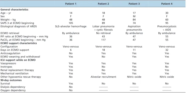
For didactic reasons, the VV-ECMO support can be modeled by two oxygenators in series: the extracorporeal membrane and the native lungs (Figure 1 - Panel A) (9). Through the first oxygenator (VV-ECMO apparatus), blood drawn from the vena cava is pumped at a set flow rate, leaving a fraction of the venous return, i.e., of the CO (13), to proceed to the heart deoxygenated. Therefore, any elevation of the CO, unaccompanied by equal elevations in the ECMO blood flow, will result in a higher fraction of the CO returning deoxygenated to the right heart and to the native lungs (Figure 1 - Panel B). In this situation, the intuitive reaction would be to increase the VV-ECMO blood flow to improve oxygenation. This increase could, however, pre-cipitate recirculation between the return and drainage cannulae, mitigating any possible benefits (9). In addition, high blood flows can cause hemolysis and collapse of the inferior vena cava over the drainage cannula, suddenly reducing blood flow and thus aggravating hypoxemia. Some authors have recognized an ECMO blood flow to cardiac output ratio greater than 0.6 as an index for ECMO efficiency (12).
The second oxygenator in the model (native lungs, Figure 1 – Panel A) will further improve blood oxygenation according to its residual function, which is inversely
Table 1 -Characteristics of patients.
Patient 1 Patient 2 Patient 3 Patient 4
General characteristics
Age – yr 14 18 17 30
Sex F F M F
Weight – kg 48 48 84 60
SAPS 3 at ECMO beginning 105 89 74 95
Etiological diagnosis of ARDS SLE+alveolar hemorrhage Lobar pneumonia +cystic fibrosis
Aspiration pneumonitis
Pneumocystosis
+AIDS
ECMO retrieval By ambulance No retrieval By ambulance By ambulance
P/F ratio at ECMO beginning – mm Hg 36 43 47 55
PaCO2at ECMO beginning – mm Hg 36 117 47 55
ECMO support characteristics
Configuration Veno-venous Veno-venous Veno-venous Veno-venous
Days on ECMO support 6 18 11 32
Anticoagulation No Yes Yes Yes
ECMO weaning and withdrawal Yes No Yes Yes
ICU support while on ECMO
Vasopressors Yes Yes Yes Yes
Inotropes Yes No Yes No
Renal replacement therapy Yes No Yes Yes
Mechanical ventilation Yes Yes Yes Yes
Other hypoxemia rescue therapy No Alveolar recruitment Nitric oxide Nitric oxide 90-day outcomes
Survival Yes No No No
Dialysis dependency No --- ---
---Oxygen dependency No --- ---
---ECMO - extracorporeal membrane oxygenation. SLE - systemic lupus erythematosus.
proportional to the pulmonary shunt (9). The pulmonary shunt can be modulated by decreasing the alveolar collapse, e.g., with the use of higher levels of end-expiratory pressure. The pulmonary shunt also depends on the CO, and it has been shown that reducing CO in healthy lungs slightly improves pulmonary shunting (14), an effect that is even more pronounced in injured lungs during hypoxemic respiratory failure (15). By the same token, the use of inotropic drugs can worsen pulmonary shunting (16). Oxygenation of mixed venous blood has been positively correlated with CO (17) and is an important modulator of the pulmonary shunt (14,15,18). In ICU patients, however, the response of the pulmonary shunt to venous hypoxemia is erratic, depending on systemic factors related to the underlying disease (19).
Blood recirculation from the return cannula to the drainage cannula can reduce the efficacy of the ECMO support because the membrane will oxygenate already
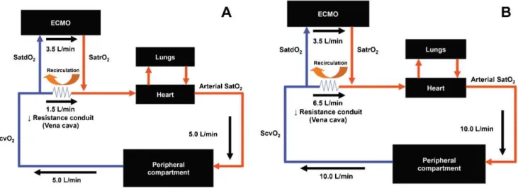
oxygenated blood (10), while the systemic venous blood will return to the heart without proper oxygenation. Finally, oxygenator dysfunction can also contribute to persistent hypoxemia. The presence of blood clots or water drops inside the membrane reduces the exchange surface and consequently the oxygenator’s efficiency. This complication can be diagnosed by the direct visualization of thrombi inside the membrane or by the presence of low post-oxygenation PO2(Figure 2B) and high PCO2.
In summary, there are four main mechanisms of hypoxemia during VV-ECMO: a high recirculation ratio, a high pulmonary shunt, a low cardiac output to ECMO blood flow ratio (,0.6), and oxygenator dysfunction. These mechanisms can occur alone or, more frequently, in combination. Figure 1 shows the equation for the prediction of arterial oxygen saturation, using all of the concepts discussed above (9).
Table 2 -Clinical characteristics at the time of severe hypoxemia diagnosis.
Patient 1 Patient 2 Patient 3 Patient 4
ECMO support
ECMO day of hypoxemia occurrence * 2 2 1 5
ECMO blood flow – mL/min 5080 6000 6500 5300
Sweep gas flow – L/min 2 5 10 7
FiO2 1 1 1 1
Drainage cannula diameter – French 20 22 21 22
Atrial cannula diameter – French 20 22 21 22
Drainage cannula SatO2- % 58 85 61 64
Drainage cannula PO2– mm Hg 30 46 32 35
Return cannula SatO2- % 100 100 99 100
Return cannula PO2– mm Hg 180 402 220 163
Blood flow/cardiac output ratio 0.57 0.61 0.56 0.50
Recirculation - % 26.3 62.5 7.3 20.0
Mechanical ventilation
Ventilatory mode PSV PCV PCV PCV
FiO2 0.3 0.6 1.0 0.6
PEEP – cm H2O 15 15 10 13
Plateau pressure – cm H2O 20 25 20 18
Tidal volume – mL 150 90 50 90
Respiratory rate – breaths/min 15 10 10 10
Patients characteristics
Arterial pH 7.421 7.435 7.500 7.370
PaCO2– mm Hg 42 41 36 47
PvCO2– mm Hg 52 51 61 59
PaO2– mm Hg 46 45 45 37
PvO2– mm Hg 23 29 35 30
Arterial Sat O2- % 82 88 80 84
Venous Sat O2- % 43 60 58 55
Hemoglobin – g/dL 7.4 7.5 8.2 8.0
Pulmonary shunt - % 36.6 44.9 62.9 47.8
Lactate – mmol/L 1.38 1.12 2.6 2.1
Lung injury score 3.75 3.50 4.00 3.75
Temperature -˚C 37.2 38.0 39.0 37.6
Total SOFA 14 17 18 12
Hemodynamics
Cardiac output – L/min 8.9 9.8 11.5 10.6
Heart rate – beats/min 129 130 128 117
Mean arterial blood pressure – mm Hg 109 75 65 105
Central venous pressure – mm Hg 10 7 5 8
Inotropes in use None None None None
Vasopressors in use None Norepinephrine Norepinephrine None
Sedation and analgesia
Analgesia in use Fentanyl Fentanyl Fentanyl None
Sedation in use None Propofol Propofol None
RASS 0 -1 -5 0
*This was the first day of severe hypoxemia
Practical approach for hypoxemia during VV-ECMO support
The rationale for VV-ECMO support is to maintain arterial oxygenation compatible with life and to prevent
further lung injury by allowing for the use of protective ventilatory settings. Accordingly, a low driving pressure (10 cm H2O), a high PEEP (10 - 15 cm H2O), a low
respiratory rate (10 breaths/minute), and a low FiO2(0.3)
have been successfully combined with VV-ECMO support
Figure 1 -VV-ECMO–supported patient model.Panel Ashows a regular patient, in whom the ECMO blood flow (3.5 L/min)/cardiac output (5.0 L/min) ratio was equal to 0.7. In this condition, it is expected that only 1.5 L/min (30% of the venous return) will pass through the vena cava without oxygenation.Panel Bexemplifies a hyperdynamic patient, in whom the ECMO blood flow (3.5 L/ min)/cardiac output (10.0 L/min) ratio was equal to 0.35. In this example, 6.5 L/min (65% of the venous return) will pass through the vena cava without oxygenation. In the former example, if the patient has a severe lung injury with a high pulmonary shunt, he or she will most likely develop severe hypoxemia. SatdO2– oxygen saturation at the drainage cannula. ScvO2– oxygen saturation at the vena cava.
SatrO2– oxygen saturation at the return cannula.
According to Mesai et al. (9):
Arterial SatO2~
Pulmonary oxygenation Residual function
ð Þ z
ECMO effective blood flow Cardiac output
|SatrO2z
1{ECMO effective blood flow Cardiac output
|ScvO2zð0:1|SatrO2Þ
ECMO effective blood flow = (1 – recirculation ratio)6ECMO blood flow.
Figure 2 - Panel Ashows the systemic arterial content of oxygen as a function of ECMO blood flow, andPanel Bshows the expected PO2
in the return cannula as a function of ECMO blood flow. Both panels were created based on a polymethylpentene oxygenator with normal function. In this graph, the original data from the swine experimental study of Park et al. were used (10). The data used presented a wide range of pre-membrane pH, drainage cannula SatO2, and drainage cannula PO2. The data were collected during a
with good outcomes (2). To maintain this protective ventilation, an arterial saturation$85% or a PaO2$50 mm
Hg has been considered sufficient while maintaining low alveolar ventilation (8,20). However, certain patients with refractory hypoxemia do not achieve this minimal safe oxygenation under protective ventilation settings. For these patients, we suggest a sequential approach (Figure 3), aiming for an arterial SatO2$85% in patients with a normal
or near normal PaCO2.
The approach presented in Figure 3 is based on the following ideas: 1) optimizing the ECMO blood flow to cardiac output ratio (initially manipulating the ECMO blood flow), 2) identifying recirculation and repositioning of the cannulae accordingly, 3) optimizing residual lung function, 4) diagnosing oxygenator dysfunction and considering its replacement, and 5) optimizing the ECMO blood flow to cardiac output ratio based on the reduction in cardiac output (avoiding fever or providing active patient cooling, decreasing oxygen consumption if necessary with neuro-muscular blockers, and possibly reducing cardiac output with the use of beta-blockers after careful consideration of the potential clinical deterioration due to cardiovascular depression) (6,8). Blood transfusions to optimize the DO2
can be considered at any time.
Permissive hypoxemia is an option, according to the patient’s clinical situation, when other interventions have failed. Severe ARDS patients without VV-ECMO support develop long-term neuropsychological impairment associated with hypoxemia in the acute phase of the disease (21). However, in VV-ECMO– supported severely injured patients, an arterial SatO2as low as
70% has been allowed in awake and participative patients with normal arterial PCO2(7), with a high survival rate (76%) and
without significant long-term sequelae in health-related quality of life (7,22). Maintaining a normal PCO2 seems to be
mandatory in permissive severe hypoxemia because the association of low PO2with high PCO2can potentially cause
severe brain injury (23).
The clinical vignettes
Patient 1 – She was awake and collaborative, with a high recirculation ratio and a low ECMO blood flow to cardiac output. The ventilator FiO2 was 0.3. In this patient, the
ECMO blood flow was raised to 6000 mL/min when the SpO2reached 85%.
Patient 2 – This patient was cannulated with a femoro-femoral approach, with a high recirculation ratio and an adequate ECMO blood flow to cardiac output ratio. Due to difficulty in repositioning the cannulae, we chose to perform
alveolar recruitment. A slight improvement in oxygenation was obtained, keeping the arterial SatO2between 85 and 89%.
Patient 3 – The patient was febrile and hyperdynamic. There was no recirculation, and the ECMO blood flow to cardiac output was low (ratio = 0.56). This patient received active interventions to control the fever, which slowly improved the hypoxemia.
Patient 4 – This patient had a low ratio of ECMO blood flow to cardiac output and a low recirculation ratio. The ECMO blood flow was raised, thereby bringing the hypoxemia to an acceptable level.
Severe hypoxemia can occur during VV-ECMO respira-tory support, and it is crucial to understand the underlying mechanisms. Knowledge of the pathophysiology of hypox-emia is important to guide specific interventions. A stepwise approach, as proposed here, can often be used to address this concerning clinical situation. When other alternatives have failed, permissive severe hypoxemia is acceptable. In this group of patients, maintaining normo-capnia is essential to attenuate the risk of associated sequelae.
& AUTHOR CONTRIBUTIONS
Nunes LB, Mendes PV, Barbosa EV, Hirota AS, and Park M were responsible for the acquisition, analysis, and interpretation of the data. Nunes LB, Mendes PV, and Park M wrote the manuscript. Costa EL, Azevedo LC, and Schettino GP critically reviewed the manuscript.
& REFERENCES
1. Park M, Azevedo LC, Mendes PV, Carvalho CR, Amato MB, Schettino GP, et al. First-year experience of a Brazilian tertiary medical center in supporting severely ill patients using extracorporeal membrane oxyge-nation. Clinics. 2012;67(10):1157-63, http://dx.doi.org/10.6061/clinics/ 2012(10)07.
2. Peek GJ, Mugford M, Tiruvoipati R, Wilson A, Allen E, Thalanany MM, et al. Efficacy and economic assessment of conventional ventilatory support versus extracorporeal membrane oxygenation for severe adult respiratory failure (CESAR): a multicentre randomised controlled trial. Lancet. 2009;374(9698):1351-63, http://dx.doi.org/10.1016/S0140-6736 (09)61069-2.
3. Davies A, Jones D, Bailey M, Beca J, Bellomo R, Blackwell N, et al. Extracorporeal Membrane Oxygenation for 2009 Influenza A(H1N1) Acute Respiratory Distress Syndrome. JAMA. 2009;302(17):1888-95. 4. Brogan TV, Thiagarajan RR, Rycus PT, Bartlett RH, Bratton SL.
Extracorporeal membrane oxygenation in adults with severe respiratory failure: a multi-center database. Intensive Care Med. 2009;35(12):2105-14, http://dx.doi.org/10.1007/s00134-009-1661-7.
5. Pham T, Combes A, Roze´ H, Chevret S, Mercat A, Roch A, et al. Extracorporeal membrane oxygenation for pandemic influenza a(h1n1) induced acute respiratory distress syndrome. a cohort study and propensity-matched analysis. Am J Respir Crit Care Med. 2013; 187(3):276-85, http://dx.doi.org/10.1164/rccm.201205-0815OC. 6. Guarracino F, Zangrillo A, Ruggeri L, Pieri M, Calabro` MG, Landoni G,
et al.b-Blockers to optimize peripheral oxygenation during extracorpor-eal membrane oxygenation: a case series. J Cardiothorac Vasc Anesth. 2012;26(1):58-63, http://dx.doi.org/10.1053/j.jvca.2011.05.013. 7. Linden V, Palmer K, Reinhard J, Westman R, Ehren H, Granholm T, et al.
High survival in adult patients with acute respiratory distress syndrome treated by extracorporeal membrane oxygenation, minimal sedation, and pressure supported ventilation. Intensive Care Med. 2000;26(11):1630-7.
8. Combes A, Bacchetta M, Brodie D, Muller T, Pellegrino V. Extracorporeal membrane oxygenation for respiratory failure in adults. CurrOpinCrit Care. 2012;18(1):99-104.
9. Messaı¨ E, Bouguerra A, Harmelin G, Di Lascio G, Cianchi G, Bonacchi M. A new formula for determining arterial oxygen saturation during venovenous extracorporeal oxygenation. Intensive Care Med. 2013; 39(2):327-34, http://dx.doi.org/10.1007/s00134-012-2756-0.
10. Park M, Costa EL, Maciel AT, Silva DP, Friedrich N, Barbosa EV, et al. Determinants of oxygen and carbon dioxide transfer during extra-corporeal membrane oxygenation in an experimental model of multiple organ dysfunction syndrome. PLoS One. 2013;8(1):e54954, http://dx.doi. org/10.1371/journal.pone.0054954.
11. Williams EM, Viale JP, Hamilton RM, McPeak H, Sutton L, Hahn CE. Within-breath arterial PO2 oscillations in an experimental model of acute respiratory distress syndrome. Br J Anaesth. 2000;85(3):456-9.
12. Schmidt M, Tachon G, Devilliers C, Muller G, Hekimian G, Bre´chot N, et al. Blood oxygenation and decarboxylation determinants during venovenous ECMO for respiratory failure in adults. Intensive Care Med. 2013;39(5):838-46, http://dx.doi.org/10.1007/s00134-012-2785-8. 13. Guyton AC. Determination of cardiac output by equating venous return
curves with cardiac response curves. Physiol Rev. 1955;35(1):123-9. 14. Smith G, Cheney FW, Winter PM. The effect of change in cardiac output
on intrapulmonary shunting. Br J Anaesth. 1974;46(5):337-42.
15. Dantzker DR, Lynch JP, Weg JG. Depression of cardiac output is a mechanism of shunt reduction in the therapy of acute respiratory failure. Chest. 1980;77(5):636-42, http://dx.doi.org/10.1378/chest.77.5.636. 16. Bryan TL, van Diepen S, Bhutani M, Shanks M, Welsh RC, Stickland MK.
The effects of dobutamine and dopamine on intrapulmonary shunt and gas exchange in healthy humans. J Appl Physiol. 2012;113(4):541-8, http://dx.doi.org/10.1152/japplphysiol.00404.2012.
17. Zampieri FG, Park M, Azevedo LC, Amato MB, Costa EL. Effects of arterial oxygen tension and cardiac output on venous saturation: a mathematical modeling approach. Clinics. 2012;67(8):897-900, http://dx. doi.org/10.6061/clinics/2012(08)07.
18. Dantzker DR, Brook CJ, DeHart P, Lynch JP, Weg JG. Ventilation-perfusion distributions in the adult respiratory distress syndrome. Am Rev Respir Dis. 1979;120(5):1039-52.
19. Rossaint R, Hahn SM, Pappert D, Falke KJ, Radermacher P. Influence of mixed venous PO2 and inspired O2 fraction on intrapulmonary shunt in patients with severe ARDS. J Appl Physiol. 1995;78(4):1531-6. 20. Sidebotham D, McGeorge A, McGuinness S, Edwards M, Willcox T, Beca
J. Extracorporeal membrane oxygenation for treating severe cardiac and respiratory failure in adults: part 2-technical considerations. J Cardiothorac Vasc Anesth. 2010;24(1):164-72, http://dx.doi.org/10. 1053/j.jvca.2009.08.002.
21. Mikkelsen ME, Christie JD, Lanken PN, Biester RC, Thompson BT, Bellamy SL, et al. The adult respiratory distress syndrome cognitive outcomes study: long-term neuropsychological function in survivors of acute lung injury. Am J Respir Crit Care Med. 2012;185(12):1307-15. 22. Linde´n VB, Lidegran MK, Frise´n G, Dahlgren P, Frenckner BP, Larsen F.
ECMO in ARDS: a long-term follow-up study regarding pulmonary morphology and function and health-related quality of life. Acta Anaesthesiol Scand. 2009;53(4):489-95, http://dx.doi.org/10.1111/j. 1399-6576.2008.01808.x.
23. Mendes PV, Moura E, Barbosa EV, Hirota AS, Scordamaglio PR, Ajjar FM, et al. Challenges in patients supported with extracorporeal membrane oxygenation in Brazil. Clinics. 2012;67(12):1511-5, http://dx. doi.org/10.6061/clinics/2012(12)27.
APPENDIX