Generation and Characterization of a
Human/Mouse Chimeric GD
2
-Mimicking
Anti-Idiotype Antibody Ganglidiximab for Active
Immunotherapy against Neuroblastoma
Christin Eger1‡, Nikolai Siebert1‡*, Diana Seidel1, Maxi Zumpe1, Madlen Jüttner1, Sven Brandt2, Hans-Peter Müller2, Holger N. Lode1
1Department of Pediatric Oncology and Hematology, University Medicine Greifswald, 17475, Greifswald, Germany,2ZIK HIKE, Center for Innovation Competence: Humoral Immune Reactions in Cardiovascular Diseases, University of Greifswald, Greifswald, Germany
‡These two authors are co-first authors of this work.
*nikolai.siebert@uni-greifswald.de
Abstract
Vaccination with proteins mimicking GD2that is highly expressed on neuroblastoma (NB) cells is a promising strategy in treatment of NB, a pediatric malignancy with poor progno-sis. We previously showed efficacy of ganglidiomabin vivo, a murine anti-idiotype (anti-Id)
IgG1. In order to tailor immune responses to variable regions, we generated a new human/mouse chimeric anti-Id antibody (Ab) ganglidiximab by replacing murine constant fragments with corresponding human IgG1 regions. DNA sequences encoding for variable regions of heavy (VH) and light chains (VL) were synthesized by RT-PCR from total RNA of ganglidiomab-producing hybridoma cells and further ligated into mammalian expres-sion plasmids with coding sequences for constant regions of human IgG1 heavy and light chains, respectively. We established a stable production cell line using Chinese hamster ovarian (CHO) cells co-transfected with two expression plasmids driving the expression of either ganglidiximab heavy or light chain. After purification from supernatants, anti-idiotypic characteristics of ganglidiximab were demonstrated. Binding of ganglidiximab to anti-GD2Abs of the 14.18 family as well as to NK-92tr cells expressing a GD2-specific chi-meric antigen receptor (scFv(ch14.18)-zeta) was shown using standard ELISA and flow cytometry analysis, respectively. Ganglidiximab binding affinities to anti-GD2Abs were further determined by surface plasmon resonance technique. Moreover, binding of anti-GD2Abs to the nominal antigen GD2as well as GD2-specific Ab-mediated cytotoxicity (ADCC, CDC) was competitively inhibited by ganglidiximab. Finally, ganglidiximab was successfully used as a protein vaccinein vivoto induce a GD2-specific humoral immune
response. In summary, we report generation and characterization of a new human/mouse chimeric anti-Id Ab ganglidiximab for active immunotherapy against NB. This Ab may be useful to tailor immune responses to the paratope regions mimicking GD2overexpressed in NB.
OPEN ACCESS
Citation:Eger C, Siebert N, Seidel D, Zumpe M, Jüttner M, Brandt S, et al. (2016) Generation and Characterization of a Human/Mouse Chimeric GD2
-Mimicking Anti-Idiotype Antibody Ganglidiximab for Active Immunotherapy against Neuroblastoma. PLoS ONE 11(3): e0150479. doi:10.1371/journal. pone.0150479
Editor:Pierre Busson, Gustave Roussy, FRANCE
Received:November 11, 2015
Accepted:February 15, 2016
Published:March 11, 2016
Copyright:© 2016 Eger et al. This is an open access article distributed under the terms of the Creative Commons Attribution License, which permits unrestricted use, distribution, and reproduction in any medium, provided the original author and source are credited.
Data Availability Statement:All relevant data are within the paper.
Introduction
NB is the most common extracranial solid tumor in early childhood. About 50% of patients show a high-risk NB phenotype, characterized by wide spread dissemination and poor long-term survival despite intensive multimodal treatments [1–3]. A successful approach to improve survival in high-risk NB patients is immunotherapy targeting the tumor-associated antigen (TAA) GD2due to its high expression on NB cells [4,5] and poor physiological expression on healthy tissues [6]. Therefore, monoclonal Abs (mAbs) directed against GD2have been devel-oped and successfully applied over the past two decades in a number of clinical trials for NB [7, 8]. However, one obstacle associated with infusion of ch14.18/CHO is pain toxicity [9] which correlates with infusion rate [10]. Recently, a novel treatment method based on a long term infusion of ch14.18/CHO in combination with interleukin-2 was initiated and showed a reduced toxicity profile [10]. Since passive immunotherapy does not induce a long lasting immune response, there is a considerable interest in extending these studies into active immu-notherapy. However, active vaccination with GD2encounters several significant drawbacks including its weak immunogenicity and T cell independence due to its glycolipid structure [11, 12]. A promising alternative strategy exploits the immune network hypothesis of Jerne [13] to present GD2as a protein epitope by an anti-Id Ab acting as a T cell-dependent surrogate with enhanced immunogenicity. Therefore, TAA-mimicking anti-Id Abs are used for protein vac-cine development and successfully applied in a number of preclinical and clinical studies for a variety of solid tumors [14,15]. For treatment of high-risk NB patients, an anti-Id Ab 1A7 bearing the internal image of GD2was developed [16] and used as protein vaccine [1]. Impor-tantly, patients vaccinated with 1A7 showed little side effects which underlines the suitability of such a vaccine for clinical application. To provide unrestricted access for clinical develop-ment in Europe, we recently generated and characterized a murine anti-Id Ab ganglidiomab which paratopes mimic GD2. Vaccination of mice resulted in an induction of a GD2-specific humoral immunity [17]. To further tailor this immune response induced by ganglidiomab to GD2-mimicking paratopes in prospective clinical trials, we generated a chimeric human/ mouse anti-Id Ab ganglidiximab by replacing murine constant regions with corresponding fragments of human IgG1.
Here, we report the generation and characterization of this new chimeric human/mouse GD2-mimicking anti-Id Ab providing an important baseline for the development of protein vaccines with clinical potential for active immunotherapy against NB.
Materials and Methods
Cell culture
A GD2-positive murine NB cell line NXS2 [18] and CHO cell line [19] (ATCC, Wesel, Ger-many) were cultured in Dulbecco's modified Eagle's medium supplemented with stable gluta-mine, 4.5 g/l glucose (DMEM; PAN-Biotech, Aidenbach, Germany), 10% (v/v) fetal calf serum (FCS), 100 U/ml penicillin and 0.1 mg/ml streptomycin (1× P/S; PAA, Pasching, Austria). Hybridoma cells producing murine anti-Id Ab ganglidiomab [17] were cultured in serum-free DMEM with stable glutamine and 4.5 g/l glucose supplemented with 1× non-essential amino acids (PAA, Pasching, Austria) and 50μMβ-mercaptoethanol (Sigma Aldrich, Steinheim,
Ger-many). The human NB cell line LA-N-1 [20] was cultured in RPMI (PAN-Biotech, Aidenbach, Germany) supplemented with 4.5 g/l glucose, 2 mM stable glutamine (PAA, Pasching, Aus-tria), 10% (v/v) FCS and 1× P/S. The genetically engineered NK-92-scFv(ch14.18)-zeta cell line (NK-92tr), expressing a GD2-specific chimeric antigen receptor derived from ch14.18, was
funders had no role in study design, data collection and analysis, decision to publish, or preparation of the manuscript.
kindly provided by Prof. W. Wels (Georg-Speyer Haus, Frankfurt, Germany) and cultured as previously described [21].
Mice
Analysis of GD2-specific humoral immune response upon vaccination with ganglidiximab was performed in female A/J mice (10 weeks of age; Charles River Laboratories, Sulzfeld, Ger-many). Mice were housed in standard animal laboratories (12 h light/dark cycle) with free access to water and standard laboratory chowad libitum. Animal experiments were approved by the animal welfare committee (Landesamt für Landwirtschaft, Lebensmittelsicherheit und Fischerei Mecklenburg-Vorpommern, Thierfelderstraße 18, 18059 Rostock, LALLF M-V/TSD/ 7221.3-1-011/11) and approved and supervised by the commissioner for animal welfare at the University Medicine Greifswald representing the Institutional Animal Care and Use Commit-tee (IACUC).
Design and construction of ganglidiximab expression plasmids
RNA isolation and RT-PCR analysis. Total RNA was isolated from 5× 10⁶hybridoma cells producing murine anti-Id Ab ganglidiomab using the RNeasy1
Mini Kit (QIAGEN GmbH, Hilden, Germany) and concentration was determined spectrophotometrically (Bio-Photometer plus, Eppendorf, Hamburg, Germany). 1μg of total RNA was used for cDNA
syn-thesis w SuperScript1II Reverse Transcriptase (Invitrogen GmbH, Darmstadt, Germany) according to the manufacturer’s guidelines. PCR amplification of ganglidomab VH and -VL was performed using gene-specific primers designed with online primer design tool Primer3 to allow amplification of a 440 bp PCR-product. For ganglidiomab VH amplification forward primer sequence5`-GGGgcggccgcCATGGCTGTCTTGGGGCTGCTCTTCT-3’including NotI-HF restriction site (italic, lowercase) and the reverse primer sequence5’-CCCctcga GACGGTGACTGAGGTTCCTTGA-3’includingXhoI restriction site (italic, lowercase) were
used. VH was amplified by 30 cycles consisting of +94°C (15 s) for denaturation, +81°C (15 s) for primer-specific annealing and +72°C (30 s) for elongation. For ganglidiomab VL amplifica-tion, gene-specific primers were designed as described above. Following primer sequences were used for amplification of a 420 bp PCR-product:5’-GGGgcggccgcCATGAAGTTGCCTGT TAGGCTGTTG -3’(forward primer includingNotI-HF restriction site; italic, lowercase) and
5’- CCCcgtacgAGCCCGTTTGATTTCCAGCTT -3’(reverse primer includingBsiWI
restriction site; italic, lowercase). VL was amplified by 30 cycles consisting of +94°C (15 s) for denaturation, +73°C (15 s) for primer-specific annealing, and +72°C (30 s) for elongation. Finally, PCR prodcucts were analyzed by agarose gel electrophoresis.
Isolation of murine ganglidiomab variable region from agarose gel. PCR products were isolated from agarose gels using a Nucleo Spin1
Extract II Kit (Macherey-Nagel, Düren, Ger-many) according to the manufacturer’s guidelines. Briefly, DNA fragments were excised from agarose gel and lysis buffer was added followed by incubation at +50°C until gel slices were completely dissolved. Then, the sample was loaded onto a Nucleo Spin1column containing a silica membrane binding DNA (centrifugation: 11,000× g, 1 min, RT). After a wash step (centri-fugation: 11,000× g, 1 min, RT), the silica membrane was dried (centri(centri-fugation: 11,000× g, 2 min, RT) and bound DNA was eluted inAqua destillata (A.dest)(50μl; centrifugation: 11,000× g, 1
min, RT). DNA concentration was determined spectrophotometrically as described above.
Cloning of murine ganglidiomab variable region into pCR1
2.1-TOPO1
plasmid.
pCR1
2.1-TOPO1
plasmid (10 ng) for 5 min at RT. Then, One Shot1
TOP 10 chemically competentE.colicells were added and incubated for 20 min on ice. For transformation,E.coli
were subjected to heat-shock for 30 s at +42°C and immediately placed back on ice. Subse-quently, S.O.C. outgrowth medium was added andE.coliwere shaken horizontally (400 rpm) for 1 h at +37°C. Then, transformedE.coliwere incubated overnight at +37°C on ampicillin (50μg/ml) containing Luria/Miller (LB) agar plates (Roth, Karlsruhe, Germany) coated with
X-gal in dimethylformamide (DMF) (40 mg/ml) for blue/white screening. Finally, 15 positive clones (white colonies) were selected and cultured overnight in 5 ml LB medium (Roth, Karls-ruhe, Germany) supplemented with 50μg/ml ampicillin (+37°C, shaking at 100 rpm).
Purification of pCR12.1-TOPO1plasmid containing murine ganglidiomab variable region. Plasmids of successfully transformed clones were isolated fromE.colicultures using NucleoSpin1Plasmid Kit (Macherey-Nagel, Düren, Germany) according to the manufac-turer's guidelines. Briefly, pelletedE.coli(centrifugation: 11,000× g, 30 s, RT) were resuspended in provided buffer and lysed for 5 min at RT followed by a neutralization step to ensure appro-priate DNA binding conditions. After centrifugation (11,000× g, 5 min, RT), the supernatant was loaded onto a Nucleo Spin1plasmid column containing a silica membrane binding plas-mid DNA (centrifugation: 11,000× g, 1 min, RT). After two wash steps (centrifugation: 11,000× g, 1 min, RT), the silica membrane was dried (centrifugation: 11,000× g, 2 min, RT) and bound DNA was eluted inA.dest(centrifugation: 11,000× g, 1 min, RT). Finally, purified plasmids containing DNA fragments of expected molecular size were sequenced (LGC Geno-mics, Berlin, Germany).
Restriction analysis of pCR12.1-TOPO1plasmids containing murine ganglidiomab variable regions. For further cloning, ganglidiomab VH and -VL were excised from purified pCR12.1-TOPO1plasmids using respective restriction enzymes. For ganglidiomab VH, plas-mid DNA (4μg) was incubated withNotI-HF (1 unit; New England Biolabs GmbH, Frankfurt/
Main, Germany) andXhoI-HF (1 unit; New England Biolabs GmbH, Frankfurt/Main, Ger-many) in CutSmart1
Buffer (1×) for 1 h at +37°C followed by enzyme heat inactivation (20 min, +65°C). Resulting plasmid DNA products were analyzed by agarose gel electrophoresis and ganglidiomab VH DNA fragment (427 bp) was purified from agarose gel and spectropho-tometrically quantified as described above. For ganglidiomab VL, double restriction digest using bothNotI-HF andBsiWI was not recommended due to different optimum incubation temperatures. Therefore, plasmid DNA (8μg) was first linearized usingNotI-HF (2 units)
fol-lowed by purification from agarose gel as described above. Then, linearized plasmid DNA (4μg) was incubated withBsiWI (1 unit; New England Biolabs GmbH, Frankfurt/Main,
Ger-many) in NEBuffer 3.1 (1×) for 1 h at +55°C followed by enzyme heat inactivation (20 min, +65°C). After agarose gel electrophoresis, ganglidiomab VL (407 bp) was purified from agarose gel and spectrophotometrically quantified as described above.
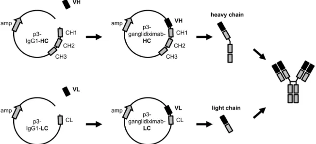
Generation of chimeric human/mouse ganglidiximab heavy and light chain. For gangli-diximab generation, purified ganglidiomab VH and -VL were cloned in frame into respective mammalian expression plasmids with coding sequences for human IgG1 heavy (p3-IgG1-HC) and light chain (p3-IgG1-LC), respectively (evitria AG, Zurich-Schlieren, Switzerland) (Fig 1). First, plasmid DNA was linearized using respective restriction enzymes as described above allowing integration of respective murine variable region into the multiple cloning site (MCS). For this, linearized plasmid DNA (50 ng) and ganglidiomab variable region DNA fragment (50 ng) were incubated with Rapid Ligation Buffer (1×; Promega GmbH, Mannheim, Germany) and T4 DNA Ligase (1 unit; Promega GmbH, Mannheim, Germany) overnight at +4°C. Subse-quently, 10μl of the ligation product were used to transform NEB 10-beta chemically
Karlsruhe, Germany) containing ampicillin (50μg/ml). ResistantE.colicolonies were
trans-ferred into 5 ml LB medium (Roth, Karlsruhe, Germany) supplemented with 50μg/ml
ampicil-lin and cultured overnight at +37°C. Plasmids of successful transformed cells were then isolated from suspension followed by determination of DNA concentration as described above. After sequencing, sufficient amounts of plasmid DNA of correct sequence were purified as described above.
Establishment of a cell line producing chimeric human/mouse
ganglidiximab
Co-transfection of CHO cells with p3-ganglidiximab-HC/p3-ganglidiximab-LC. To allow permenant Ab production, a new cell line stably producing chimeric ganglidiximab was established. For this purpose, we employed CHO cells, a mammalian cell line most commonly used for industrial recombinant protein production [9]. Co-transfection of CHO cells was per-formed using the newly generated plasmids encoding for ganglidiximab heavy (p3-ganglidixi-mab-HC) or light chain (p3-ganglidiximab-LC). One day prior to co-transfection, 3× 10⁵CHO cells were seeded in 2 ml/well of Opti-MEM1I Reduced Serum Medium (Life Technologies, Darmstadt, Germany) supplemented with 3% (v/v) FCS, 10 U/ml penicillin and 0.01 mg/ml streptomycin in each well (6-well plate; Sarstedt, Nümbrecht, Germany). 2μg of each plasmid
were mixed with Opti-MEM1
I Reduced Serum Medium, Lipofectamine1
3000 transfection reagent and P3000™reagent (Life Technologies, Darmstadt, Germany) according to the manu-facturer's guidelines and incubated for 5 min at RT. The entire transfection mixture was added to CHO cells followed by incubation at +37°C, 5% CO2for 24 h. CHO cells co-transfected with empty plasmid controls or incubated with transfection reagent without DNA plasmids as well as non-transfected cells were utilized as negative controls. To confirm ganglidiximab produc-tion in co-transfected CHO cells, supernatants were collected 24 h after co-transfecproduc-tion and analyzed for ganglidiximab production using standard ELISA. First, 96-well plates (Sarstedt, Fig 1. Schematic overview of generation of human/mouse chimeric anti-Id Ab ganglidiximab.The human/mouse chimeric anti-Id Ab ganglidiximab is composed of GD2mimicking variable regions (VH, VL) of murine anti-Id ganglidiomab and human IgG1 constant regions. Coding sequences of VH and VL
were synthesized and inserted into mammalian expression plasmids containing DNA sequences for human IgG1 heavy (p3-IgG1-HC) and light chain (p3-IgG1-LC), respectively. For ganglidiximab production, CHO cells were stably co-transfected with the two generated expression plasmids (p3-ganglidiximab-HC and p3-ganglidiximab-LC).
Nümbrecht, Germany) were coated with 250 ng/well of murine anti-GD2Ab 14G2a (100μl
per well, 0.1 M carbonate/hydrogen carbonate buffer, pH 9.6, 1 h, +37°C). After three wash steps (200μl per well; 0.05% (v/v) Tween-20 in phosphate buffered saline (PBS, pH 7.4; PAA,
Pasching, Austria)), wells were blocked with 1% (w/v) bovine serum albumin (BSA; Sigma Aldrich, Steinheim, Germany) in PBS (pH 7.4) (200μl per well; 1 h, +37°C) and washed three
times (200μl per well; 0.05% (v/v) Tween-20 in PBS, pH 7.4). Then, samples were added and
incubated for 1 h at +37°C (100μl per well). Supernatants of CHO cells co-transfected with
empty plasmid controls or incubated with transfection reagent without DNA plasmids as well as non-transfected cells served as negative controls. After three wash steps (200μl per well;
0.05% (v/v) Tween-20 in PBS, pH 7.4) ganglidiximab binding to 14G2a was detected by horse-radish peroxidase-(HRP)-conjugated goat anti-human IgG (Fc-specific) mAb as a secondary Ab (100μl per well, 1:10,000; 1 h, +37°C) (# A0170-1ML, Sigma Aldrich, Steinheim, Germany).
Plates were washed three times (200μl per well; 0.05% (v/v) Tween-20 in PBS, pH 7.4) and
75μl of detection reagent (R&D Systems Inc, Minneapolis, MN, USA) were added according
to the manufacturer's guidelines. After 5 min, 50μl of 2 N H2SO4were added to stop the
reac-tion. Absorption was determined in a Synergy HT multimode microplate reader (BioTek Ger-many, Bad Friedrichshall, Germany) at 450 nm.
Selection of a cell line stably producing ganglidiximab. To establish a cell line for perma-nent Ab production, CHO cells were harvested 24 h after co-transfection with p3-ganglidixi-mab-HC/p3-ganglidiximab-LC and limited dilutions were performed (1 cell/well in 200μl of
DMEM supplemented with stable glutamine, 4.5 g/l glucose, 10% (v/v) FCS and 1× P/S) fol-lowed by culturing for 14 days. Next, supernatants were collected and analyzed for ganglidixi-mab production using ELISA as described above. For each further subcloning step, clones with the highest ganglidiximab-production rate were used for limited dilutions. To establish a cell line stably producing ganglidiximab, a selected cell clone was analyzed for ganglidiximab pro-duction rate after 15 passages as well as two freeze-thaw cycles. For this, isolated RNA was used for RT-PCR analysis as described above.
Isolation of ganglidiximab from stably transfected CHO cell line
CHO cells stably producing ganglidiximab were cultured in DMEM supplemented with stable glutamine, 4.5 g/l glucose, 10% (v/v) FCS and 1× P/S for ten days. CHO supernatant was fil-tered in two steps using 1.2μm and 0.22μm filter systems (Merck Millipore Corporation,
Darmstadt, Germany). Then, Ab was concentrated with a Centrifugal Concentrator (regener-ated cellulose membrane, molecular weight cut off: 30 kDa) (PALL Life Sciences, Dreieich, Germany) according to the manufacturer's guidelines resulting in a ganglidiximab concentra-tion of 21μg/ml determined by ELISA using 14G2a as a capture mAb (as described above).
For quantification seven standard samples of known ganglidiximab concentration (25.0, 12.5, 6.25, 3.13, 1.56 and 0.78μg/ml) were prepared in PBS (pH 7.4). Prior to incubation
Binding analysis of ganglidiximab to anti-GD
2antibodies and NK-92tr
To evaluate anti-idiotypic characteristics, ganglidiximab binding to anti-GD2Abs and NK-92tr expressing a GD2-specific CAR was analyzed by ELISA and flow cytometry, respectively. For binding analysis of ganglidiximab to anti-GD2Abs, 96-well plates were coated with 250 ng ganglidiximab per well (100μl, 0.1 M carbonate/hydrogen carbonate buffer, pH 9.6, 1 h,
+37°C). After three wash steps (200μl per well; 0.05% (v/v) Tween-20 in PBS, pH 7.4), wells
were blocked with 1% (w/v) BSA in PBS (pH 7.4) (200μl per well; 1 h, +37°C) and washed
three times (200μl per well; 0.05% (v/v) Tween-20 in PBS, pH 7.4). Following anti-GD2Abs
were diluted in PBS (pH 7.4) to final concentrations of 1.0, 0.5, 0.25, 0.13, 0.06, 0.03 and 0.015μg/ml and incubated overnight (100μl per well, +4°C): murine Ab 14G2a [22], chimeric
Ab ch14.18/CHO containing human IgG1 constant regions and variable regions of murine 14G2a [23], ch14.18-dCH2 generated by deletion of heavy chain CH2 domain [24], humanized hu14.18K322A containing a single point mutation (K322A) in the constant region which nearly abrogates complement activation [25] as well as Ab-cytokine fusion proteins named immunocytokines consisting of a GD2-specific Ab linked to interleukin-2 (IL-2) (ch14.18-IL-2 and hu14.18-IL-2 [26]). After five wash steps (200μl per well; 0.05% (v/v) Tween-20 in PBS,
pH 7.4), ganglidiximab binding to anti-GD2Abs was detected using biotinylated ganglidiomab as a secondary Ab diluted in 1% (w/v) BSA in PBS (pH 7.4) (100μl per well; 1:5,000; 2 h,
+37°C). Biotinylation of ganglidiomab was performed using EZ-Link1Sulfo-NHS-LC-Biotin (Thermo Scientific, Erlangen, Germany) as previously described [17]. After three wash steps (200μl per well; 0.05% (v/v) Tween-20 in PBS, pH 7.4) 100μl of Pierce High Sensitivity
Neu-trAvidin-HRP (Thermo Scientific, Erlangen, Germany) diluted 1:10,000 in 1% (w/v) BSA in PBS (pH 7.4) were added and incubated for 20 min at +37°C. Then, binding of ganglidiximab to anti-GD2Abs was detected as described above. ELISA signals (optical density; OD) deter-mined with 1μg/ml ch14.18/CHO were defined as 100% binding. Rituximab and IgG2a served
as negative controls. Binding of anti-GD2Abs to ganglidiximab were calculated according to the formula: binding in % = (OD 450 nm × 100%) / OD 450 nm ch14.18/CHO (1μg/ml). For
flow cytometric analysis of ganglidiximab binding to NK-92tr, 1× 10⁶cells were harvested and washed with fluorescence-activated cell sorting (FACS) buffer (PBS (pH 7.4) supplemented with 1% (w/v) BSA, 0.1% (w/v) EDTA and 0.1% (w/v) NaN3; Roth, Karlsruhe, Germany) (cen-trifugation: 300× g, 5 min, RT). Then, cells were incubated with ganglidiximab (1μg) for 30
min on ice (dark). Ganglidiomab served as a positive control and murine IgG1 and rituximab as isotype controls. After a further wash step, cells were incubated with 1μg biotinylated
ch14.18/CHO and washed again. PE-labeled streptavidin (BioLegend, San Diego, CA, USA) was used for detection (1:1,300; diluted in FACS buffer; 20 min, on ice, dark). Exclusion of dead cells was based on staining with DAPI (4μl, 0.1μg/ml, 5 min) prior to analysis. Sample
acquisition was performed with a FACSCanto II flow cytometer and FACSDiva software (BD Biosciences, Heidelberg, Germany). FlowJo (Treestar, Ashland, OR, USA) was used for data analysis.
Binding affinity of ganglidiximab to anti-GD
2antibodies
surface followed by injection of the respective Ab. Ganglidiximab and ganglidiomab were diluted in acetate buffer (10 mM, pH 5.5, Biacore, Uppsala, Sweden) to a final concentration of 2.5μg/ml. Chimeric mAb rituximab and murine IgG1 isotype control (R&D Systems Inc,
Min-neapolis, MN, USA) served as negative controls. Immobilization target level was defined as 300 response units (RU). Free reactive ester groups on the surface were subsequently blocked using ethanolamine hydrochloride buffer (1 M, pH 8.5, 10μl/min for 60 s). Serial dilutions of each
analyte (14G2a, ch14.18/CHO, hu14.18K322A, ch14.18-IL-2 and hu14.18-IL-2) was prepared in PBS (pH 7.4, 10 mM phosphate buffer, 2.7 mM KCl, 137 mM NaCl; Biacore, Uppsala, Swe-den) (final concentrations: 128.0, 32.0, 8.0, 2.0 and 0.5μg/ml). Between injections the sensor
surface was regenerated for 30 s with 10 mM Glycine/HCL (pH 3, 30μl/min). Binding affinity
was analyzed using the“Single-Cycle-Kinetics”method. The dissociation constant (KD) was calculated for each Ab with BiaEvaluation software (Biacore, Uppsala, Sweden) using a steady-state fit model. Affinity measurements of both anti-Id Abs were performed in triplicates.
Ganglidiximab-dependent competitive inhibition of anti-GD2
antibody
binding to GD
2To confirm anti-Id characteristics of ganglidiximab, the previously described competitive ELISA was used to inhibit binding of anti-GD2Abs (14G2a, ch14.18/CHO, ch14.18-dCH2, hu14.18K322A, ch14.18-IL-2 and hu14.18-IL-2) to the nominal antigen GD2[17]. Briefly, 96-well plates were coated with 50 ng GD2(Sigma Aldrich, Steinheim, Germany) per well (50μl; 99.9% methanol (Roth, Karlsruhe, Germany); 1 h, +50°C) as a capture antigen. After
methanol evaporation wells were blocked with 1% (w/v) BSA in PBS (pH 7.4) (100μl per well;
1 h, +37°C) and washed three times (200μl per well; 0.1% (w/v) BSA in PBS, pH 7.4).
Gangli-diximab was diluted in PBS (pH 7.4) to a final concentration of 1.68μg/ml followed by serial
1:2 dilution yielding 0.840μg/ml (1:2), 0.420μg/ml (1:4), 0.210μg/ml (1:8), 0.105μg/ml
(1:16), 0.053μg/ml (1:32), 0.026μg/ml (1:64) and 0.013μg/ml (1:128). Then, anti-GD2Abs
were applied to each dilution of ganglidiximab at a final concentration of 0.330μg/ml and
mixed. 200μl of each dilution was added per well and incubated (2 h, +37°C). After five wash
steps (200μl per well; 0.1% (w/v) BSA in PBS, pH 7.4) anti-GD2antibodies bound to GD2
were analyzed using biotinylated ganglidiomab as a secondary Ab diluted in 1% (w/v) BSA in PBS (pH 7.4) (100μl per well; 1:5,000; 2 h, +37°C). After plates were washed three times
(200μl per well; 0.1% (w/v) BSA in PBS, pH 7.4), 75μl detection reagent (R&D Systems Inc,
Minneapolis, MN, USA) were added according to the manufacturer's guidelines. After 5 min, 50μl of 2 N H2SO4were added to stop the reaction. Absorption was determined in a Synergy
HT multimode microplate reader (BioTek Germany, Bad Friedrichshall, Germany) at 450 nm. ELISA signals (OD) determined using anti-GD2Abs (0.330μg/ml) without
ganglidixi-mab were defined as 100% binding, that is, 0% inhibition OD. Rituxiganglidixi-mab served as a negative control. Percent of binding inhibition was then calculated according to the formula: inhibition of binding % = 100%—[experimental OD / (0% inhibition OD × 100%)].
Competitive inhibition of GD2-specific cytotoxicity against NB by
ganglidximab
in heat-inactivated 12.5% (v/v) FCS in RPMI and incubated with 10 mM calcein-AM (Sigma Aldrich, Steinheim, Germany) for 30 min at +37°C shaking at 100 rpm under CO2-free atmo-sphere (dark). After two wash steps, supernatants were collected for background calculation. For analysis of Ab-mediated NB cell lysis, target cells were then resuspended in RPMI (comple-ment-dependent cytotoxicity; CDC) or RPMI supplemented with heat-inactivated 12.5% (v/v) FCS (antibody-dependent cellular cytotoxicity; ADCC). For ADCC, 5×10³LA-N-1 cells labeled with calcein-AM were incubated with ch14.18/CHO (1μg/ml) for 30 min at +37°C in CO2-free
atmosphere (dark). Then, effector cells of a healthy donor were incubated using an effector-to-target cell ratio (E:T) 40:1 as previously described [28] for further 4 h at +37°C in CO2-free atmosphere (dark). For CDC, 5×10³calcein-AM-labeled LA-N-1 cells were incubated with ch14.18/CHO (1μg/ml) and 50μl serum of a healthy donor without effector cells for 4 h at
+37°C in CO2-free atmosphere (dark). For evaluation of GD2-specific NB cell lysis mediated by NK-92tr cells, 5×10³LA-N-1 cells labeled with calcein-AM as described above were incu-bated with NK-92tr cells (E:T 6:1) for 4 h at +37°C in CO2-free atmosphere (dark). Respective cell culture medium was added to achieve a final volume of 200μl per well. To analyze
gangli-diximab-dependent inhibition of GD2-specific target cell lysis, samples was pre-incubated with excess of ganglidiximab (5μg/ml) for 20 min at RT. After incubation, supernatants (50μl) of
each well were transferred into a black 96-well plate (PAA, Pasching, Austria) for determina-tion of fluorescence at 495 nm excitadetermina-tion and 515 nm emission wavelengths using a Synergy HT multimode microplate reader (BioTek Germany, Bad Friedrichshall, Germany). Analyses of spontaneous release (target cells only) and maximum release were further included [17]. Experiments were analyzed using six replicate wells. Cytotoxicity was calculated according to the formula: lysis % = (experimental release—spontaneous release) / (maximum release—
spontaneous release) × 100%. Finally, inhibition of GD2-specific NB cell lysis was calculated as follows: inhibition of lysis % = 100%—[(lysis % × 100%) / (lysis % w/o anti-Id Ab)].
Analysis of GD
2-specific humoral immune response after vaccination
with anti-idiotype ganglidiximab
Mice (n = 8) were immunized intraperitoneally (i.p.) by injection of 100μg ganglidiximab in
combination with 400μg of adjuvant aluminum hydroxide (Invivogen, Toulouse, France).
Alu-minum hydroxide is a clinically approved highly effective adjuvant. It forms a long-lasting depot maintaining local antigen concentrations and promotes antigen uptake by antigen-pre-senting cells. Furthermore, aluminum hydroxide mainly induces a polarized TH2 cell (T helper cell subtype 2) response to nearly all protein antigens [29]. Control groups received Al(OH)3 or 0.9% NaCl. Immunization was repeated six times at 2-week intervals and serum samples were taken before and after the last immunization step.
Next, serum of immunized mice was analyzed for Abs directed against the GD2-mimicking paratopes of ganglidiximab based on our optimized and recently reported ELISA method [17]. In this case, 96-well plates were coated with ganglidiximab. For quantification, seven standard samples containing known concentration of murine anti-GD2Ab 14G2a were prepared. Bind-ing was detected usBind-ing HRP-conjugated goat anti-mouse IgG (Fc-specific) (# A0168-1ML, Sigma Aldrich, Steinheim, Germany) as secondary mAb. To avoid quantification of Abs directed against IgG1 human constant regions, data were normalized to signals obtained using chimeric isotype rituximab as a capture Ab.
Statistics
level of<0.05 was considered significant. All data are given as means ± SD or means ± SEM. Analysis was performed using the software SigmaStat (Jandel, San Rafael, CA).
Results
Generation of chimeric anti-idiotype antibody ganglidiximab
The newly generated chimeric anti-Id Ab ganglidiximab is composed of murine variable regions (VH, VL) derived from previously reported anti-Id ganglidiomab [17] and human instead of mouse IgG1 constant regions. First, coding sequences of murine GD2-mimicking VH and VL were amplified by RT-PCR using RNA isolated from hybridoma cells producing murine anti-Id ganglidiomab (Fig 2A). PCR products were cloned into pCR12.1-TOPO1 plasmids for sequencing. The sequences of frameworks and complementary-determining regions of VH and VL were found to be identical to the previously reported sequences of gang-lidiomab [17]. Next, VH and VL were excised from pCR12.1-TOPO1plasmids by restriction enzyme digest revealing DNA fragments of expected size of 427 bp for VH and 407 bp for VL (Fig 2B). Further, VH and VL were successfully ligated in frame into respective mammalian expression plasmids with coding sequences for human IgG1 heavy and light chain, respectively. After transformation inE.coli, plasmids from 14 selected clones were isolated and subjected to further experimental steps. Sequence analysis revealed identical VH and VL sequences com-pared to the respective variable regions of ganglidiomab [17]. Finally, the two generated plas-mids encoding for ganglidiximab heavy (p3-ganglidiximab-HC) and light chain
(p3-ganglidiximab-LC) were used for further Ab production.
Establishment of a cell line stably producing ganglidiximab
To establish a cell line allowing permanent ganglidiximab production, CHO cells were stably co-transfected with the two generated expression plasmids (p3-ganglidiximab-HC, p3-ganglidiximab-LC). After five rounds of subcloning one cell clone (“VH-VL-1”) was found to produce high levels of ganglidiximab. Then, analysis of permanent Ab production was performed. For this purpose, the selected cell clone was subjected to 15 passages as well as two freeze-thaw cycles, which are procedures known to provoke shedding of
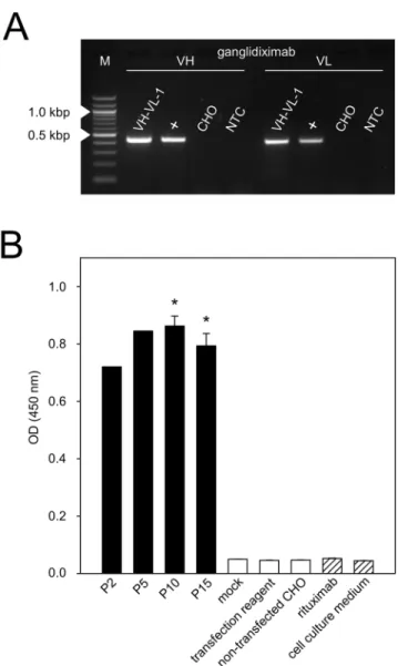
Fig 2. Amplification of DNA fragments encoding for GD2-mimicking paratopes of ganglidiximab.(A) Visualization of coding sequences of GD2-mimicking variable heavy (VH; 440 bp) and light chain (VL; 420 bp)
amplified by RT-PCR. RNA was isolated from hybridoma cells producing murine anti-Id ganglidiomab. PCR products were analyzed by agarose gel electrophoresis. Representative image is shown. NTC—no-template-control. M—Marker (100-bp). (B) PCR products were cloned into pCR12.1-TOPO1plasmids and analyzed
by restriction enzyme digest to excise DNA sequences encoding for VH and VL (product sizes 427 bp and 407 bp, respectively). Resulting DNA fragments were analyzed by agarose gel electrophoresis.
Representative image is shown. M—Marker (2-log, 0.1–10.0 kbp).
plasmids not stably integrated into the host genome. RT-PCR analysis performed after 15 passages and the second freeze-thaw cycle revealed stable mRNA expression of both murine heavy and light chain variable region in“VH-VL-1”clone (Fig 3A). ELISA analysis of supernatants collected in early (P2 and 5) and late passages (P10 and15) revealed similar levels of Ab concentration (P<0.05vs. negative controls;Fig 3B) indicating a stable Ab production over time. Importantly, two freeze-thaw cycles performed between passages 10 and 15 did not affect ganglidiximab production rate confirming stable integration of plasmid DNA into the host genome of selected cell clone. Finally, ganglidiximab production was quantified in supernatants collected after 15 cell clone passages and two freeze-thaw cycles showing about 3μg/ml ganglidiximab prior to and 21μg/ml after the Ab concentration
pro-cedure. The selected cell clone named“VH-VL-1”was used for Ab purification and further analysis.
Analysis of anti-idiotypic characteristics of ganglidiximab
Binding of anti-GD2antibodies to ganglidiximab. Binding of anti-GD2Abs of the 14.18 family to ganglidiximab was analyzed relative to the negative control rituximab using our previously described ELISA protocol [17]. Similar concentration-dependent binding of anti-GD2Abs 14G2a, ch14.18/CHO, ch14.18-delta-CH2, hu14.18K322A as well as immuno-cytokines ch14.18-IL-2 and hu14.18-IL-2 to ganglidiximab (P<0.001vs. rituximab;Fig 4A) was observed, indicating anti-idiotypic properties of the newly generated Ab. These results could be confirmed by flow cytometric analysis using a NK-92tr cell line expressing a GD2-specific chimeric antigen receptor which consists of a ch14.18-derived single chain vari-able region and a CD3-zeta chain [21] (Fig 5A). Similar binding of ganglidiximab and murine parental mAb ganglidiomab to NK-92tr was observed. As expected, neither ganglidiximab nor ganglidiomab bound to the parental NK-92 control cell line lacking CAR expression (Fig 5A).
Binding affinity of ganglidiximab to anti-GD2Abs. Binding affinity of ganglidiximab to anti-GD2Abs of 14.18 family (14G2a, ch14.18/CHO, ch14.18-IL-2, hu14.18K322A and hu14.18-IL-2) was further investigated by surface plasmon resonance analysis using a steady-state fit model for calculation of dissociation constant (KD). KDof ganglidiximab from anti-GD2Abs were in the range of 21.12 ± 3.18 to 166.30 ± 16.82 nM and found to be similar to binding affinities of ganglidiomab to anti-GD2Abs ranging from 19.87 ± 3.44 to 134.92 ± 32.66 nM (Table 1).
Ganglidiximab-dependent inhibition of binding of anti-GD2antibodies to GD2. Bind-ing of anti-GD2Abs (14G2a, ch14.18/CHO, ch14.18-IL-2, hu14.18K322A and hu14.18-IL-2) to the nominal antigen GD2was competitively inhibited by ganglidiximab in a concentration-dependent manner (P<0.001vs. rituximab;Fig 4B), again clearly indicating anti-idiotypic characteristics of ganglidiximab.
Ganglidiximab-dependent inhibition of anti-GD2Ab-mediated GD2-specific
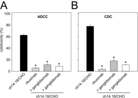
neuroblas-toma cell lysis. GD2-mimicking characteristics of ganglidiximab were further evaluated with a functional calcein-AM-based cytotoxicity assay as previously described [17,27,28]. Healthy donor leukocytes or NK-92tr cells and serum of a healthy donor were used for ADCC and CDC, respectively. Pre-incubation with excess of ganglidiximab resulted in a significant inhibi-tion of cytotoxic activity mediated by healthy donor leukocytes (“+ ganglidiximab”11.4 ± 1.6%
vs.“ch14.18/CHO”62.8 ± 1.6%;P0.05;Fig 6A) and NK-92tr. cells (“+ ganglidiximab”
69.2 ± 3.1%vs.“+ rituximab”0.9 ± 5.8%;P0.05;Fig 5B) or complement proteins (“+
gangli-diximab”18.1 ± 2.0%vs.“ch14.18/CHO”78.1 ± 2.1%;P0.05;Fig 6B). In summary, these
Fig 3. Establishment of a cell line stably producing ganglidiximab.CHO cells were stably co-transfected with two generated expression plasmids (p3-ganglidiximab-HC/ p3-ganglidiximab-LC) and a cell clone “VH-VL-1”stably producing high levels of ganglidiximab was selected for further Ab production. Permanent ganglidiximab expression was confirmed after 15 passages and two freeze-thaw cycles by RT-PCR (A) and standard ELISA (B). (A) To amplify coding sequences of ganglidiximab VH (440 bp) and -VL (420 bp), RNA of “VH-VL-1”was used for RT-PCR followed by agarose gel electrophoresis. RNA of ganglidiomab-producing hybridoma cells served as a positive control (+) and RNA of non-transfected CHO cells as a negative control. One representative image is shown. NTC—no-template-control. M—Marker (100-bp). (B) Ganglidiximab production by“VH-VL-1”was analyzed in supernatants collected during 15 passages and after two freeze-thaw cycles (P2, 5, 10 and 15). Supernatants of non-transfected CHO cells or cells incubated with control plasmids (mock) or transfection reagent only were utilized as negative controls (white columns). Human/ mouse chimeric mAb rituximab and cell culture medium were included as additional negative controls (white-striped columns). Data are shown as mean values±SEM of three independent experiments performed at least in triplicates. One-way ANOVA on ranks followed by appropriate post hoc comparison test;*P0.05vs. negative controls.
Induction of a GD
2-specific humoral immunity following vaccination with
ganglidiximab
Induction of a GD2-specific humoral immune responsein vivowas investigated after vaccina-tion of mice with the chimeric anti-Id Ab ganglidiximab. For this purpose, A/J mice received ganglidiximab in combination with the adjuvant Al(OH)3. Serum samples were obtained before (baseline) and after the last immunization and analyzed for induction of GD2-specific Abs by ELISA as described in“Material and methods”section. Mice immunized with the com-bination of ganglidiximab and Al(OH)3clearly showed induction of a GD2-specific humoral immunity compared to control groups which received Al(OH)3or 0.9% NaCl (“ganglidiximab + Al(OH)3”1.35 ± 0.02μg/mlvs.“Al(OH)3”0.25 ± 0.0μg/mlvs.“0.9% NaCl”0.18 ± 0.0μg/ ml;P0.05;Fig 7). Importantly, we detected a significant increase of Abs directed against the
GD2-mimicking paratopes compared to the baseline (Fig 7), demonstrating the efficacy of pro-tein vaccination with ganglidiximabin vivo.
Fig 4. Binding of anti-GD2antibodies to ganglidiximab and competitive inhibition of binding to nominal antigen GD2.(A) Concentration-dependent binding of ganglidiximab to anti-GD2Abs of the 14.18
family was analyzed by ELISA. Chimeric mAb rituximab and murine IgG2a were utilized as isotype controls. Data are expressed relative to binding of 1μg/ml of ch14.18/CHO to ganglidiximab (100%) and are presented as mean values±SEM of four independent experiments performed in duplicates. One-way ANOVA followed
by appropriate post hoc comparison test;***P0.001vs. rituximab. (B) Inhibition of anti-GD2Ab binding to
GD2by ganglidiximab was analyzed using competitive ELISA. Chimeric mAb rituximab and murine IgG2a
served as isotype controls. Data are expressed as percentage of binding inhibition and presented as mean values±SEM of four independent experiments performed in duplicates. One-way ANOVA followed by
appropriate post hoc comparison test;***P0.001vs. rituximab.
Discussion
Immunotherapy targeting GD2is an encouraging approach to improve survival in high-risk NB patients. Therefore, GD2-specific mAbs have been developed and successfully applied in several clinical trials [1;7;9;10]. Since passive immunotherapy generally does not induce nological memory, there is a considerable interest in extending these studies into active immu-notherapy. However, due to its poorly immunogenic properties, the nominal antigen GD2used as a vaccine elicited only low-titer IgM immune responses with decreased anti-GD2activity over time [17,30]. Hence, the use of GD2surrogate protein vaccines provides an alternative approach against GD2-expressing neuroectodermal tumors given that they are chemically Fig 5. Binding of ganglidiximab to NK-92tr and ganglidiximab-dependent inhibition of NK-92tr-mediated GD2-specific cytotoxicity against NB.(A) Binding of ganglidiximab to NK-92tr expressing a GD2
-specific chimeric antigen receptor was analyzed by flow cytometry. Cells were stained with chimeric ganglidiximab (black solid line), murine anti-Id ganglidiomab (positive control; grey solid line), chimeric rituximab (isotype control; black dashed line) or murine IgG1 (isotype control; grey dashed line) followed by incubation with biotinylated ch14.18/CHO and PE-labeled streptavidin. Staining of the parental NK-92 cell line lacking GD2-specific CAR expression was further included as negative control. Results from one
representative experiment are shown. (B) Inhibition of GD2-specific NK-92tr-mediated NB cell lysis (w/o Ab;
white-striped column) was analyzed after pre-incubation with excess of ganglidiximab (black column) using a calcein-AM-based cytotoxicity assay. Murine anti-Id ganglidiomab served as a positive control (grey column). Rituximab (white column) and murine IgG1 (white column) were utilized as negative controls. Results are expressed as percentage of lysis inhibition (mean values±SEM) of two independent experiments performed using six replicates. One-way ANOVA on ranks followed by appropriate post hoc comparison test;*P0.05vs. w/o Ab.
stable, can be economically produced and do not contain oncogenic or toxic material [14]. In previous studies, GD2epitopes translated into immunogenic peptides such as GD2peptide mimotopes have shown promising results in experimental models [31] and are currently under development for the use in patients. Here, we report another promising alternative strategy which exploits the immune network hypothesis of Jerne [13] to mimic GD2as a protein epitope by an anti-Id Ab. Acting as a TAA surrogate, anti-Id Abs were found to be associated with improved patient survival as they can induce generation of anti-anti-Id Abs recognizing the nominal TAA and may therefore facilitate host recognition of the tumor [14,32]. Recently, successful vaccination with anti-Id Ab has been shown in treatment of melanoma, lung cancer, B cell lymphoma and leukemia [14]. For NB treatment, Yuet al. conducted a clinical trial of GD2-mimicking anti-Id Ab 1A7 and showed little toxicity of anti-Id protein vaccines and effec-tive induction of biologically aceffec-tive GD2-specific immune responses [1]. To provide
Table 1. Dissociation constants (KD) of ganglidiximab and ganglidiomab from anti-GD2antibodies of the 14.18 family.
Ligand ganglidiximab KD (nM) ganglidiomab KD (nM)
14G2a 95.43±5.27 99.43±6.52
ch14.18/CHO 38.40±2.74 39.04±3.14
ch14.18-IL-2 166.30±16.82 134.92±32.66
hu14.18K322A 32.58±4.36 33.36±4.16
hu14.18-IL-2 21.12±3.18 19.87±3.44
Dissociation constants (KD) of ligands 14G2a, ch14.18/CHO, ch14.18-IL-2, hu14.18K322A and hu14.18-IL-2 from ganglidiximab and ganglidiomab were
determined in a Biacore T200 system by surface plasmon resonance method using a steady-statefit model. Values are presented as mean values±SEM
of three independent experiments.
doi:10.1371/journal.pone.0150479.t001
Fig 6. Ganglidiximab-dependent inhibition of GD2-specific ch14.18/CHO-mediated ADCC and CDC.
GD2-specific ch14.18/CHO-mediated ADCC (A) and CDC (B). Ch14.18 induced ADCC and CDC of NB cells
LA-N-1 (black column) was compared to rituximab used as negative control (white-striped column). Pre-incubation of ch14.18 with excess of ganglidiximab (grey column) or ganglidiomab (white column) resulted in inhibition of both ADCC and CDC. Results are expressed as percentage of cytotoxicity (mean values±SEM) of three independent experiments performed at least in triplicates. One-way ANOVA on ranks followed by appropriate post hoc comparison test;*P0.05vs. ch14.18/CHO.
unrestricted access of a GD2-mimicking anti-Id Ab in Europe, we previously generated a new murine anti-Id Ab ganglidiomab and successfully used it as a protein vaccine to induce GD2-specific humoral immunity in mice [17]. Once applied in humans, the immune response induced against a mouse IgG1 mAb will be directed against the entire xenogeneic protein. In order to tailor humoral immunity towards GD2-mimicking paratopes for future clinical trials, we exchanged murine with human constant regions and generated the chimeric human/mouse anti-Id Ab ganglidiximab, consisting of human IgG1 constant regions, which are not immuno-genic in humans, and murine variable regions derived from anti-Id ganglidiomab [17]. To allow permanent ganglidiximab production, two expression plasmids encoding for heavy and light chains were designed and stably co-transfected into CHO cells, the most widely used mammalian cell line for recombinant protein production [9,33].
After Ab purification, we demonstrated GD2-mimicking properties of ganglidiximab which were found to be comparable to a previously reported murine anti-Id Ab ganglidiomab [17]. Similar to GD2, ganglidiximab specifically binds to clinically used Abs of the 14.18 family (14G2a, ch14.18/CHO, ch14.18-dCH2, hu14.18K322A as well as immunocytokines
ch14.18-IL-2 and hu14.18-IL-2) (Fig 4A) as well as to a GD2-specific CAR expressed on geneti-cally engineered NK-92 cells (NK-92tr) [21] (Fig 5A). Furthermore, GD2-mimicking properties were confirmed by ganglidiximab-dependent competitive inhibition of binding of anti-GD2 Abs to GD2. Then, medium binding affinities of ganglidiximab to anti-GD2Abs of 14.18 family were determined using a surface plasmon resonance technique (between 10−7and 10−8M; Tab. 1) and were found to be similar to the previously generated murine anti-Id ganglidiomab [17]. To further confirm ganglidiximab function as a GD2surrogate, we applied a recently Fig 7. Induction of GD2-specific humoral immunity after vaccination with ganglidiximab.Female A/J
mice (n = 8) were immunized six times with ganglidiximab combined with the adjuvant Al(OH)3every two
weeks. Control groups received Al(OH)3or 0.9% NaCl. Serum samples were taken before (baseline; white
column) and after the last immunization step (black column) and analyzed for Abs directed against GD2
-mimicking paratopes of ganglidiximab using ELISA. Mann-Whitney Rank Sum test or One-way ANOVA followed by appropriate post hoc comparison test;***P0.001vs. baseline (prior to the first immunization);
§P<0.05 vs. NaCl control group after six immunizations.
reported functional cytotoxicity assay [17,28], demonstrating ganglidiximab-dependent inhi-bition of a GD2-specific NB cell lysis mediated by effector cells (ADCC) and serum of a healthy donor (CDC;Fig 6). These data are in line with our previous results clearly showing similar anti-idiotypic properties of both anti-Id Abs [17,28]. Finally, as it has already been shown for murine anti-Id Absin vivo[16,17], ganglidiximab was used as a GD2surrogate protein vaccine to induce a humoral immunity towards the GD2-mimicking paratopes in mice (Fig 7). For future clinical application, we plan a combined two-phase anti-Id-based vaccination strategy starting with murine and followed by human/mouse chimeric anti-Id Abs. In humans, the murine ganglidiomab Fc portion probably serves as an immunogenic carrier [14] thereby priming a GD2-specific humoral response upon repeated vaccination. In the second phase, vac-cination with the chimeric ganglidiximab, containing non-immunogenic human constant regions, may tailor humoral immunity towards the therapy-relevant paratopes mimicking GD2.
In summary, we describe the generation and characterization of a new chimeric anti-Id Ab ganglidiximab used as a protein vaccine to induce humoral immunity against NB in mice. Our results are an important baseline for further development of new anti-Id Ab-based vaccination approaches against GD2-expressing malignancies.
Acknowledgments
We would like to acknowledge Maria Asmus, Manuela Brueser and Theodor Koepp (Univer-sity Medicine Greifswald, Pediatric Hematology and Oncology, Greifswald) for excellent tech-nical assistance.
Author Contributions
Conceived and designed the experiments: HL NS CE. Performed the experiments: HL NS CE DS MZ MJ SB HPM. Analyzed the data: HL NS CE DS MZ MJ SB HPM. Contributed
reagents/materials/analysis tools: HL NS CE DS MZ MJ SB HPM. Wrote the paper: HL NS CE DS MZ MJ SB HPM.
References
1. Alice L.Yu. Progress in Treatment of High-risk Neuroblastoma with Immunotherapy. American Society of Clinical Oncology, American Society of Clinical Oncology.
2. Maris JM, Hogarty MD, Bagatell R, Cohn SL. Neuroblastoma. Lancet. 2007 Jun 23; 369(9579):2106– 20. PMID:17586306
3. Matthay KK, Reynolds CP, Seeger RC, Shimada H, Adkins ES, Haas-Kogan D, et al. Long-term results for children with high-risk neuroblastoma treated on a randomized trial of myeloablative therapy fol-lowed by 13-cis-retinoic acid: a children's oncology group study. J Clin Oncol. 2009 Mar 1; 27(7):1007– 13. doi:10.1200/JCO.2007.13.8925PMID:19171716
4. Cheung NK, Neely JE, Landmeier B, Nelson D, Miraldi F. Targeting of ganglioside GD2 monoclonal antibody to neuroblastoma. J Nucl Med. 1987 Oct; 28(10):1577–83. PMID:3655911
5. Schulz G, Cheresh DA, Varki NM, Yu A, Staffileno LK, Reisfeld RA. Detection of ganglioside GD2 in tumor tissues and sera of neuroblastoma patients. Cancer Res. 1984 Dec; 44(12 Pt 1):5914–20. PMID: 6498849
6. Svennerholm L, Bostrom K, Fredman P, Jungbjer B, Lekman A, Mansson JE, et al. Gangliosides and allied glycosphingolipids in human peripheral nerve and spinal cord. Biochim Biophys Acta. 1994 Sep 15; 1214(2):115–23. PMID:7918590
7. Matthay KK, George RE, Yu AL. Promising therapeutic targets in neuroblastoma. Clin Cancer Res. 2012 May 15; 18(10):2740–53. doi:10.1158/1078-0432.CCR-11-1939PMID:22589483
9. Ladenstein R, Weixler S, Baykan B, Bleeke M, Kunert R, Katinger D, et al. Ch14.18 antibody produced in CHO cells in relapsed or refractory Stage 4 neuroblastoma patients: a SIOPEN Phase 1 study. MAbs. 2013 Sep; 5(5):801–9. doi:10.4161/mabs.25215PMID:23924804
10. Lode HN. Immune activation, clinical response and survival following long-term infusion of anti-GD2 antibody ch14.18/CHO in combination with interleukin-2 in high-risk neuroblastoma patients. Christian Jensen, Nikolai Siebert, Silke Kietz, Karoline Ehlert, Ina Müller, Ruth Ladenstein et al., editors. 2015. Cancer Research.
11. Becker R, Eichler MK, Jennemann R, Bertalanffy H. Phase I clinical trial on adjuvant active immuno-therapy of human gliomas with GD2-conjugate. Br J Neurosurg. 2002 Jun; 16(3):269–75. PMID: 12201397
12. Mond JJ, Lees A, Snapper CM. T cell-independent antigens type 2. Annu Rev Immunol. 1995; 13:655– 92. PMID:7612238
13. Jerne NK. Towards a network theory of the immune system. Ann Immunol (Paris). 1974 Jan; 125C(1– 2):373–89.
14. Bhattacharya-Chatterjee M, Chatterjee SK, Foon KA. Anti-idiotype antibody vaccine therapy for cancer. Expert Opin Biol Ther. 2002 Dec; 2(8):869–81. PMID:12517266
15. Herlyn D, Somasundaram R, Li W, Maruyama H. Anti-idiotype cancer vaccines: past and future. Can-cer Immunol Immunother. 1996 Oct; 43(2):65–76. PMID:8954140
16. Sen G, Chakraborty M, Foon KA, Reisfeld RA, Bhattacharya-Chatterjee MB. Induction of IgG antibod-ies by an anti-idiotype antibody mimicking disialoganglioside GD2. J Immunother. 1998 Jan; 21(1):75– 83. PMID:9456440
17. Lode HN, Schmidt M, Seidel D, Huebener N, Brackrock D, Bleeke M, et al. Vaccination with anti-idio-type antibody ganglidiomab mediates a GD(2)-specific anti-neuroblastoma immune response. Cancer Immunol Immunother. 2013 Jun; 62(6):999–1010. doi:10.1007/s00262-013-1413-yPMID:23591980
18. Lode HN, Xiang R, Varki NM, Dolman CS, Gillies SD, Reisfeld RA. Targeted interleukin-2 therapy for spontaneous neuroblastoma metastases to bone marrow. J Natl Cancer Inst. 1997 Nov 5; 89 (21):1586–94. PMID:9362156
19. PUCK TT, CIECIURA SJ, ROBINSON A. Genetics of somatic mammalian cells. III. Long-term cultiva-tion of euploid cells from human and animal subjects. J Exp Med. 1958 Dec 1; 108(6):945–56. PMID: 13598821
20. Seeger RC, Rayner SA, Banerjee A, Chung H, Laug WE, Neustein HB, et al. Morphology, growth, chro-mosomal pattern and fibrinolytic activity of two new human neuroblastoma cell lines. Cancer Res. 1977 May; 37(5):1364–71. PMID:856461
21. Esser R, Muller T, Stefes D, Kloess S, Seidel D, Gillies SD, et al. NK cells engineered to express a GD2 -specific antigen receptor display built-in ADCC-like activity against tumour cells of neuroectodermal origin. J Cell Mol Med. 2012 Mar; 16(3):569–81. doi:10.1111/j.1582-4934.2011.01343.xPMID: 21595822
22. Mujoo K, Kipps TJ, Yang HM, Cheresh DA, Wargalla U, Sander DJ, et al. Functional properties and effect on growth suppression of human neuroblastoma tumors by isotype switch variants of monoclonal antiganglioside GD2 antibody 14.18. Cancer Res. 1989 Jun 1; 49(11):2857–61. PMID:2720646
23. Gillies SD, Lo KM, Wesolowski J. High-level expression of chimeric antibodies using adapted cDNA variable region cassettes. J Immunol Methods. 1989 Dec 20; 125(1–2):191–202. PMID:2514231
24. Mueller BM, Reisfeld RA, Gillies SD. Serum half-life and tumor localization of a chimeric antibody deleted of the CH2 domain and directed against the disialoganglioside GD2. Proc Natl Acad Sci U S A. 1990 Aug; 87(15):5702–5. PMID:2198570
25. Sorkin LS, Otto M, Baldwin WM III, Vail E, Gillies SD, Handgretinger R, et al. Anti-GD(2) with an FC point mutation reduces complement fixation and decreases antibody-induced allodynia. Pain. 2010 Apr; 149(1):135–42. doi:10.1016/j.pain.2010.01.024PMID:20171010
26. Gillies SD, Reilly EB, Lo KM, Reisfeld RA. Antibody-targeted interleukin 2 stimulates T-cell killing of autologous tumor cells. Proc Natl Acad Sci U S A. 1992 Feb 15; 89(4):1428–32. PMID:1741398
27. Seidel D, Shibina A, Siebert N, Wels WS, Reynolds CP, Huebener N, et al. Disialoganglioside-specific human natural killer cells are effective against drug-resistant neuroblastoma. Cancer Immunol Immun-other. 2015 May; 64(5):621–34. doi:10.1007/s00262-015-1669-5PMID:25711293
28. Siebert N, Seidel D, Eger C, Juttner M, Lode HN. Functional bioassays for immune monitoring of high-risk neuroblastoma patients treated with ch14.18/CHO anti-GD2 antibody. PLoS One. 2014; 9(9): e107692. doi:10.1371/journal.pone.0107692PMID:25226154
30. Basak S, Birebent B, Purev E, Somasundaram R, Maruyama H, Zaloudik J, et al. Induction of cellular immunity by anti-idiotypic antibodies mimicking GD2 ganglioside. Cancer Immunol Immunother. 2003 Mar; 52(3):145–54. PMID:12649743
31. Fest S, Huebener N, Weixler S, Bleeke M, Zeng Y, Strandsby A, et al. Characterization of GD2 peptide mimotope DNA vaccines effective against spontaneous neuroblastoma metastases. Cancer Res. 2006 Nov 1; 66(21):10567–75. PMID:17079481
32. DeNardo GL, Bradt BM, Mirick GR, DeNardo S. Human antiglobulin response to foreign antibodies: therapeutic benefit? Cancer Immunol Immunother. 2003 May; 52(5):309–16. PMID:12700946


![Tab. 1) and were found to be similar to the previously generated murine anti-Id ganglidiomab [17]](https://thumb-eu.123doks.com/thumbv2/123dok_br/18387378.357126/16.918.303.663.109.474/tab-similar-previously-generated-murine-anti-id-ganglidiomab.webp)
