online | memorias.ioc.fiocruz.br Angiostrongyluscantonensis, the rat lungworm, is the
primary causative agent of eosinophilic meningitis and eosinophilic meningoencephalitis in humans (Ramirez-Avila et al. 2009). This species was first discovered in rats from China in 1935 (Chen 1935) and the first hu-man case was reported in Taiwan in 1945 (Nomura & Lin 1945). Although this infection is mainly prevalent in Southeast Asia and the Pacific Basin (Asato et al. 2004, Hochberg et al. 2011, Qu et al. 2011, Tsai et al. 2011), many new cases have been reported among travellers returning from endemic areas (Bärtschi et al. 2004, Ali et al. 2008, Malvy et al. 2008, Luessi et al. 2009) and, by 2008, more than 2,800 cases had been recorded worldwide (Wang et al. 2008). The major clinical manifestations of this dis-ease include headache, neck stiffness, muscle weakness, nausea, vomiting and paraesthesia. Although the clini-cal course of infection is usually benign and self-limited, severe infections may lead to irreversible outcomes and even death (Punyagupta et al. 1975).
The life cycle of A. cantonensis is complex, requiring both an intermediate host and a final definitive host. The
giant African snail Achatina fulica and the channelled apple snail Pomaceacanaliculata are the two most com-mon intermediate hosts for A. cantonensis worldwide (Kliks & Palumbo 1992, Lv et al. 2009). Following entry into the intermediate host through the oral or percuta-neous route, the first-stage larvae undergo two moults and develop into the infective larvae within two-three weeks. Humans are an accidental host for this parasite, becoming infected with the parasite by ingesting raw or uncooked individuals of intermediate host species (A. fu-lica, P. canaliculata, Hyla aurea), paratenic host species (Macrobrachium lar, Ocypode ceratophthalma, Hoplo- batrachus rugulosus, Rana plancyi, Tilapia mossam-bica, Vaginulus plebeius, Laevicaulis alte) (freshwater crustaceans, frogs, fish and planarians) or contaminated vegetables carrying the third-stage larvae (Wallace & Rosen 1966, 1967, Ash 1968, Tsai et al. 2004). These larvae then penetrate the intestinal tract and migrate to the central nervous system, where they moult twice and develop into young adults, approximately one week after infection (Alicata 1965).
The available genomic information for A. canto- nensis is limited to data from small-scale transcriptomic investigations and cloning studies. Following the sub-mission of 1,226 expressed sequence tags (ESTs) from young adult A. cantonensis (5th-stage larva) to the Na-tional Center for Biotechnology Information dbEST by our laboratory in 2005, these and some additional se-quences were analysed and were found to encode pro-teins participating in metabolism, cellular development, immune evasion and host-parasite interactions (Xu et al. 2009) and were subsequently grouped into 13 categories (Fang et al. 2010). Among proteins encoded by cDNA doi: 10.1590/0074-0276108062013005
Financial support: NSC, Executive Yuan, ROC (NSC96-2320-B-182-020, NSC100-2320-B-182-013), Chang Gung Memorial Hospital Research (CMRPD150121, CMRPD1A0111)
+ Corresponding author: wanglc@mail.cgu.edu.tw Received 22 November 2012
Accepted 13 June 2013
Identification and characterisation of microRNAs in young adults
of
Angiostrongylus
cantonensis
via a deep-sequencing approach
Shih-Hsin Chang1, Petrus Tang1,2,3, Cheng-Hung Lai4, Ming-Ling Kuo1,5, Lian-Chen Wang1,2/+
1Graduate Institute of Biomedical Sciences 3Bioinformatics Center 2Department of Parasitology
5Department of Microbiology and Immunology, College of Medicine, Chang Gung University, Kwei-Shan, Taoyuan County, Taiwan 4Department of Veterinary Medicine, College of Veterinary Medicine, National Chung Hsing University, Taichung City, Taiwan
Angiostrongylus cantonensis is an important causative agent of eosinophilic meningitis and eosinophilic menin-goencephalitis in humans. MicroRNAs (miRNAs) are small non-coding RNAs that participate in a wide range of biological processes. This study employed a deep-sequencing approach to study miRNAs from young adults of A. cantonensis. Based on 16,880,456 high-quality reads, 252 conserved mature miRNAs including 10 antisense miRNAs that belonging to 90 families, together with 10 antisense miRNAs were identified and characterised. Among these sequences, 53 miRNAs from 25 families displayed 50 or more reads. The conserved miRNA families were divided into four groups according to their phylogenetic distribution and a total of nine families without any members show-ing homology to other nematodes or adult worms were identified. Stem-loop real-time polymerase chain reaction analysis of aca-miR-1-1 and aca-miR-71-1 demonstrated that their level of expression increased dramatically from infective larvae to young adults and then decreased in adult worms, with the male worms exhibiting significantly higher levels of expression than female worms. These findings provide information related to the regulation of gene expression during the growth, development and pathogenesis of young adults of A. cantonensis.
clusters found in the fourth-stage larvae, an enrichment of binding and catalytic activity was also revealed (He et al. 2009). The transcripts found in pepsin-activated infective larvae (3rd-stage) encode proteins that partici-pate in a wide range of biological processes (Chang et al. 2011). Although cloning of galectin (Hao et al. 2007), cystatin (Liu et al. 2010), Ac16 (Li et al. 2011), cathe-psin B (Han et al. 2011), a cathecathe-psin B-like cysteine proteinase (Cheng et al. 2012) and a protein disulphide isomerase (Liu et al. 2012) has been accomplished in A. cantonensis, the characterisation of these proteins is far from complete. Moreover, the genome of this parasite has not yet been sequenced.
MicroRNAs (miRNAs) are a type of small non-coding RNAs, approximately 21-23 nucleotides (nt) in length, that have been discovered in diverse organisms, including animals, plants, viruses and fungi (Ambros 2004, Lee et al. 2010, Khan et al. 2011, Plaisance-Bon-staff & Renne 2011). Because the first two miRNAs, lin-4 and let-7, were discovered and demonstrated to be involved in the regulation of developmental timing in Caenorhabditis elegans (Lee et al. 1993, Reinhart et al. 2000), more than 15,000 microRNA gene loci in over 140 species have been reported in miRBase v16 (Kozo-mara & Griffiths-Jones 2011). MiRNAs regulate a wide range of biological processes, including development, metabolism, cell proliferation and differentiation (Bar-tel 2009, Krol et al. 2010). In animals, miRNAs usu-ally silence their target mRNAs through degradation or translational repression (Nilsen 2007). With respect to experimental methods, computational approaches are considered a useful strategy for identifying miRNAs, even in species whose genomes have not been fully sequenced (Berezikov et al. 2006). Computational ap-proaches can also be utilised to quantify miRNA ex-pression (Luo 2012).
As parasitic helminths usually exhibit a complex life cycle, investigations involving miRNAs enable us to not only understand the roles of these riboregula-tors in the physiology, development and evolution of these organisms, but also the mechanisms of host inva-sion and pathogenesis (Xue et al. 2008, Hao et al. 2010, Poole et al. 2010, Chen et al. 2011a, Winter et al. 2012). MiRNA expression in adult worms of A. cantonensis was recently identified and compared between the sex-es (Chen et al. 2011b). The adult worms may only cause severe obstruction of the pulmonary arteries and respi-ratory failure (Wang et al. 2012). In contrast, because the young adults of A. cantonensis are at a pathogenic developmental stage associated with the central ner-vous system, miRNA expression profiling may provide further information on the pathogenesis of eosinophilic meningitis and eosinophilic meningoencephalitis. In the present study, we employed a deep-sequencing ap-proach to identify conserved miRNAs among young adults of A. cantonensis. Following a phylogenic dis-tribution analysis, the expression profiles of select-ed miRNAs at different developmental stages were analysed via stem-loop real-time polymerase chain reaction (RT-PCR).
MATERIALS AND METHODS
Parasites - A strain of A. cantonensis has been main-tained in our laboratory in Biomphalariaglabrata snails and Sprague-Dawley rats since 1980 (Wang et al. 1989). Young adults and adult worms were collected on days 21 and 50 post-infection, respectively, from the cere-bral tissues and pulmonary arteries of rats. The sex of these worms was determined based on their morpho-logical characteristics: male worms are usually shorter and exhibit copulatory bursa and long spicules. Infective larvae were collected from the tissues of infected snails through digestion with 0.6% (w/v) pepsin-HCl (pH 2-3) for 1 h. These worms were washed with normal saline, phosphate buffered saline and distilled water and then stored at -80ºC for further analyses (Hwang et al. 2010). This experiments performed in this study followed the recommendations of the Institutional Animal Care and Use Committee of Chang Gung University.
SmallRNAlibrary constructionand high- through-put sequencing - Total RNA was isolated from young adults of A. cantonensis (500 worms of each sex) using the TRI Reagent, according to the instructions of the manufacturer (Molecular Research Center, Cincinnati, OH, USA). RNA integrity was assessed via 1% (w/v) agarose gel electrophoresis, while RNA purity was de-termined based on the absorbance recorded at 260/280 nm using an SMA1000 UV Spectrophotometer (Merin-ton Technology, Beijing, China). The RNA preparations were then stored at -80ºC for further experiments.
RNA fragments of 18-30 nt in length were separated from total RNA using the Small RNA Sample Prep Kit (Illumina, San Diego, CA, USA). Following 15% Novex TBE-urea polyacrylamide gel electrophoresis (PAGE),
the purified fragments were ligated to 5′ and 3′ RNA
adaptors, reverse transcribed to produce single-stranded cDNAs and amplified via PCR. RNA fragments (ap-proximately 92 bp) were isolated from the PCR products and sequenced using the Illumina HiSeq 2000 system (Illumina, San Diego, CA, USA).
Bioinformaticanalysis - The small RNA library was analysed through a deep-sequencing small RNA analysis pipeline (DSAP) (dsap.cgu.edu.tw) (Huang et al. 2010). Following the removal of adaptors and poly-A/T/C/G/N nt, sequences longer than 16 nt were considered clean sequence tags. Using the clustering module of DSAP, re-dundant clean reads were merged into unique tags. The reads for each unique tag were counted as its expression abundance.
To identify transfer RNAs (tRNAs), ribosomal RNAs (rRNAs), small nucleolar RNAs (snoRNAs), small nuclear RNAs (snRNAs) and other annotated non-coding RNAs (ncRNAs), the unique tags were subjected to searches against the transcribed sequence library of the ncRNA (Rfam) database (version 10.0). The remain-ing sequences were compared with mature miRNA se-quences in miRBase (version 16) using BLASTN.
sex). The expression levels of 1-1 and aca-miR-71-1 were determined via modified stem-loop RT-PCR (Chen et al. 2005). The stem-loop reverse transcrip-tion primer and forward primer for aca-miR-1-1 were
5′-GTCGTATCCAGTGCAGGGTCCGAGGTATTC-GCACTGGATACGACTACATAC-3′ and 5′-CGCGGC-TGGAATGTAAAGAAGT-3′, respectively, and those for aca-miR-71-1 were 5′-GTCGTATCCAGTGCAGGGT-CCGAG-GTATTCGCACTGGATACGACCGTCTCA-3′ and 5′-GCGCGGTGAAAGACATGG-GTAGTG-3′, re -spectively. The reverse primer employed in these assays
was 5′-GTGCAGGGTCCGAGGT-3′.
Small RNAs from A. cantonensis at different de-velopmental stages were extracted using an miRNA isolation kit (Geneaid, Sijhih City, Taipei County, Tai-wan) according to the instructions of the manufacturer. First-strand cDNAs were synthesised using SuperScript III Reverse Transcriptase (Invitrogen, Carlsbad, CA, USA). Briefly, reaction mixtures (20 µL) containing the purified small RNAs (160 ng), 5X First-Strand buffer (4 µL), individual specific stem-loop RT primers (0.5 µM), dNTP mix (0.5 mM), DTT (5 mM), SuperScript III Re-verse Transcriptase (10 units) and RNaseOUT (2 units) were incubated at 55ºC for 50 min, then inactivated by heating at 70ºC for 15 min and subsequently incubated with two units of RNase H at 37ºC for 20 min.
Quantitative RT-PCR was performed using RealQ PCR Master Mix (Ampliqon A/S, Skovlunde, Denmark) in the Stratagene Mx3000P QPCR System (Agilent Tech-nologies, Santa Clara, CA, USA). The PCR mixtures (20 µL) included the reverse transcription product for each miRNA (3 µL), RealQ-PCR 2X Master Mix (10 µL), a specific forward primer (0.5 µM) and the reverse primer (0.5 µM). The RT-PCR assays were carried out with an initial step at 95ºC for 10 min, followed by 40 cycles of amplification, with denaturation at 95ºC for 30 s, primer annealing at 58ºC for 1 min and elongation at 72ºC for 30 s. The specificity of this assay was confirmed via 15% PAGE. Finally, the expression levels of the targeted miR-NAs were determined through three rounds of stem-loop RT-PCR amplification and analysed according to the 2−ΔΔCt method (Livak & Schmittgen 2001).
Statistical analysis - The expression levels of the miRNAs were expressed as the mean ± standard devia-tion. Means were compared via one-way ANOVA and the least significance difference test was used for post-hoc multiple range comparisons. p < 0.05 was consid-ered to be statistically significant.
RESULTS
AnalysisofshortRNAs - A total of 22,484,156 raw sequence reads were obtained through high-throughput sequencing from the small RNA library generated for young adultsof A. cantonensis. The removal of adapters, contaminated nt and low-quality sequences resulted in 16,880,456 (75.1%) high-quality, clean reads of 16-31 nt in length, averaging 22.8 nt. A majority of the sequences were 23 nt in length (46.1%), followed by lengths of 22 nt (26.3%) and 24 nt (12.1%) sequences. Among the clean reads, 10,766,590 (63.8%) sequences were found to match
entries in the Rfam database. They included 7,736,727 tRNAs (45.8%), 1,191,869 rRNAs (7.1%), 228,262 snoR-NAs (1.4%), 174,633 snRsnoR-NAs (1%) and 1,435,099 ncR-NAs (8.5%) (Fig. 1).
Identificationof conserved miRNAs - From the re-maining 7,548,965 clean reads, 252 conserved mature miRNAs including 10 antisense miRNAs were identi-fied based on comparison with entities in miRbase. All of the identified miRNAs were homologous to miRNAs from Metazoa and their lengths ranged from 20-24 nt, with most being 22 nt in length (117), followed by lengths of 21 nt (54), 23 nt (49), 20 nt (19) and 24 nt (13). These miRNAs belonged to 90 families, in addition to which 10 antisense miRNAs were discovered. The number of reads obtained was 50 or more for 53 of these miRNAs, 21-49 for 28 miRNAs, 11-20 for 14 miRNAs, two-10 for 82 miRNAs and only a single read was observed in the remaining 75 miRNAs.
Supplementary data lists the 53 miRNAs with 50 or more reads, which belonged to 25 families. More than one miRNA was identified from the following families: let-7, mir-1, mir-34, mir-87, mir-71 mir-99, mir-2, mir-9, mir-31, mir-50 and mir-103. Only one miRNA was found in the remaining 14 families. Nine miRNAs displayed more than 1,000 reads. Aca-miR-1-1 exhibited the high-est number of reads, at 184,946, followed by aca-miR-71-1 (92,433). These two highly expressed miRNAs constituted 50.7% and 25.3% of the total reads (365,112), respectively, among this group of 53 miRNAs.
Phylogenicdistribution - To analyse the evolutionary features of A. cantonensis miRNAs, the identified miR-NAs showing 50 or more reads, from 25 families, were compared with those from other nematodes in miR-Base. The families let-7, 1, 2, 9, 31, mir-34, mir-44, mir-50, mir-67, mir-71, mir-81, mir-87 and mir-124 contained members that were homologous to parasitic nematodes (Ascarissuum and Brugia malayi) and free-living nematodes (C.elegans, Caenorhabditis
Fig. 1: classification of small RNAs of Angiostrongyluscantonensis
briggsae, Caenorhabditisremanei and Pristionchus pa-cificus). Members of the mir-60 and mir-235 families were homologous to sequences from free-living nema-todes, but not parasitic nematodes. The family mir-99 was homologous only to A. suum, while members of the 21, 29, 30, 103, 140, 146, mir-185, mir-191 and mir-320 families did not exhibit homol-ogy with sequences from other nematodes (Table).
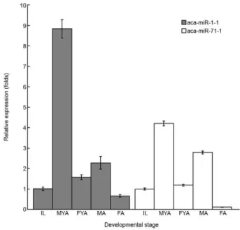
Expression of miRNAs at different developmental stages - To confirm the expression of the miRNAs from A. cantonensis young adults that were identified through the applied high-throughput approach and to determine the expression levels of these miRNAs at different devel-opmental stages, the levels of 1-1 and aca-miR-71-1 transcripts were validated in a modified stem-loop quantitative RT-PCR analysis. Significant differences in the expression levels of these miRNAs were observed among different developmental stages (aca-miR-1-1: F = 521.55, p < 0.001; aca-miR-71-1: F = 1585.86, p < 0.001). In male worms, the expression of the two miRNAs was sig-nificantly higher in young adults than in infective larvae or adult worms (p < 0.05). Additionally, the expression in adult worms was significantly higher than in infective larvae (p < 0.05). In female worms, significantly higher expression was also observed in young adults (p < 0.05). However, there was no significant difference in aca-miR-1-1 expression observed between infective larvae and adult worms (p > 0.05) and the infective larvae exhibited significantly higher aca-miR-71-1 expression than adult worms (p < 0.05). Moreover, male worms displayed sig-nificantly higher expression than female worms in both the young adult and adult stages (p < 0.05) (Fig. 2).
DISCUSSION
Using a deep-sequencing approach, based on 7,548,965 reads, we identified and characterised 252 conserved mature miRNAs including 10 antisense miR-NAs that belonging to 90 families from young adults of A. cantonensis. Previous authors identified miRNAs in adult male and female worms from 592,899 and 458,447 reads, respectively (Chen et al. 2011b). However, no novel miRNAs were discovered in either the present or previous studies. The sequences obtained from the adult male and female worms were matched to the C. elegans genome, although the percentage of perfect matches was quite low (18.94% in females and 22.58% in males). Al-though C. elegans and A. cantonensis are both nema-todes, the former species is free-living in soil or water, whereas the latter is parasitic, being found in the pulmo-nary arteries of its definitive host and requiring a mollusc as an intermediate host (Chen et al. 2011b). In the pres-ent study, a search for sequences in miRBase discovered only conserved mature miRNAs. We hypothesise that novel miRNAs will be identified only when a reference genome for A. cantonensis becomes available.
In the present study, we identified nine miRNAs in young adults of A. cantonensis displaying more than a 1,000 reads: two in the let-7 family, four in the mir-1 family, one in the mir-44 family, one in the mir-71 fam-ily and one in the mir-99 famfam-ily. In male adult worms, seven miRNAs exhibited more than 1,000 reads: miR-1,
miR-228, miR-44, miR-45, miR-71, miR-72 and miR-81. In the female worms, 10 miRNAs showed more than a 1,000 reads: miR-1, miR-2, miR-228, miR-44, miR-45, miR-60, miR-71, miR-72, miR-81 and miR-87 (Chen et al. 2011b). MirR-1, mirR-71 and miR-44 show high expression in both the young adult and in adult stages. In contrast, let-7 and miR-99 are highly expressed only in young adults, whereas miR-45 and miR-81 are ex-pressed only in adult worms. These findings indicate that a stage-specific expression of miRNAs occurs in A. cantonensis. Moreover, the regulatory functions of miR-99 require further investigation.
Let-7 and lin-4 are two important miRNA families associated with the lifespan of C. elegans. These miR-NAs have been characterised as playing an essential role in the developmental timing of the worm by downregu-lating specific targets, such as the TRIM protein lin-41 and the transcription factor lin-14 (Ibáñez-Ventoso et al. 2006, Ambros 2011). Let-7 is an miRNA families that was discovered in ancient animals (Christodoulou et al. 2010). The let-7 family is expressed in a wide range of animals and the sequences of its members are highly con-served. However, significant variations in the size of the let-7 family occur among organisms: of the 55 organisms known to express members of the let-7 family, 13 express only one let-7 miRNA, including the nematodes P. paci-ficus, C. remanei, C. elegans, C. briggsae and B. malayi, whereas 19 mature let-7 sequences have been identified in the zebrafish (Daniorerio) (Pasquinelli et al. 2003). In the present study, we identified 19 members of the let-7 fam-ily in young adults of A. cantonensis, 11 of which exhib-ited 50 or more reads. In adult worms, the copy number of this miRNA was determined to be less than 50 in both sexes. These finding suggests the importance of let-7 in gene regulation in young adults of A. cantonensis. How-ever, understanding the role of let-7 in the development of this parasite will require further studies.
Among the miRNA families found to be expressed in young adults of A. cantonensis, mir-1 displayed the highest number of total reads. Members of this family have been found in a wide range of organisms, includ-ing worms, flies, fishes, mice and humans (Bentwich et al. 2005, Sokol & Ambros 2005, Wienholds et al. 2005, Zhao et al. 2005). They are evolutionarily conserved and have been characterised as playing essential roles in regulating proliferation and the differentiation of muscle development via the regulation of synaptic transmission (Simon et al. 2008, Jones et al. 2011). In adult worms, the mir-71 family presented the highest number of reads in both sexes (Chen et al. 2011b). This family has been reported to promote longevity and stress resistance in worms (Pincus et al. 2011) and is involved in the sexual maturation of female worms (Gomes et al. 2011). These expression patterns suggest the different roles of miR-NAs at different developmental stages.
families were homologous to sequences of other para-sitic nematodes, two to sequences of free-living nema-todes, but not parasitic nemanema-todes, and one to A. suum sequences. Nine families did not exhibited any member that was homologous to either a free-living or parasitic nematode. Moreover, these miRNAs observed in young adults of A. cantonensis were not found in adult worms (Chen et al. 2011b). It is possible that the first two groups regulate general biological or physiological functions in nematodes. The miRNAs showing homology to A. suum may be specific to parasitic nematodes. As adult worms of A. cantonensis live within the central nervous system, the last group may regulate adaptive functions of worms related to this special environment, which may also cause pathological changes in the central nervous system. Fur-ther studies are required to confirm these hypotheses.
Analysis of the levels of 1-1 and aca-miR-71-1 expression via the stem-loop RT-PCR revealed dif-ferent expression patterns based on developmental stag-es and sex. Thstag-ese two miRNAs were selected because they are highly expressed not only in young adults, but also in adult worms of both sexes. In both male and fe-male worms, the level of expression of these miRNAs
increased dramatically from the levels observed in in-fective larvae and peaked in young adults, subsequently declining to a low level in adult worms. Overall, the ex-pressions levels of these miRNAs were found to be high-est in male adult males. Similar expression patterns have been reported for 18 miRNAs in adult worms (Chen et al. 2011b). These miRNAs may be important in regulat-ing sex differentiation, rather than developmental stages. The lower expression levels of these miRNAs observed in female worms indicate that females may require a lower degree of post-transcriptional regulation than male worms. Moreover, the higher expression levels of the miRNAs detected in young adults suggest that more significant changes may occur during the young adult stage than in the adult stage.
Replicate analyses were difficult in the present study because of the technical difficulties involved in obtain-ing sufficient sample sizes from infected animals. This limitation may restrict the quantitation of our find-ings. However, we identified 53 miRNAs, belonging to 25 families, that displayed 50 or more reads. These findings provide reliable information about the global miRNA expression profiles found in A. cantonensis.
Al-TABLE
Phylogenic distributions of conserved microRNAs families of Angiostrongyluscantonensis young adults with 50 or more reads
Family A.cantonensis Ascarissuum Brugiamalayi
Caenorhabditis elegans
Caenorhabditis briggsae
Caenorhabditis remanei
Pristionchus pacificus
let-7 + + + + + + +
mir-1 + + − + − + +
mir-2 + + + + − + −
mir-9 + + + + + + +
mir-21 + − − − − − −
mir-29 + − − − − − −
mir-30 + − − − − − −
mir-31 + − + − − + +
mir-34 + + + + + + −
mir-44 + + − + + + +
mir-50 + + + + + + −
mir-60 + − − + + + −
mir-67 + + − + + + −
mir-71 + + + + + + −
mir-81 + + − + + + −
mir-87 + + + + + + +
mir-99 + + − − − − −
mir-103 + − − − − − −
mir-124 + + + + + + +
mir-140 + − − − − − −
mir-146 + − − − − − −
mir-185 + − − − − − −
mir-191 + − − − − − −
mir-235 + − − + − + −
though Northern blotting is considered a gold-standard approach for detecting miRNAs, this method is limited by its low sensitivity and difficulties in distinguishing homologous miRNAs from highly similar sequences (van Rooij 2011, Pritchard et al. 2012). Moreover, we succeeded in confirming the expression of two miRNAs initially identified via the high-throughput approach in A. cantonensis young adults through the more sensitive and specific technique of stem-loop quantitative RT-PCR. These findings demonstrated the reliability of the results obtained herein.
Based on the results of the present study, there are significant differences in the expression of miRNAs between young adults and adult worms of A. canto- nensis. These differences are not only qualitative, but also quantitative. In the present study, we identified nine miRNA families without homologous members in the available sequences of other nematodes in the adult stage. Moreover, the expression levels of miR-1 and miR-71 increase from a low expression level in infec-tive larvae to a peak in young adults and subsequently decrease in adult worms. These results suggest that miRNAs play a more important role in the regulation of biological functions in young adults than in adult worms of A. cantonensis.
REFERENCES
Ali AB, Van den Enden E, Van Gompel A, Van Esbroeck M 2008. Eosinophilic meningitis due to Angiostrongyluscantonensis in a Belgian traveller. Travel Med Infect Dis 6: 41-44.
Alicata JE 1965. Biology and distribution of the rat lungworm, An-giostrongylus cantonensis and its relationship to eosinophilic meningoencephalitis and other neurological disorders of man and animals. Adv Parasitol 3: 223-248.
Ambros V 2004. The functions of animal microRNAs. Nature 431: 350-355.
Ambros V 2011. MicroRNAs and developmental timing. Curr Opin Genet Dev 21: 511-517.
Asato R, Taira K, Nakamura M, Kudaka J, Itokazu K, Kawanaka M 2004. Changing epidemiology of angiostrongyliasiscantonensis in Okinawa prefecture, Japan. Jpn J Infect Dis 57: 184-186.
Ash LR 1968. The occurrence of Angiostrongylus cantonensis in frogs of New Caledonia with observations on paratenic hosts of metastrongyles. J Parasitol 54: 432-436.
Bartel DP 2009. MicroRNAs: target recognition and regulatory func-tions. Cell 136: 215-233.
Bärtschi E, Bordmann G, Blum J, Rothen M 2004. Eosinophilic men-ingitis due to Angiostrongylus cantonensis in Switzerland. Infec-tion 32: 116-118.
Bentwich I, Avniel A, Karov Y, Aharonov R, Gilad S, Barad O, Bar-zilai A, Einat P, Einav U, Meiri E, Sharon E, Spector Y, Bentwich Z 2005. Identification of hundreds of conserved and noncon-served human microRNAs. Nat Genet 37: 766-770.
Berezikov E, Cuppen E, Plasterk RH 2006. Approaches to microRNA discovery. Nat Genet 38 (Suppl.): S2-S7.
Chang SH, Tang P, Wang LC 2011. A transcriptomic study on the pepsin-activated infective larvae of Angiostrongylus cantonen-sis. Mol Biochem Parasitol 179: 47-50.
Chen C, Ridzon DA, Broomer AJ, Zhou Z, Lee DH, Nguyen JT, Bar-bisin M, Xu NL, Mahuvakar VR, Andersen MR, Lao KQ, Livak KJ, Guegler KJ 2005. Real-time quantification of microRNAs by stem-loop RT-PCR. Nucleic Acids Res 33: e179.
Chen HT 1935. Un nouveau nematode pulmonaire, PulmonemA. can-tonensis n.g., n. sp. des rats de Canton. Ann Parasitol 13: 312-317.
Chen MX, Ai L, Xu MJ, Chen SH, Zhang YN, Guo J, Cai YC, Tian LG, Zhang LL, Zhu XQ, Chen JX 2011a. Identification and char-acterization of microRNAs in Trichinellaspiralis by comparison with Brugiamalayi and Caenorhabditiselegans. Parasitol Res 109: 553-558.
Chen MX, Ai L, Xu MJ, Zhang RL, Chen SH, Zhang YN, Guo J, Cai YC, Tian LG, Zhang LL, Zhu XQ, Chen JX 2011b. Angiostrongy-luscantonensis: identification and characterization of microR-NAs in male and female adults. Exp Parasitol 128: 116-120.
Cheng M, Yang X, Li Z, He H, Qu Z, He A, Wu Z, Zhan X 2012. Cloning and characterization of a novel cathepsin B-like cysteine proteinase from Angiostrongyluscantonensis. Parasitol Res 110: 2413-2422.
Christodoulou F, Raible F, Tomer R, Simakov O, Trachana K, Klaus S, Snyman H, Hannon GJ, Bork P, Arendt D 2010. Ancient ani-mal microRNAs and the evolution of tissue identity. Nature 463: 1084-1088.
Fang W, Xu S, Wang Y, Ni F, Zhang S, Liu J, Chen X, Luo D 2010. ES proteins analysis of Angiostrongyluscantonensis: products of the potential parasitism genes? Parasitol Res 106: 1027-1032.
Gomes MS, Muniyappa MK, Carvalho SG, Guerra-Sá R, Spillane C 2011. Genome-wide identification of novel microRNAs and their target genes in the human parasite Schistosomamansoni. Geno- mics 98: 96-111.
Han YP, Li ZY, Li BC, Sun X, Zhu CC, Ling XT, Zheng HQ, Wu ZD, Lv ZY 2011. Molecular cloning and characterization of a cathepsin B from Angiostrongyluscantonensis. Parasitol Res 109: 369-378.
Hao L, Cai P, Jiang N, Wang H, Chen Q 2010. Identification and char-acterization of microRNAs and endogenous siRNAs in Schisto-somajaponicum. BMC Genomics 11: 55.
Hao L, Wu K, Chen XG, Wang Q 2007. Cloning, prokaryotic expres-sion and immunoreactivity evaluation of Angiostrongylus can-tonensis galectin. Nan Fang Yi Ke Da Xue Xue Bao 27: 584-587.
He H, Cheng M, Yang X, Meng J, He A, Zheng X, Li Z, Guo P, Pan Z, Zhan X 2009. Preliminary molecular characterization of the human pathogen Angiostrongyluscantonensis. BMC Mol Biol 10: 97.
Hochberg NS, Blackburn BG, Park SY, Sejvar JJ, Effler PV, Herwaldt BL 2011. Eosinophilic meningitis attributable to Angiostrongylus cantonensis infection in Hawaii: clinical characteristics and po-tential exposures. Am J Trop Med Hyg 85: 685-690.
Huang PJ, Liu YC, Lee CC, Lin WC, Gan RR, Lyu PC, Tang P 2010. DSAP: deep-sequencing small RNA analysis pipeline. Nucleic Acids Res 38: W385-W391.
Hwang KP, Chang SH, Wang LC 2010. Alterations in the expression level of a putative aspartic protease in the development of An-giostrongyluscantonensis. Acta Trop 113: 289-294.
Ibáñez-Ventoso C, Yang M, Guo S, Robins H, Padgett RW, Driscoll M 2006. Modulated microRNA expression during adult lifespan in Caenorhabditis elegans. Aging Cell 5: 235-246.
Jones AK, Rayes D, Al-Diwani A, Maynard TP, Jones R, Hernando G, Buckingham SD, Bouzat C, Sattelle DB 2011. A Cys-loop mu-tation in the Caenorhabditiselegans nicotinic receptor subunit UNC-63 impairs but does not abolish channel function. J Biol Chem 286: 2550-2558.
Khan GA, Declerck M, Sorin C, Hartmann C, Crespi M, Lelandais-Brière C 2011. MicroRNAs as regulators of root development and architecture. Plant Mol Biol 77: 47-58.
Kliks MM, Palumbo NE 1992. Eosinophilic meningitis beyond the Pacific Basin: the global dispersal of a peridomestic zoonosis caused by Angiostrongylus cantonensis, the nematode lungworm of rats. Soc Sci Med 34: 199-212.
Kozomara A, Griffiths-Jones S 2011. miRBase: integrating microR-NA annotation and deep-sequencing data. Nucleic Acids Res 39: D152-D157.
Krol J, Loedige I, Filipowicz W 2010. The widespread regulation of microRNA biogenesis, function and decay. Nat Rev Genet 11: 597-610.
Lee HC, Li L, Gu W, Xue Z, Crosthwaite SK, Pertsemlidis A, Lewis ZA, Freitag M, Selker EU, Mello CC, Liu Y 2010. Diverse path-ways generate microRNA-like RNAs and Dicer-independent small interfering RNAs in fungi. Mol Cell 38: 803-814.
Lee RC, Feinbaum RL, Ambros V 1993. The C. elegans heterochronic gene lin-4 encodes small RNAs with antisense complementarity to lin-14. Cell 75: 843-854.
Li ZY, Lv ZY, Wei J, Liao Q, Zheng HQ, Wu ZD 2011. Cloning and characterization of a novel gene encoding 16 kDa protein (Ac16) from Angiostrongyluscantonensis. Parasitol Res 110: 2145-2153.
Liu Q, Yang X, Zhang M, Wang L, Liu J, Chen J, He A, Li Z, Wu Z, Zhan X 2012. Molecular characterization and immunolocaliza-tion of a protein disulfide isomerase from Angiostrongylus can-tonensis. Parasitol Res 110: 2501-2507.
Liu YH, Han YP, Li ZY, Wei J, He HJ, Xu CZ, Zheng HQ, Zhan XM, Wu ZD, Lv ZY 2010. Molecular cloning and characterization of cystatin, a cysteine protease inhibitor, from Angiostrongylus cantonensis. Parasitol Res 107: 915-922.
Livak KJ, Schmittgen TD 2001. Analysis of relative gene expression data using real-time quantitative PCR and the 2(-Delta Delta C(T)) Method. Methods 25: 402-408.
Luessi F, Sollors J, Torzewski M, Müller HD, Siegel E, Blum J, Sommer C, Vogt T, Thömke F 2009. Eosinophilic meningitis due to An-giostrongylus cantonensis in Germany. J Travel Med 16: 292-294.
Luo S 2012. MicroRNA expression analysis using the Illumina mi-croRNA-Seq Platform. Method Mol Biol 822: 183-188.
Lv S, Zhang Y, Liu HX, Hu L, Yang K, Steinmann P, Chen Z, Wang LY, Utzinger J, Zhou XN 2009. Invasive snails and an emerging infectious disease: results from the first national survey on An-giostrongylus cantonensis in China. PLoS Negl Trop Dis 3: e368.
Malvy D, Ezzedine K, Receveur MC, Pistone T, Crevon L, Lemarde-ley P, Josse R 2008. Cluster of eosinophilic meningitis attribut-able to Angiostrongylus cantonensis infection in French police-men troop returning from the Pacific Islands. Travel Med Infect Dis 6: 301-314.
Nilsen TW 2007. Mechanisms of microRNA-mediated gene regula-tion in animal cells. Trends Genet 23: 243-249.
Nomura S, Lin PH 1945. First case report of human infection with
Hamostrongylus ratti Yokogawa. Formosan Medical World 3: 589-592.
Pasquinelli AE, McCoy A, Jiménez E, Saló E, Ruvkun G, Martindale MQ, Baguñà J 2003. Expression of the 22 nucleotide let-7 het-erochronic RNA throughout the Metazoa: a role in life history evolution? Evol Dev 5: 372-378.
Pincus Z, Smith-Vikos T, Slack FJ 2011. MicroRNA predictors of lon-gevity in Caenorhabditiselegans. PLoS Genet 7: e1002306.
Plaisance-Bonstaff K, Renne R 2011. Viral miRNAs. Method Mol Biol 721: 43-66.
Poole CB, Davis PJ, Jin J, McReynolds LA 2010. Cloning and bioin-formatic identification of small RNAs in the filarial nematode,
Brugiamalayi. Mol Biochem Parasitol 169: 87-94.
Pritchard CC, Cheng HH, Tewari M 2012. MicroRNA profiling: ap-proaches and considerations. Nat Rev Genet 13: 358-369.
Punyagupta S, Juttijudata P, Bunnag T 1975. Eosinophilic meningitis in Thailand. Clinical studies of 484 typical cases probably caused by
Angiostrongylus cantonensis. Am J Trop Med Hyg 24: 921-931.
Qu ZY, Yang X, Cheng M, Lin YF, Liu XM, He A, Wu ZD, Zhan XM 2011. Enzootic angiostrongyliasis, Guangdong, China, 2008-2009. Emerg Infect Dis 17: 1335-1336.
Ramirez-Avila L, Slome S, Schuster FL, Gavali S, Schantz PM, Sejvar J, Glaser CA 2009. Eosinophilic meningitis due to Angiostrongy-lus and Gnathostoma species. Clin Infect Dis 48: 322-327.
Reinhart BJ, Slack FJ, Basson M, Pasquinelli AE, Bettinger JC, Rougvie AE, Horvitz HR, Ruvkun G 2000. The 21-nucleotide let-7 RNA regulates developmental timing in Caenorhabditis el-egans. Nature 403: 901-906.
Simon DJ, Madison JM, Conery AL, Thompson-Peer KL, Soskis M, Ruvkun GB, Kaplan JM, Kim JK 2008. The microRNA miR-1 regulates a MEF-2-dependent retrograde signal at neuromuscular junctions. Cell 133: 903-915.
Sokol NS, Ambros V 2005. Mesodermally expressed Drosophila mi-croRNA-1 is regulated by Twist and is required in muscles during larval growth. Genes Dev 19: 2343-2354.
Tsai HC, Lai PH, Sy CL, Lee SS, Yen CM, Wann SR, Chen YS 2011. Encephalitis caused by Angiostrongylus cantonensis after eating raw frogs mixed with wine as a health supplement. Intern Med 50: 771-774.
Tsai HC, Lee SS, Huang CK, Yen CM, Chen ER, Liu YC 2004. Out-break of eosinophilic meningitis associated with drinking raw veg-etable juice in southern Taiwan. Am J Trop Med Hyg 71: 222-226.
Wallace GD, Rosen L 1966. Studies on eosinophilic meningitis. 2. Ex-perimental infection of shrimp and crabs with Angiostrongylus cantonensis. Am J Epidemiol 84: 120-131.
Wallace GD, Rosen L 1967. Studies on eosinophilic meningitis. IV. Experimental infection of fresh-water and marine fish with An-giostrongylus cantonensis. Am J Epidemiol 85: 395-402.
Wang LC, Chao D, Chen ER 1989. Acquired immunity in rats against Angiostrongyluscantonensis infection. Int J Parasitol 19: 617-620.
Wang LC, Yen CM, Liu CL, Chen ER, Chao D 2012. Effects of age and splenectomy on heavy infection of Angiostrongyluscanto- nensis in rats. Exp Parasitol 131: 210-214.
Wang QP, Lai DH, Zhu XQ, Chen XG, Lun ZR 2008. Human an-giostrongyliasis. Lancet Infect Dis 8: 621-630.
Wienholds E, Kloosterman WP, Miska E, Alvarez-Saavedra E, Be-rezikov E, de Bruijn E, Horvitz HR, Kauppinen S, Plasterk RH 2005. MicroRNA expression in zebrafish embryonic develop-ment. Science 309: 310-311.
Winter AD, Weir W, Hunt M, Berriman M, Gilleard JS, Devaney E, Britton C 2012. Diversity in parasitic nematode genomes: the microRNAs of Brugiapahangi and Haemonchuscontortus are largely novel. BMC Genomics 13: 4.
Xu MJ, Liu Q, Nisbet AJ, Cai XQ, Yan C, Lin RQ, Yuan ZG, Song HQ, He XH, Zhu XQ 2010. Identification and characterization of microRNAs in Clonorchissinensis of human health significance.
BMC Genomics 11: 521.
Xu SS, Ni F, Luo DM 2009. Expressed sequence tags (ESTs) analysis of Angiostrongyluscantonensis. Zhongguo Ji Sheng Chong Xue Yu Ji Sheng Chong Bing Za Zhi 27: 248-250.
Xue X, Sun J, Zhang Q, Wang Z, Huang Y, Pan W 2008. Identifica-tion and characterizaIdentifica-tion of novel microRNAs from Schistosoma japonicum. PLoS ONE 3: e4034.
Identification of conserved microRNAs (miRNAs) of Angiostrongyluscantonensis young adults with 50 or more reads
Family MiRNAs Sequence [nucleotides (n)] Reference miRNAs (accession)
Family MiRNAs Sequence [nucleotides (n)] Reference miRNAs (accession)
Reads (n)
mir-191 aca-miR-191-1 CAACGGAAUCCCAAAAGCAGCUG (23) ppy-miR-191 (MIMAT0015785) 50 mir-235 aca-miR-235-1 UAUUGCACUCGCCCCGGCCUG (21) crm-miR-235 (MIMAT0011558) 143 mir-320 aca-miR-320-1 AAAAGCUGGGUUGAGAGGGCGA (22) ppy-miR-320a (MIMAT0015821) 78
aga: Anopheles gambiae;ame: Apis mellifera;api: Acyrthosiphon pisum; asu: Ascaris suum;bfl: Branchiostoma floridae;bma: