Proteomics in sepsis: a pilot study
Proteômica na sepse: estudo piloto
INTRODUCTION
Sepsis is the main cause of death in non-coronary intensive care units. It may occur at any age (although it is more frequent in the elderly), gender (5% more common in men) and may be associated to any infectious process. It is responsible for more than 200,000 deaths a year in the United States (US).(1)
In sepsis, an acute inlammatory response is associated to the infectious in-sult. Some released proteins, like cytokines, may present an autocrine efect (when the target-cell is the cytokine secreting cell itself ), paracrine efects (when the target-cell is a neighboring cell) or endocrine efects, that lead to reactions beyond local inlammatory limits and result in systemic manifestations.(2) he complex interaction between cytokines and cytokine-neutralizing molecules is likely to determine the clinical presentation and the course of sepsis.
Rita Azevedo de Paiva1, Cid Marcos David2, Gilberto Barbosa Domont3
1. MSc, Collaborating Professor at the Pontifícia Universidade Católica do Rio de Janeiro – PUC – Rio de Janeiro (RJ), Brazil.
2. PhD, Associate Professor II for the Internal Medicine Department of the Faculdade de Medicina da Universidade Federal do Rio de Janeiro – UFRJ – Rio de Janeiro (RJ), Brazil.
3. PhD, Professor Emeritus at Biochemistry Department of the Universidade Federal do Rio de Janeiro – UFRJ – Rio de Janeiro (RJ), Brazil.
ABSTRACT
Gene expression is disrupted by sep-sis. Genetic markers can only reveal a pa-tient’s genotype, and they are not afected by environmental biological processes. hese processes are expressed by proteins.
his study was aimed to advance the insight into the molecular foundations of sepsis. It employed proteomic techniques to identify and analyze diferential serum protein expressions taken from a patient throughout the stages of sepsis (sepsis, se-vere sepsis and septic shock).
Serum samples were collected at each stage of sepsis and submitted to one-dimensional electrophoresis, on gradient strips of immobilized pH, followed by two-dimensional 12.5% polyacrylamide gel electrophoresis. he gels obtained were stained, scanned and analyzed by the Im-ageMasterPlatinum program. Proteins that were diferentially expressed in the gels were excised, digested with trypsin and identiied through mass spectrometry.
Fourteen diferentially expressed proteins were identiied throughout the stages of sepsis, as well as a protein that was not expressed in all stages, suggesting the po-tential existence of a biomarker. he difer-entially expressed proteins identiied were: serum amyloid A, apolipoprotein A-1 (2 isoforms), zinc inger protein 222, human albumin, PRO 2619, immunoglobulin kappa light chain VLJ region, monoclo-nal immunoglobulin M cold agglutinin, 7 proteinase inhibitors – alpha-1 antitrypsin. he indings of this pilot study dem-onstrate the involvement of the comple-ment and coagulation pathways, of the lipid metabolism and of genetic informa-tion in sepsis. he vast majority of pro-teins identiied are involved in the im-mune system and the proteinase inhibitor proteins are predominant.
Keywords: Shock, septic; Human genome; Mass spectrometry; Electro-phoresis, polyacrylamide gel; Proteomics; Sepsis; Case reports
Study developed at the Chemistry Institute’s Protein Chemistry Laboratory of the Universidade Federal do Rio de Janeiro – UFRJ – Rio de Janeiro (RJ), Brazil.
Submitted on May 28, 2010 Accepted on October 29, 2010
Author for correspondence:
Rita Azevedo de Paiva
Rua Visconde Pirajá nº330 / 1011 – Ipanema
Zip Code: 22410-001 – Rio de Janeiro (RJ), Brazil.
Phone/ Fax: (21) 2521-0368
In sepsis, success and survival depend on early and ap-propriate therapy. Protein expression analysis may provide a means of achieving faster sepsis diagnosis and prediction of therapeutic efects.
In the search for a sepsis biomarker, several molecules were investigated, such as procalcitonin (PCT);(3) several interleukins, including interleukin (IL)-6, IL-8, TNF;(4) and C-reactive protein (CRP).(5) High levels of macro-phage migration inhibiting factor (MIF) are found both in sepsis and septic shock and, thus, have been consid-ered mortality predictors in intensive care unit (ICU) in-fections.(6) As a patient’s condition progresses from sepsis, or severe sepsis, to septic shock, a signiicant increase of sTREM-1 (soluble triggering receptor expressed on my-eloid cells-1) (7) occurs. he HMGB-1 (High Mobility Group B-1) protein seems to be a late mediator in sepsis, and has been considered a severity marker.(8) To date, an ideal sepsis marker has yet to be found; the best way to foresee and monitor sepsis is probably through a combina-tion of biomarkers.
Recent advances in genome sequencing have signii-cantly impacted on scientiic research, however very little has been learned in relation to the gene products, proteins. Protein analysis is needed, since a genomic study would not relect the proteins’ dynamic structure, which is where dis-ease processes initiate.(9) Among the proteomic tools, the classic strategy consists of separating and quantifying the proteins of a sample (a cell, tissue, or luid) by means of two-dimensional (2D) electrophoresis and, subsequently, identifying each of the proteins through mass spectrometry (MS).(10) Proteomics reveals itself promising in promoting a more comprehensive understanding of complex biological systems as well as leading towards the development of new biomarkers and therapeutic targets.(11)
his study objective was to employ proteomic tech-niques to analyze the variations in expression of serum proteins throughout the diferent stages of septic develop-ment – sepsis, severe sepsis and septic shock – in a single patient, by: identifying the proteins expressed in each stage of sepsis, evaluating the change in expression of these proteins from one stage to the next and searching for a protein or proteins that may serve as a marker/markers of severity or protection (biomarkers).
his original study contributes to medical science and raises issues for further investigation.
CASE REPORT
A prospective observational longitudinal study of a sep-tic patient admitted to the Intensive Care Unit (ICU) of the
Hospital Central do Exército (HCE), Rio de Janeiro, Brazil, after an Informed Consent Form (ICF) was obtained from his legal guardian and approved by the Ethics Committee (EC).his ICU is comprised of 20 beds. It was necessary for us to collect serum samples from 11 diferent patients over three consecutive months before encountering a pa-tient that evolved sequentially through the three described stages. his study was performed in the Protein Chemis-try Lab of the Rio de Janeiro Federal University’s (UFRJ) Chemistry Institute. he institution’s previously installed infrastructure and existing resources were suicient for the research. he patent’s treatment was conducted in compli-ance with the routine established by the ICU. No change or intervention was made by the researchers.
Study Design
Sepsis was deined in three stages according to the fol-lowing:
– Sepsis – Systemic Inlammatory Response Syndrome – ( SIRS) associated with a identiied or suspected infective focus. his was deined as Stage A.
– Severe Sepsis – Sepsis associated with manifestations of tissue hypoperfusion and organ dysfunction, charac-terized by lactic acidosis, oliguria or change in level of consciousness, or, also, arterial hypotension with systolic blood pressure below 90 mmHg, however not requiring vasopressor agents. his phase was deined as Stage B.
– Septic Shock – Hypotension or hypoperfusion in-duced by sepsis refractory to appropriate volemic resus-citation, and followed by the need for vasopressor agents. his phase was deined as Stage C.
he patient progressed, respectively, from Stage A to B, B to C and then on to death. Blood samples were col-lected sequentially as the patient changed from one stage to the next, totaling three samples. he patient was his own comparator. here was no control case. Cultures were prepared from patient samples (blood, urine, and tracheal secretion), proteins were dosed, and the Acute Physiology and Chronic Health Evaluation (APACHE II) score(12) was calculated within the irst 24 hours from admission, and the Sequential Organ Failure Assessment (SOFA)(13) score, was calculated daily.
Preparation for the 1st dimension
After the sample proteins were dosed, about 1,000 μg of proteins (16.1 μL of the sample A serum and 16.7 μL of the B and C samples) were diluted in a re-hydration bufer – 2.7 g urea 9.0M, 0.1g 2% 3-cholamidopropyl dimethyl-ammonio-1-propane (CHAPS), 3.5 mg 1% dithiothreitol (DTT), 1.75 μL immobilized pH gradients bufer (IPG bufer), 0.5% 3-10 non-linear, 10 μL 1% bromophenol blue/ Tris and 134 μL MilliQ water – yielding a inal vol-ume of 350 μL. Each sample was vortexed for 30 minutes, to shake and denature the proteins. Insoluble materials were removed by 5 minutes of 14,000 rpm centrifugation.
1st dimension
Immobilized pH gradient (IPG) isoelectric focus-ing (IEF) promotes protein separation accordfocus-ing to the proteins’ isoelectric points (pI). he EttanTM IPGPhor IITM (GE-Healthcare) system used for isoelectric focus-ing, with temperature control and programmable supply. Isoelectric focusing was performed in accordance with the following schedule: IEF 20ºC 50 μA/strip and several se-quential cycles up to a total of 80,000Vh: 1st step, 30 V per 12:00 hours; 2nd step, 200 V for 1:00 hour, and 3rd step, 500 V for 1:00 hour; 4th step, 1,000 V for 1:00 hour; run: 8,000 V; 82,060 Vht in 25:00 hours. he IPG gel strips were sealed in plastic and stored at -80ºC.
2nd dimension
Polyacrylamide sodium dodecyl sulface gel (SDS-PAGE) 12.5% electrophoresis performed with Amer-sham Biosciences® equipment and reagents. Proteins were split according to their molecular weight (MW) using the Ettan DALTsix system – a vertical electropho-resis system with water circulator for temperature con-trol (Multitemp III®). Before proceeding to the second dimension, each strip was removed from the freezer at -80ºC, balanced in the presence of SDS and submitted to reduction with 100 mg dithiothreitol (DTT) for 20 minutes. Next, strips were alkylated with 250 mg IAA in equilibration solution (urea 6M, 30% glycerol, 2% SDS, 0.05 M Tris-HCL, pH 8.8) for 20 minutes. Following equilibration, the strips were immersed in running buf-fer (0.25 M Tris, 1.92 M glycine, 1% SDS and distilled water to complete 1,000 mL) for 10 seconds to remove excessive balance solution. Next they were placed on top of the second dimension gel and ixed with running buf-fer with 0.5% agar, and then submitted to the second dimension on the vertical SDS-PAGE Ettan DALTsix (GE-HealthCare®) system, at 20ºC. he run was per-formed in two phases. During the irst we used 1.5 W/
gel for 30 minutes and during the second 16.6 W/gel for 03h30 minutes, totaling 299 V, 96 W, 312 mA.
At the conclusion of the runs, each gel was placed in an ethanol ixing solution: acetic acid: water (4 hours and 10 minutes: 50 v/v/v) for three hours, stained for 24 hours in 1% Coomassie Blue R-350 solution and discolored with methanol: acetic acid: water (40: 10: 50 v/v/v) in three 30 minute washes. Next, the gels were left in a 5% acetic acid water solution for 24 hours until the spots were revealed, where, potentially, each spot should correspond to a poly-peptide chain species.
he nine gels were digitized with Amersham Bio-sciences Labscan v 3.0 software on a Umax scanner, with an integrated transparency system and stored in 5% acetic acid solution in sealed plastic. he images were digitized and analyzed to determine the molecu-lar mass and isoelectric point (pIs) with reference to standards and an analysis of the pH bufer range of the immobilized pH gradients that were employed. he gels were analyzed with the ImageMaster 2D Platinum (Amersham Biosciences) software using a combination of automatic (performed by the program) and manual spot detection. Six hundred and thirty six spots were detected. Only proteins presenting a volume percent above 0.5 were considered for analysis. Proteins with increased or reduced intensity were selected for iden-tiication. Selection was based on the degree of varia-tion seen between them. hose presenting a ratio of at least a twofold increase or reduction (in % of vol-ume) were selected. he comparison between gels was performed automatically, after several gel reference points had been identiied. Following digitization, the speciic proteins in each gel were compared to the cor-responding proteins in the reference gel. After editing and comparing the 9 gels (3 for each stage of sepsis), we proceeded to prepare a single synthetic gel to repre-sent each phase. Each of the 3 Stage A gels generated a single synthetic gel to represent Stage A, the same oc-curring with the 3 from Stage B and from Stage C. his composition of three gels to refer to a single stage was performed as proof of the experiment’s reproducibility. Again a comparison (diferential analysis) between gels was conducted, this time, between each of the synthetic gels that represented a discrete stage (A, B or C).
remaining solvent was removed from the gel fragments by vacuum centrifugation (29). he proteins were rehy-drated in ice for 15 minutes in a 15 μL solution of 25 mM ammonium bicarbonate containing 0.2 μg modi-ied trypsin. Gel fragments were covered with a 20 μL bufer and digestion was performed in a 16 hour bath at 37ºC. he peptides were then extracted from the gel by a 50% acetonitrile solution and triluoroacetic acid (5%) in water, and vacuum centrifuged until concentrated to a volume of approximately 5 μL.
Mass spectrometry
Following protein selection, excision and hydrolysis, the mixture containing the digested peptides was blended 1:1.5 with an α-cyano-4-hydroxycinnamic acid saturated solution in 50% acetronile/ 0.3% triluoroacetic acid in water. One μL of the mixture was added to the MALDI plate, and matrix crystallization occurred at room tem-perature prior to mass spectrometer analysis.
he mass spectra were obtained using an ABI 4700 TOF/TOF (Applied Biosystems) device. he interactive mode was used, in which all samples were automatically analyzed in the MS relector mode, after which the six most intensive peaks underwent further MS/MS analysis. Laser intensity was of 4800 in MS mode and 5200 for MS/MS, and the collision cell was set to 1 kV pressure 1 x 10⁻⁶ torr with atmospheric gas.
he combined analysis spectra (MS and MS/MS) were iltered by the peaks’ signal to noise ratio, which was 20 for the MS data and 10 for MS/MS, and sub-mitted to peptide mass homology and amino acid se-quencing comparison searches in the non-redundant NCBI (National Center for Biotechnology Information – www.ncbi.nlm.nih.gov/NCBInr) protein database. he search was performed with the MASCOT (Matrix Science Ltd.) software interface, that employs algorithms to test the identiications statistical signiicance. A mini-mum ion score of 30 and minimini-mum protein score of 70 were considered as statistical values for identiication. he MS-BLAST software was also used for searches in case of doubtful identiication redoing the sequence.(15)
DISCUSSION
he pathogen identiied in the blood cultures drawn at each of the protocol deined stages; the SOFA score, calculated by the sampling times; the protein contents and other patient data, are described in chart 1.
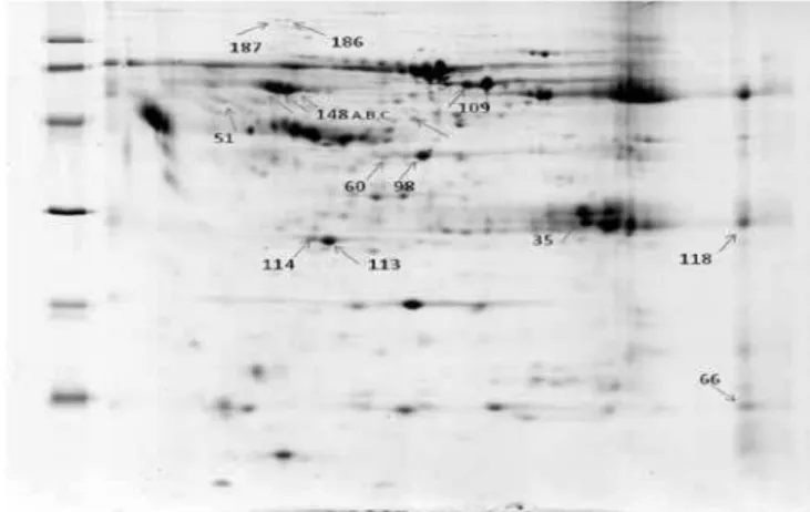
he 15 proteins that presented a range ratio above 2 in this analysis were selected for identiication. he diferen-tially expressed proteins are identiied in igure 1.
Figure 1 – Phase B 2D Gel with identiied excised spots.
he proteins with diferential expressions that were considered signiicant were: serum amyloid A, two apo-lipoprotein A1 isoforms, seven serpin family members, zinc inger protein 222, serum albumin, PRO 2619 (al-buminoid super-family), immunoglobulin kappa light chain VLJ region, immunoglobin M monoclonal of cold aglutination. he identiied serpin family proteins were, respectively, three alpha-1-antitrypsin isoforms, serpin, peptidase inhibitor, class A (antiproteinase alpha-1-anti-trypsin), member 1, two serine proteinase inhibitor iso-forms, class A (antiproteinase alpha-1, antitrypsin), mem-ber 1 and crystalized structure of serpin-protease complex, chain A.
Chart 2 shows proteins identiied with the spot num-ber, isoelectric point, and theoretical and experimental molecular weight, protein score (minimum 70), ion score (minimum 30), best Peptide Sequencing, NCBI database
Chart 1 – Studied patient demographic and clinical characteristics
Gender Age APACHE II Samples Pathogen Phase SOFA Serum protein (g/dL)
Male 57 18 D1 E. coli A 2 6.2
D11 E. coli B 4 6.0
D15 E. coli C 6 6.0
Chart 2 – Diferentially expressed proteins identiication and description
Identiication group
Proteins score/ Ions score
Peptides sequencing Access
number Protein description Function
Expression variation
66 196/174 GPGGAWAAEVISNAR
SFFSFLGEAFDGAR gi|337743 Serum amyloid A Scavenger C<B 0A
113 449/370
EQLGPVTQEFWDNLEK THLAPYSDELR
WQEEMELYR DEPPQSPWDR
gi|90108664 Apolipoprotein A-1 Innate
immunity A>B>C
114 448/378
EQLGPVTQEFWDNLEK THLAPYSDELR
WQEEMELYR
gi|90108664 Apolipoprotein A-1 Innate
immunity A>B>C
98 342/291
ITPNLAEFAFSLYR
LYHSEAFTVNFGDTEEAKK VFSNGADLSGVTEEAPLK
gi|11514321
Serpin-Proteinase complex crystallized structure, chain A
Proteinase
inhibitor C<B>A
133 168/100 VFDEFKPLVEEPQNLIK
FQNALLVR gi|23307793 Serum albumin
Amino acid
metabolism A>B>C
109 481/342
HPYFYAPELLFFAK HPDYSVVLLLR
VFDEFKPLVEEPQNLIK RPCFSALEVDETYVPK
gi|11493459 PRO 2619 (albuminoid super-family)
Amino acid
metabolism A>B>C
60 100/- EKPFQGENCK
SFCLRSSLNR gi|20988840 Zinc inger 222 protein
Transcription
factor C<B>A
118 200/191
SGTASVVCLLNNFYPR VYACEVTHQGLSSPVTK TVAAPSVFIFPPSDEQLK
gi|21669353 Immunoglobulin kappa
light chain VLJ region Host defense C>B>A
186 209/142 VFSNGADLSGVTEEAPLK
DTEEEDFHVDQVTTVK gi|50363217
Serin proteinase inhibitor, class A (alpha 1 anti-proteinase, antitrypsin), member 1
Proteinase
inhibitor C>B>A
187 191/143 ITPNLAEFAFSLYR
LYHSEAFTVNFGDTEEAKK gi|50363217
Serin proteinase inhibitor, class A (alpha-1 anti-proteinase, antitrypsin), member 1
Proteinase
inhibitor C>B>A
35 150/140
SGTASVVCLLNNFYPR VYACEVTHQGLSSPVTK TVAAPSVFIFPPSDEQLK
gi|10835792
Cold agglutination immunoglobulin M monoclonal
Host defense C>B>A
51 172/143 ITPNLAEFAFSLYR
VFSNGADLSGVTEEAPLK gi|15080499
Serpin, peptidase inhibitor, class A (anti-proteinase alpha-1, antitrypsin), member 1
Proteinase
inhibitor C>B>A
148 A 229/181 LYHSEAFTVNFGDTEEAKK
LYHSEAFTVNFGDTEEAK gi|177831 Alpha-1- antitrypsin
Proteinase
inhibitor C>B>A
148 B 376/318
LYHSEAFTVNFGDTEEAKK LQHLENELTHDIITK VFSNGADLSGVTEEAPLK LQHLENELTHDIITK
gi|6137432 Alpha-1- antitrypsin Proteinase
inhibitor C>B>A
148 C 396/355
LYHSEAFTVNFGDTEEAKK LQHLENELTHDIITK TDTSHHDQDHPTFNK GKWERPFEVK
gi|224224 Alpha-1- antitrypsin Proteinase
entry number, protein function and diferential expression in sepsis stages.
Each identiied protein was categorized into one of the established functional categories: metabolism, ge-netic information processing, environmental informa-tion processing, cell processes and human diseases. he Kyoto Encyclopedia of Genes and Genomes (KEGG - www.genome.jp/kegg/) classiication was adopted. Proteins were classiied as follows: cell processes/im-mune system (66.6%), cell processes/endocrine system (13.3%), metabolism (13.3) and genetic information/ transcription factor (6.6%), as can be seen in igure 2.
Figure 2 – Identified proteins relative functional distribu-tion. The reported categories were previously reported by the Kyoto Encyclopedia of Genes and Genomes.
he relative abundance of proteins involved in im-mune system cell processes may be related to the body’s reaction against the infectious process. hese proteins are found in the coagulation cascade pathways and complement system. he results indicate a signiicant focus on these pathways, known for their signiicant role in sepsis.(16) Other systems involved were the endo-crine, with its signaling pathways PPAR (13.3%), the metabolism (13.3%) and genetic information process-ing/transcription factor (6.6%). he PPAR signaling pathway is involved in the lipid mechanism (KEGG).
Identified proteins
Apolipoprotein A1 (APO A-1) is the main protein component of HDL (high density lipoprotein). APO A-1 binds to lipopolysaccharides or endotoxins, with a relevant role in HDL anti-endotoxin function.(17) It interacts with LBP, the LPS-binding protein, and cir-culates in plasma modulating LPS binding to HDL and acting as a cofactor for LPS neutralization. Lipopro-teins could have a role in innate immunity, as they re-duce cytokine response in animal sepsis models.(18) APO A-1 expression decreased with the progression of sepsis severity. his may indicate a reduction of innate
immu-nity action and anti-endotoxin efects.
Serum amyloid A (SAA) is an HDL complex apo-lipoprotein. SAA acts by redirecting LPS neutralized HDL from the CD14 macrophage activation pathway to the liver, with fast clearance and, consequently, re-duced HDL levels. herefore, during the acute phase LPS neutralizing ability is reduced, perpetuating sys-temic inlammatory response and contributing to or-gan failure and death.(19) In this study, SAA was not detected during the initial sepsis stage, and was most expressed during Stage B (severe sepsis), with decreased expression in the stage C of the study. hese data may relect a lower expression due to Stage C’s decreased need for APO A-1 displacement or a reduction of the liver’s ability to secrete it.
Also identiied in the study within the immune sys-tem category, and a participant in the coagulation and complement cascade, is immunoglobulin kappa light chain VLJ region. Immunoglobulins integrate the hu-moral immune system. heir contribution to immuni-ty may occur in three diferent forms: by binding to a pathogen to prevent its entry to the cell; by covering a pathogen to stimulate its removal by macrophages; and they may also trigger direct pathogen destruction by stimulating other immune responses such as comple-ment pathway activation.(20) Another immunoglobulin identiied in this study was immunoglobin M mono-clonal of cold aglutination. Cold agglutinins are auto- immunoglobulin M antibodies, characterized by their ability to agglutinate at low temperatures with eryth-rocytes in vitro (4-22ºC).(21) Both of the diferent im-munoglobins identiied in the study presented greater expression in stage C, decreasing towards stage A, thus exhibiting greater expression in the more advanced stages of the infectious process.
would apply to tissue regeneration, which is also among its functions.
Albumin is the most abundant plasma protein, comprising 50% of the human serum proteins.(23) his study identiied a reduction of albumin expression as sepsis worsened. One of albumin’s important functions is to maintain the volume of circulating plasma, due to its relatively low molecular weight and high concentra-tion. Albumin’s role also comprises: the maintenance of the acid-base balance; the transportation of a series of physiological substances; the distribution and metabo-lism of several diferent substances, both endogenous and exogenous; and, additionally, acting as an amino acid reserve.(24)
he PRO 2619 protein belongs to the albuminoid family, and is a serum albumin precursor. herefore, its expression was also decreased as the sepsis worsened.
Several proteins related to the serpin family were also identified. “Serpins” is a name given to the pro-teinase inhibitors family. Serpins share a complex and well preserved tertiary structure. The following ser-pin family proteins were found: serine proteinase in-hibitor, class A (alpha 1 anti-proteinase, antitrypsin), member 1; alpha-1-antitrypsin and serpin A chain present in a crystalline serpin-proteinase complex. Alpha-1-antitrypsin is a glycoprotein known to be a trypsin inhibitor, and plays a central role in homeosta-sis by neutralizing the detrimental effects of elastase, a powerful enzyme found in white blood cells. As pro-teinase inhibitors, serpins have an array of functions including blood clotting regulation, cell remodeling and motility, blood pressure regulation and angiogen-esis.(25) The serpin identified in the database, as part of a serpin-proteinase complex, displayed increasing ex-pression from the early stage to severe sepsis, and then dropped. Since this complex involves proteinases, this may suggest that these enzymes were reduced in septic shock, or that the enzyme amount would remain the same having less expression of serpins on the other hand. The remaining serpin expression increased as the patient’s infection worsened, i.e., a greater expres-sion from stage A to B and B to C, respectively. This profile demonstrates the involvement of the immune system and of the coagulation and complement cas-cade in sepsis progression, encompassing tissue re-pair and lung protection mechanisms. This condition probably arose as a consequence of the Acute Respi-ratory Distress Syndrome (ARDS) episode developed by the patient with a concomitant increase in elastase concentration.
Alpha-1-antitrypsin isoforms were isolated. he usually low level of isoform concentration, increases up to a hundredfold under inlammatory conditions,(26) in agreement with this study results. he increase may have resulted from alpha-1-antitrypsin fragmentation, which has an important lung function role. he frag-mentation of this protein may be a factor in the ARDS pathogenesis, indicating that its increase may be rel-evant and a useful biological marker for ARDS diag-nosis.
Apolipoprotein A-1 isoforms were similarly identi-ied. Variations in the percentage of diferent APO A-1 isoforms may occur as a result of an alteration in the fractional catabolic rate of this apolipoprotein.(27) he decreasing expression of these isoforms as sepsis wors-ened in this study, may indicate reduced innate immu-nity action, with regard to anti-endotoxin efects and cardiovascular protection.
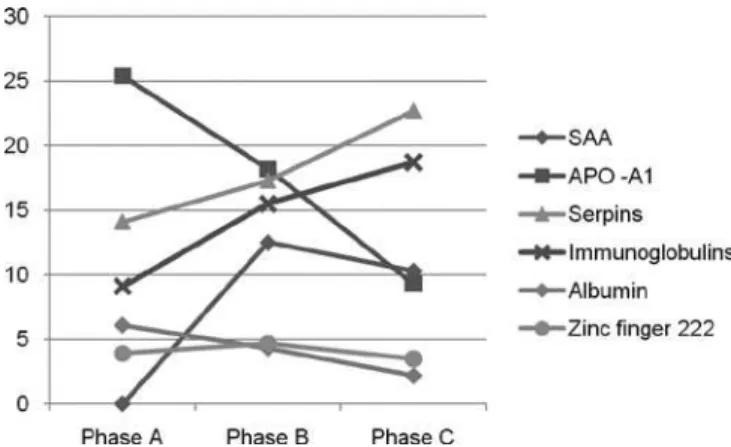
he functional meaning of these proteins was con-sidered. While many of them are inlammation and acute response biomarkers, the list of over- or under-expressed proteins in this study shows that sepsis has signiicant efects on coagulation, the immune system, the lipid metabolism, apoptosis and genetic informa-tion, conirming the indings of previous studies. Fig-ure 3 shows the proile presented by the proteins identi-ied in this study, according to their range of expression throughout the stages.
Figure 3 – Plot of the percent proteins expression in pha-ses A, B and C.
in-hibit the increasing amounts of elastase expressed dur-ing the ARDS episode.
SAA was not seen in Stage A, it would only appear during Stage B and reduced expression during Stage C. his behavior could likely be explained by the reduced need of APO A-1 displacement during phase C, when its presence was lessened, or the liver’s capacity of se-creting it was diminished.
he zinc inger 222 protein increased its expres-sion from Stage A to Stage B, then decreased in Stage C. Being a transcription repression factor, its reduced expression and, therefore, diminished repression of a harmful protein may have contributed to the onset of septic shock phase.
Stage A was characterized by a predominance of APO A-1 and albumins, representing the protective re-sponse and maintenance of homeostasis in sepsis. Dur-ing Stage B, the zinc Dur-inger 222 transcription factor and SAA were predominant, demonstrating intense inlam-matory activity by APO A-1 displacement and protein repression attempts. Stage C was characterized by in-creased expression of proteinase inhibitors and immu-noglobulins, proteins that are functionally accountable for the body’s defense.
The proteins identified in this study are related to those described in previous proteomic studies of sep-sis. In this study, however, a longitudinal approach of the same patient was performed. While this study’s analysis of a single patient may be considered a limita-tion, that does not allow results to be inferred, on the other hand, the study of a single patient, without a mixture of samples, presents greater value, due to the absence of confounding factors that would arise from mixing serums from patients of different genders, ages, ethnicity (African, Asian, European, etc), infect-ing organisms, immune responses towards insultinfect-ing agents, nutritional habits – which are all factors that can influence protein expression. Several previous tri-als employed this mixed approach, potentially weak-ening their findings.
One of the major opportunities we envision through proteomic studies is the characterization of protein ex-pression diferences to identify potential therapeutic targets and disease biomarkers, thus widening our un-derstanding of biological processes. A functional pro-teomic approach could provide data on sepsis’ inlam-matory signaling network, identifying critical regula-tion nodes. But how can proteomic data help us direct our therapy to a speciic signaling pathway or critical crossroad between the connection pathways responsible
for maintaining the septic state? he future challenge lies in translating what this research data can convey to clinical diagnosis, to greatly impact the decision pro-cess regarding patient therapy.
his investigation reveals the potential that pro-teomic techniques – two-dimensional electrophoresis and mass spectrometry – hold for advancing research into sepsis.
his is the irst proteomic-based description, of sep-sis’ progressive changes in a single patient. Despite this study’s limitations, it holds signiicant value as a start-ing point for other studies along this line that may vali-date the conclusions reached herein regarding protein expression patterns in sepsis.
RESUMO
Na sepse ocorre desregulação da expressão gênica. Os marcadores genéticos revelam apenas o genótipo do indi-víduo, não sendo afetados pelos processos biológicos decor-rentes da ação do ambiente, estes expressos nas proteínas. Este estudo teve como objetivo alcançar maior compreensão sobre as bases moleculares da sepse. Para tal realizou a iden-tiicação e análise da expressão diferencial de proteínas no soro de paciente séptico em diferentes estágios de gravidade (sepse, sepse grave e choque séptico) através de técnicas pro-teômicas. Amostras de soro referentes a cada estágio da sepse foram colhidas e submetidas à eletroforese unidimensional em itas com gradiente de pH imobilizado seguida de ele-troforese bidimensional em gel de poliacrilamida 12,5%. Os géis obtidos foram corados, escaneados e analisados através do programa ImageMasterPlatinum. As proteínas expressas diferencialmente nos géis foram excisadas, digeridas com tripsina e identiicadas através de espectrometria de massa. Foram identiicadas 14 proteínas expressas diferencialmente entre os estágios da sepse, assim como uma proteína não ex-pressa em todos os estágios, sugerindo a existência de um possível biomarcador. Foram elas: amilóide sérico A, apoli-poproteína A-1 (2 isoformas), proteína dedo de zinco 222, albumina humana, PRO 2619, imunoglobulina de cadeia leve kappa região VLJ, imunoglobulina M monoclonal de aglutinação a frio e 7 inibidoras de proteases - alfa-1 anti-tripsina. Os resultados obtidos neste estudo piloto demon-stram a participação das vias do complemento e coagulação, do metabolismo lipídico e da informação genética na sepse. A grande maioria de proteínas identiicadas está envolvida no sistema imune com predomínio das proteínas inibidoras de proteases.
REFERÊNCIAS
1. Cohen J. he immunopathogenesis of sepsis. Nature. 2002;420(6917):885-91.
2. Bone RC, Grodzin CJ, Balk RA. Sepsis: a new hypothesis for pathogenesis of the disease process. Chest. 1997;112(1):235-43. Review.
3. Becker KL, Snider R, Nylen ES. Procalcitonin assay in systemic inflammation, infection, and sepsis: clinical utility and limitations. Crit Care Med. 2008;36(3):941-52.
4. Tili E, Michaille JJ, Cimino A, Costinean S, Dumitru CD, Adair B, et al. Modulation of 155 and miR-125b levels following lipopolysaccharide/TNF-alpha stimulation and their possible roles in regulating the response to endotoxin shock. J Immunol. 2007;179(8):5082-9.
5. Müller B, Schuetz P, Trampuz A. Circulating biomarkers as surrogates for bloodstream infections. Int J Antimicrob Agents. 2007;30 Suppl 1:S16-23.
6. Calandra T, Echtenacher B, Roy DL, Pugin J, Metz CN, Hültner L, et al. Protection from septic shock by neutralization of macrophage migration inhibitory factor. Nat Med. 2000;6(2):164-70.
7. Phua J, Koay ES, Zhang D, Tai LK, Boo XL, Lim KC, Lim TK. Soluble triggering receptor expressed on myeloid cells-1 in acute respiratory infections. Eur Respir J. 2006;28(4):695-702.
8. Wang H, Bloom O, Zhang M, Vishnubhakat JM, Ombrellino M, Che J, et al. HMG-1 as a late mediator of endotoxin lethality in mice. Science. 1999;285(5425):248-51.
9. Ciero L, Bellato CM. Proteoma: avanços recentes em técnicas de eletroforese bidimensional e espectrometria de massa. Biotecnol Ciênc Desenvolv. 2002;5(29):158-64.
10. Mann M, Hendrickson RC, Pandey A. Analysis of proteins and proteomes by mass spectrometry. Annu Rev Biochem. 2001;70:437-73. Review.
11. Hoehn GT, Sufredini AF. Proteomics. Crit Care Med. 2005;33(12 Suppl):S444-8. Review.
12. Knaus WA, Draper EA, Wagner DP, Zimmerman JE. APACHE II: a severity of disease classiication system. Crit Care Med. 1985;13(10):818-29.
13. Vincent JL, Moreno R, Takala J, Willatts S, De Mendonça A, Bruining H, et al. he SOFA (Sepsis-related Organ Failure Assessment) score to describe organ dysfunction/failure. On behalf of the Working Group on Sepsis-Related Problems of the European Society of Intensive Care Medicine. Intensive Care Med. 1996;22(7):707-10.
14. Jenö P, Mini T, Moes S, Hintermann E, Horst M. Internal sequences from proteins digested in polyacrylamide gels. Anal Biochem. 1995;224(1):75-82.
15. Habermann B, Oegema J, Sunyaev S, Shevchenko A. he power and the limitations of cross-species protein identiication by mass spectrometry-driven sequence similarity searches. Mol Cell Proteomics. 2004;3(3):238-49.
16. Shen Z, Want EJ, Chen W, Keating W, Nussbaumer W, Moore R, Gentle TM, Siuzdak G. Sepsis plasma protein profiling with immunodepletion, three-dimensional liquid chromatography tandem mass spectrometry, and spectrum counting. J Proteome Res. 2006;5(11):3154-60.
17. Harris HW. Apolipoprotein E: from Alzheimer’s to sepsis. Crit Care Med. 2005;33(11):2696-7.
18. van Leeuwen HJ, Heezius EC, Dallinga GM, van Strijp JA, Verhoef J, van Kessel KP. Lipoprotein metabolism in patients with severe sepsis. Crit Care Med. 2003;31(5):1359-66.
19. van Leeuwen HJ, van Beek AP, Dallinga-hie GM, van Strijp JA, Verhoef J, van Kessel KP. he role of high density lipoprotein in sepsis. Neth J Med. 2001;59(3):102-10.
20. Ravetch JV, Bolland S. IgG Fc receptors. Annu Rev Immunol. 2001;19:275-90.
21. Ciejka JZ, Cook EB, Lawler D, Martin J, Woodson RD, Graziano F. Severe cold agglutinin disease and cryoglobulinemia secondary to a monoclonal anti-Pr2 IgM lambda cryoagglutinin. Clin Exp Rheumatol. 1999;17(2):227-31.
project: the Mammalian Gene Collection (MGC). Genome Res. 2004;14(10B):2121-7. Erratum in: Genome Res. 2006;16(6):804. Morrin, Ryan [corrected to Morin, Ryan].
23. Doweiko JP, Nompleggi DJ. Role of albumin in human physiology and pathophysiology. JPEN J Parenter Enteral Nutr. 1991;15(2):207-11. Review.
24. Whicher J, Spence C. When is serum albumin worth measuring? Ann Clin Biochem. 1987;24(Pt 6):572-580. Review.
25. Barret AJ, Salvesen G, editors. Proteinase inhibitors. Amsterdam: Elsevier; 1986.
26. Ferrero S, Gillott D, Anserini P, Remorgida V, Price K, Ragni N, Grudzinskas J. 168: Alpha1-Antitrypsin isoforms in endometriosis: a proteomic study. J Minim Invasive Gynecol. 2005; 12(5 Suppl 1):70.