Biopolymers and Cell. 2015. Vol. 31. N 2. P. 123–130 doi: http://biopolymers.org.ua/doi/bc.0008D6
UDC 575.1+577.11+577.21
Novel gene
PUS3
c.A212G mutation
in Ukrainian family with intellectual disability
R. V. Gulkovskyi
1, 2, S. Y. Chernushyn
1, L. A. Livshits
1, 21 Institute of Molecular Biology and Genetics, NAS of Ukraine
150, Akademika Zabolotnoho Str., Kyiv, Ukraine, 03680
2 Educational and Scienti
fi c Center "Institute of Biology", Taras Shevchenko National University of Kyiv
64/13, Volodymyrska Str., Kyiv, Ukraine, 01601 livshits@imbg.org.ua
Aim. To evaluate a possible role of a novel c.A212G substitution in the PUS3 gene at intellectual dis-ability (ID). Methods. The observed group consisted of the ID Ukrainian family members (parents and two affected children) and the control group – of 300 healthy individuals from general population of Ukraine. Sanger sequencing of the PUS3 gene exon 1 was performed for the family members. Polymorphic variants of c.A212G were analyzed using ARMS PCR. The homology models of wild type and p.Y71C mutant catalytic domains of human Pus3 were generated using the crystal structure of the human Pus1 catalytic domain (PDB ID: 4NZ6) as a template. Results. It was shown that the father of the affected siblings was the c.A212G substitution heterozygous carrier whereas the mother was a wild type allele homozygote, and the exom sequencing result was confi rmed – the affected children are 212G homozy-gotes. We supposed de novo mutation in the maternal germ line. A low frequency of 212G allele (0.0017) was shown in the population of Ukraine. Homology modelling of the wild type and p.Y71C mutant cata-lytic domain of human Pus3 revealed that substitution p.Y71C is located in close proximity to its active site. Conclusions. The absence of hypoproteinemia in our patients, homozygous for the 212C allele al-lows us to assume that the mutation c.A212G PUS3 is rather neutral and cannot be the major cause of ID. However, considering a low frequency of the 212G allele in the population and close localization of p.Y71C substitution to the active site of hPus3 we cannot exclude that the c.A212G mutation in PUS3 may be a modifi er for some pathologies including syndromic ID.
K e y w o r d s: PUS3 gene, intellectual disability, mutation, population, pseudouridine.
© 2015 R. V. Gulkovskyi et al.; Published by the Institute of Molecular Biology and Genetics, NAS of Ukraine on behalf of Biopolymers and Cell. This is an Open Access article distributed under the terms of the Creative Commons Attribution License (http://creativecommons.org/licenses/by/4.0/), which permits unrestricted reuse, distribution, and reproduction in any medium, provided the original work is properly cited
Introduction
In the frame of CHERISH project (no. 223692) de-voted to the genetic basis of intellectual disability, the next generation exome sequencing was conduct-ed for two affectconduct-ed children (proband and his young-er sibling) from the Ukrainian family (UKR 094) with healthy non–consanguineous parents. The pro-band (UKR 263) is a 12-year-old boy with non-syn-dromic ID (IQ 43), hypermobility of joints, hyperac-tivity and mild dysmorphic features. His brother (UKR 264) is a 4-year-old boy with non-syndromic ID,
epilepsy, hypermobility of joints and mild dysmor-phic features. The biochemical analysis of blood re-vealed no deviations from the normal ranges for the main groups of blood plasma proteins, aminoacids and acylcarnitines examined by TANDEM MS in both probands. The previous extensive genetic in-vestigations (karyotype analysis and array–CGH analysis (400K resolution) have not found out any abnormalities as well.
Among these candidates, we decided to concentrate on the PUS3 gene, where novel homozygous SNP c.A212G (NM_031307:c.A212G:p.Y71C) was de-tected in both ID-patients. The PUS3 gene is located at chromosome 11q24.2 and conserved from Es che-richia coli to human [2].
The human pseudouridine synthase 3 (hPus3) is a member of the tRNA pseudouridine synthase truA family and involved in the formation of pseudourid-ine (Ψ) at position 39 in the anticodon stem and loop (ASL) of many transfer RNAs [3, 4]. Ψ is found in almost all tRNAs, Ψ’s at positions 38–40 in the An-ticodon Stem Loop of tRNAs play an important role in maintaining translational effi ciency and accuracy [5]. Pseudouridine appears to be necessary for the correct codon–anticodon interactions [6, 7] and to prevent mischarging of the tRNA [8]. It was shown that the mouse Pus3 (mPus3) can also serve as a nu-clear receptors (NR) coactivator (as well as mPus1 known to form pseudouridine at positions 27, 28, 34, and 36 in tRNAs), except that it does not enhance the sex steroid receptor activity [4]. A mutation in the PUS1 gene (another truA family member) has been linked to the mitochondrial myopathy and siderob-lastic anemia [4, 9–11].
To evaluate a possible involvement of the PUS3 gene c.A212G mutation in intellectual disability, as the fi rst step, we analyzed this mutation in the healthy parents of the affected children and in general popu-lation of Ukraine and modeled 3D structure of the hPus3 catalytic domain to defi ne if the change in p.Y71C position infl uences the 3D structure of hu-man Pus3 protein.
Materials and Methods
The observed group consisted of 300 adult (25–30 years-old) individuals including 164 (54.6 %) males and 136 (45.3 %) females. This group is representa-tive for the estimation of DNA polymorphism fre-quency in autosomal genes [12, 13].
The DNA–samples were extracted from peripheral blood leucocytes of unrelated volunteer donors from different regions of Ukraine by the standard phe nol– chloroform method. Informed consents were obtained from all the individuals participating in our study.
The polymorphic variants c.A212G found thro-ugh exome sequencing in the affected children (from UKR 094 family) were analyzed in the DNA sam-ples of their parents by Sanger sequencing. This ana-lysis was performed by the standard dideoxynucle-otide chain–termination method using [35S]–dATP or
[35S]–dCTP (ICN), the Sequenase version 2.0 DNA
se-quencing kit (USB), «ABI Prism Big Dye Terminator Cycle Sequincing Ready Reaction Kits» and ABI Prism 3110 Genetic Analyser (Applied Biosystems Ana lysis of the c.A212G substitution was performed by ARMS (amplifi cation refractory mutation sys-tem). The primers were designed using the web–based PRIMER 3.0 program (http://workbench.sdsc.edu) and the «BLAST» (http://www.ncbi.nlm.nih.gov/ blast) (Table 1).
Amplifi cation of the allele 212A is accomplished using a complementary primer «wild type» paired with a «forward» primer (Table 1). On the other hand, the 212G allele will be amplifi ed if the 3’ resi-due of primer is complementary to the «mutant»
se-Table 1. Sequences of ARMS-PCR primers used in genotping reactions
Substitution Nucleotide sequence Amplicon size, bp
CTGGAGCAAGAGGTGCAAAGACT – «forward» 320
ACCTGTCCAAAGGCACTAACTCC – «reverse»
c.A212G CTGGAGCAAGAGGTGCAAAGACT – «forward» 193
GACACGTAGCCCTAAGAATAGCCTa – «wild type»
c.A212G CCTGGTATCCCCAGCCCATAc – «mutant» 172
quence – PCR product fl anked by «mutant» and «re-verse» primers (Table 1). Thus, as a result a normal individual generates PCR product (193 bp) only in the normal reaction; a heterozygote gives products (193 bp and 172 bp) in both reactions, and a ho-mozygous mutant individual gives amplifi cation (172 bp PCR product) only in the case of mutant variant. The PCR product fl anked by «forward» and «reverse» primers (320 bp) will be amplifi ed in all reactions (Fig.1).
Thereby three different patterns could be observed for the c.A212G variant after the amplifi cation: 320 bp and 193 bands (for A/A); 320 bp, 193 bp and 172 bp bands (for A/G); 320 bp and 172 bp bands (for G/ G) (Fig. 3).
The PCR amplifi cation was performed in one tube in a fi nal volume of 25 l containing 1 PCR buffer, 1.5 mM MgCl2, 200 M of each dNTP, 1 M of each prim-er, 0.2 units of Taq–DNA polymerase and 200 ng of the DNA template. The cycling conditions were as follows: initial denaturation at 94 C for 5 min, 30 cycles con-sisting of denaturation at 94 C for 30s, annealing at 64
C for 30s, extension at 72C for 30s, and a fi nal elon-gation step at 72C for 3 min. The amplifi ed fragments were analyzed by electrophoresis in 2 % agarose gel. The sample that previously underwent Sanger sequenc-ing was used as a positive control.
Multiple sequence alignment was performed ac-cording to the Homologene program with default settings and the sequences: NP_079491.2 (Homo sa-piens), NP_112597.3 (Homo sapiens), XP_001148378.1 (Pan troglodytes), XP_001111887.1 (Macaca mulat-ta), NP_075781.3 (Mus musculus), NP_001101604.1 (Rattus norvegicus), XP_536533.1 (Canis lupus fa mi-liaris), NP_001029684.1 (Bos taurus), XP_004948004.1 (Gallus gallus), NP_956361.1 (Danio rerio), NP_9 88969.1 (Xenopus tropicalis), NP_611646.1 (Dro so-phila melanogaster), XP_318500.4 (Anopheles gam-biae str.), NP_496062.3 (Caenorhabditis elegans), NP_116655.1 (Saccharomyces cerevisiae), XP_454596.1 (Kluyveromyces lactis) and P07649.1 (Escherichia co li). The sequences of the Homo sapiens hPus3 and hPus1 proteins and homologous proteins from other species were obtained from the NCBI database (http://www.ncbi.nlm.nih.gov/).
In silico modeling. The homology models of wild type and mutant (p.Y71C) catalytic domains of hu-man Pus3 were generated by Swiss Model server (http://swissmodel.expasy.org) using as a template a crystal structure of the human (mitochondrial) Pus1 catalytic domain (PDB ID: 4NZ6).
Results and Discussion
The Sanger sequencing of the gene PUS3 exon 1 re-vealed that probands UKR 263 and UKR 264 are homozygotes for the c.A212G substitution that con-fi rms the exome sequencing results. In turn, the fa-ther (UKR 266) of the affected siblings is a hetero-zygous carrier for the c.A212G substitution and, surprisingly, the mother (UKR 265) is a wild type allele homozygote (Fig. 2).
The possible explanation of such results is the de novo mutation in the maternal germ line.
Of the 300 analyzed samples, we found one hete ro-zygous carrier for the c.A212G mutation only. Thus, the genotypes distribution was as follows: A/A – 99.7 %, A/G – 0.3 % and GG – 0 %. The observed
Fig. 1. Electrophoregram for the ARMS analysis of c.A212G PUS3 gene variants (2 % agarose gel electrophoresis): 1 – mo-lecular weight marker (50 bp ladder); 2, 3 – individuals with homozygous genotype AA; 4 – individual with heterozygous genotype AG; 5 – individual with homozygous genotype GG and 6 – negative control
1 2 3 4 5 6
320 bp
genotype distribution showed no deviations from Har-dy–Weinberg expectations. The PUS3 212G (mu-tant) allele frequen cy was 0.0017 and 212A (wild type) – 0.9983. To our knowledge, this is the fi rst published data on the c.A212G PUS3 gene variants distribution in the populations.
Pseudouridylate synthase 3 is a 481 amino acid (aa) protein that belongs to the highly conserved tRNA pseudouridine synthase truA family and mostly is lo-calized to cytoplasm and nucleus [3, 4, 14]. The mem-bers of this family have been shown to modify mul-ti ple posimul-tions in cytoplasmic and mitochondrial tRNAs
[11, 15], as well as non–tRNA substrates: U2 snRNA [16] and SRA [17]. The TruA family includes Pus1 from E. coli (also called truA or hisT) which modi-fi es positions 38, 39, and 40 in the ASL of bacterial tRNAs [18]. Other members are Pus3 from mouse [1]; Pus3 from yeast (also known as Deg1) – modi-fi es positions 38 and 39 [14] and human mitochon-drial Pus1 modifi es positions 27, 28, 34, and 36 in tRNAs in vitro and in vivo [19, 20]. The atomic mod-els for various members of the PUS families (TruA, TruB, TruD, RluA, RsuA, and Pus10) have been sol-ved and have shown a consersol-ved catalytic core that presents a high degree of structural similarity and is the most stable region of the protein. [21–28].
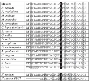
The c.A212G mutation results in substitution of aromatic Tyrosine 71 to sulfur–containing Cysteine located in the catalytic domain of human Pus3. The analyses of site orthologs, using the NCBI Homo lo-Gene database, revealed that the Tyr71 amino acid position is conserved from Escherichia coli to hu-man, indicating that there may be an evolutionarily conserved function (Fig. 3).
To defi ne if the change in p.Y71C position infl u-ences the 3D structure of human Pus3 protein, we modeled the hPus3 catalytic domain 3D structure us-ing the crystal structures of the human (mitochondrial) Pus1 catalytic domain as a template (Fig. 4, C and D). The mitochondrial hPus1, as well as hPus3, belongs to the tRNA pseudouridine synthase TruA family and is mostly localized in the mitochondria [16, 17, 29].
From the crystal structure of the bacterial TruA (Fig. 4, A) it is known that the active site of the TruA family members is populated by four strictly conser-ved amino acids including a catalytic aspartate (D60 in TruA), two arginines (R58 and R205), and a tyro-sine (Y118) [22]. These residues correspond to D146, R144, R295, Y201 in hPus1 (Fig. 4, B) and D118, R116, R280 and Y195 in hPus3 (see Fig. 4, C and 4, D) [22, 27, 28, 30]. Tyrosine 71 of hPus3, cor-responding to Y18 in bacterial TruA and Y92 – in hPus1, is another strictly conserved amino acid (Fig. 3). As can be seen from Fig. 4, C and 4, D, the substi-tution of highly conserved aromatic Tyrosine 71 to sulfur–containing Cysteine is located in close proxim-ity to the active site of hPus3 and may directly cause
Fig. 2. Representative sequence chromatogram of the PUS3 gene loci with c.A212G substitution PCR products: A – UKR 265 (mother) – wild type allele homozygote (AA); B – UKR 266 (fa-ther) – heterozygous carriers of the c.A212G substitution; C – UKR 263 and UKR 264 – homozygous GG
G
G
G
G
G
G C
C
C C
C
C
T A
B
C T
T
T
T
T
T
T
T A
A
G
A
A
the changes in catalytic domain conformational fl exi-bility and spatial organization. Nevertheless, we still cannot suppose that the p.Y71C mutation may result in the change of pseudouridylate synthase effi cien-cy of hPus3, Tyr71 (shown in black boxes) from Escherichia coli to human. Tyrosine 71 of hPus3 cor-responds to Y92 in hPus1 (shown in black boxes).
This is true considering the fact that Sibert et al., interpreting the site–directed mutagenesis experi-ments with hPus1, indicate that Y173 (corresponds to Y195 in hPus3), and R267 (corresponds to R280 in hPus3) known to compose the active site of TruA of the enzyme near a critical aspartate (position 118), do not play any essential role in the catalysis [30]. They changed Tyr201 and Arg295 to several other amino acids and found that many variants had sig-nifi cant activity [30].
However, Ψ’s at positions 38–40 in the Anticodon Stem Loop of tRNAs plays an important role in maintaining translational effi ciency and accuracy. These modifi cations of uridines were shown to in-crease the thermal stability of the ASL, which could affect the anticodon–codon interaction or conforma-tional changes of the tRNA during translation and spliceosome assembly [5, 32]. Thus, the PUS3 muta-tions, that result in loss of pseudouridine in a wide range of tRNAs, may affect the protein synthesis.
The strong relationship between the reduced growth rate of E. coli or S. typhimurium and the ab-sence of pseudouridines 38–40 in anticodon stem– loop of several tRNAs was reported more than two decades ago [33]. The HisT mutant E. coli strains display a 20–25 % reduction in the rate of polypep-tide chain elongation and exhibit pleiotropic abnor-malities in the cell division processes, resulting in an increase in doubling time of more than 30 % [34]. When the PUS3 gene was disrupted in yeast, it was not lethal, but the growth rate of the yeast consider-ably reduced, especially at 37 C [14]. Since there is an effect on the growth of prokaryotes and yeast when the pseudouridine 38–40 synthase activity is deleted, what might be the effect of the PUS3 gene missense mutations in humans?
The physiological importance of the appropriate pseudouridine synthase activity is illustrated by the
disorders such as DKC (dyskeratosis congenital) and MLASA (mitochondrial myopathy and siderob-lastic anemia) [9–11, 35]. A missense mutation in the human PUS1 gene affecting a highly conserved amino acid (Arg144–to–Trp mutation in the active site of the enzyme and a mutation of Glu220, which leads to C–terminally truncated protein) has been associated with mitochondrial myopathy and si-deroblastic anemia, a rare autosomal recessive dis-order of oxidative phosphorylation and iron metab-olism [9–11]. The X–linked form of the only other known human disease of pseudouridylation dysk-eratosis congenita is caused by the mutations in the gene encoding dyskerin [35].
The previously mentioned PUS1 mutations result in the loss of pseudouridine in some tRNAs that may affect protein synthesis [9–11]. Furthermore, the mam-malian pseudouridine synthase 1 (Pus1) was report-ed to modulate the class I and class II nuclear recep-tor responses through its ability to modify the Steroid receptor RNA Activator (SRA) [4, 17, 28] and it was suggested that other abnormalities in these MLASA patients, such as facial dysmorphisms, may be due to
Fig. 3. Pseudouridylate synthase 3 (481 aa) conservation anal-ysis. Conserved amino acid positions are shown in grey boxes. Protein alignment showed conservation of residue Tyr71 (shown in black boxes) from Escherichia coli to human. Ty-rosine 71 of hPus3 corresponds to Y92 in hPus1 (shown in black boxes)
Mutated AFDFSAHGRRHVALRIACMGWGYQGFASQE
H. sapiens AFDFSAHGRRHVALRIACMGWGYQGFASQE
P. troglodytes AFDFSAHGRRHVALRIAYMGWGYQGFASQE
M. mulatta AFDFSAHGRRHVALRIAYMGWGYQGFASQE
M. musculus AFDFSAHGRRHVALKIAYLGWGYQGFASQE
R. norvegicus AFDFSAHGRRHVALKIAYLGWGYQGFASQE
C. lupus familiaris AFDFSAHGRRHVALKIAYLGWGYQGFASQE
B. taurus AFDFSAHGQRHVALKIAYLGWGYQGFASQE
G. gallus PFDFSAHGRRHVALRIAYLGWGYQGFASQE
D. rerio PFDFSAHPRRHVALRLAYLGWQYQGFAVQE
X. tropicalis AFDFSAHPKQHVALRLAYLGWGYQGFASQE
D. melanogaster KFDWSSAHKRHVLLKITYLGWDYQGFACQE
A. gambiae str. PFDFAKCFKRHILLRFYYLGWGYQGFAAQE
C. elegans TLDFLAHPRRKIAIQFFYLGWEHDGLVQQP
S. cerevisiae KFDFSKHNTRFIALRFAYLGWNYNGLAVQK
K. lactis EFDFSKYNTRFVAFKFAYLGWNYNGLAIQK
E. coli MSDQQQPPVYKIALGIEYDGSKYYGWQRQN
H. sapiens AFDFSAHGRRHVALRIAYMGWGYQGFASQE
the loss of this activity of Pus1 [10]. Recently it has been shown that Pus3 (as well as Pus1) acts as a reg-ulator of the nuclear receptors activity [4]. Therefore, it cannot be excluded that some symptoms observed in the affected children from Ukrainian family (UKR 094), such as facial dysmorphisms, may be the con-sequence of defective hSRA–NR signaling.
However, in some known human pseudouridyla-tion diseases, the loss of pseudouridine that results in a decrease of the protein synthesis effi ciency, has a pleiotropic effect which causes syndromic patholo-gies. The similar pleiotropic effect may take place in case of the c.A212G mutationin the PUS3 gene in-dentifi ed in this study. Nevertheless, we did not ob-serve syndromic intellectual disability in the affected children from Ukrainian family (UKR 094). Fur-thermore, in our patients we did not detect any
evi-dence of hypoproteinemia, which is a common indi-cator for both MLASA and DKC and may be an evi-dence of the defi ciency in protein synthesis.
Thus, we assume that the c.A212G PUS3 muta-tion is rather neutral and cannot be the major cause of intellectual disability. However, considering a low frequency of the 212G allele (0.0017) in the popula-tion of Ukraine and the locapopula-tion of p.Y71C substitu-tion in close proximity to the active site of hPus3 protein we cannot exclude that the c.A212G muta-tionin the PUS3 gene may still be considered as a modifying factor for some pathologies including syndromic intellectual disability.
Further studies will be carried out to evaluate the infl uence of the c.A212G PUS3 gene mutation on the pseudouridylate synthase effi ciency and its in-volvement in the ID development.
Fig. 4. Overall views of the crystal structure of bacterial TruA (PDB ID: 2NQP) (A), hPus1 (PDB ID: 4NZ6) (B) and homology models of wild type (C) and p.Y71C mutant (D) hPus3 catalytic domain. Crystal structure of pseudoudirinde synthase TruA mono-mer in complex with leucyl tRNA (A) – uracil 39 shown in ball and stick model and colored in black, the catalytic amino acid residues D146, R144, R295 and Y201 are shown as sticks and colored in green, Y92 shown in stick model and colored in blue. The hPus1 monomer (B) – catalytic amino acid residues D60, R58, R205 and Y118 are shown as sticks and colored in green, Y18 shown in stick model and colored in blue. Homology models of wild-type (C) and p.Y71C mutant (D) catalytic domain of human Pus3 generated by Swiss Model server. The images were created by ViewerLite v.4.2 with Y71 mutation sites shown in blue, residues D118, R116, R280 and Y195 are shown in green [28, 31]
A
C
B
Acknowledgements
We thank the family for its cooperation. We thank M. Noukas, M. Sauk, L. Milani, T. Pippucci, F. Ba-lombo for their kind assistance in the next generation exome sequencing of the UKR 094 family members.
REFERENCES
1. Gulkovskyi RV, Chernushyn SY, Kravchenko SA, Bychkova GM, Livshits LA. EPHA1 gene SNPs analysis in population of Ukraine. Biopolym Cell. 2013;29(6):506–10.
2. Satoh J, Kawana N, Yamamoto Y. Pathway analysis of ChIP-Seq-Based NRF1 target genes suggests a logical hypothesis of their involvement in the pathogenesis of neurodegenera-tive diseases. Gene Regul Syst Bio. 2013;7:139–52. 3. Chen J, Patton JR. Pseudouridine synthase 3 from mouse
modifi es the anticodon loop of tRNA. Biochemistry. 2000;
39(41):12723–30.
4. Zhao X, Patton JR, Ghosh SK, Fischel-Ghodsian N, Shen L, Spanjaard RA. Pus3p- and Pus1p-dependent pseudouridyla-tion of steroid receptor RNA activator controls a funcpseudouridyla-tional switch that regulates nuclear receptor signaling. Mol En do-crinol. 2007;21(3):686–99.
5. Agris PF. Decoding the genome: a modifi ed view. Nucleic Acids Res. 2004;32(1):223–38.
6. Tomita K, Ueda T, Watanabe K. The presence of pseudouri-dine in the anticodon alters the genetic code: a possible me-chanism for assignment of the AAA lysine codon as aspar-agine in echinoderm mitochondria. Nucleic Acids Res. 1999;
27(7):1683–9.
7. Zerfass K, Beier H. Pseudouridine in the anticodon G psi A of plant cytoplasmic tRNA(Tyr) is required for UAG and UAA suppression in the TMV-specifi c context. Nucleic Acids Res. 1992;20(22):5911–8.
8. Perret V, Garcia A, Grosjean H, Ebel JP, Florentz C, Giegé R. Relaxation of a transfer RNA specifi city by removal of modifi ed nucleotides. Nature. 1990;344(6268):787–9. 9. Bykhovskaya Y, Casas K, Mengesha E, Inbal A,
Fischel-Gho-dsian N. Missense mutation in pseudouridine synthase 1 (PUS1) causes mitochondrial myopathy and sideroblastic anemia (MLASA). Am J Hum Genet. 2004;74(6):1303–8.
10. Fernandez-Vizarra E, Berardinelli A, Valente L, Tiranti V, Zeviani M. Nonsense mutation in pseudouridylate synthase 1 (PUS1) in two brothers affected by myopathy, lactic aci-dosis and sideroblastic anaemia (MLASA). J Med Genet. 2007;44(3):173–80.
11. Patton JR, Bykhovskaya Y, Mengesha E, Bertolotto C, Fi-schel-Ghodsian N. Mitochondrial myopathy and sideroblas-tic anemia (MLASA): missense mutation in the pseudouridi-ne synthase 1 (PUS1) gepseudouridi-ne is associated with the loss of tRNA pseudouridylation. J Biol Chem. 2005;280(20):19823–8. 12. Cordell HJ, Clayton DG. Genetic association studies.
Lan-cet. 2005;366(9491):1121-31.
13. Balding DJ. A tutorial on statistical methods for population association studies. Nat Rev Genet. 2006;7(10):781–91. 14. Lecointe F, Simos G, Sauer A, Hurt EC, Motorin Y, Grosjean H.
Characterization of yeast protein Deg1 as pseudouridine syn-thase (Pus3) catalyzing the formation of psi 38 and psi 39 in tRNA anticodon loop. J Biol Chem. 1998;273(3):1316–23. 15. Chen J, Patton JR. Mouse pseudouridine synthase 1: gene
structure and alternative splicing of pre-mRNA. Biochem J. 2000;352 Pt 2:465–73.
16. Behm-Ansmant I, Massenet S, Immel F, Patton JR, Motorin Y, Branlant C. A previously unidentifi ed activity of yeast and mouse RNA:pseudouridine synthases 1 (Pus1p) on tRNAs. RNA. 2006;12(8):1583–93.
17. Zhao X, Patton JR, Davis SL, Florence B, Ames SJ, Span ja-ard RA. Regulation of nuclear receptor activity by a pseu do-u ridine synthase throdo-ugh posttranscriptional modifi cation of steroid receptor RNA activator. Mol Cell. 2004;15(4):549–58. 18. Kammen HO, Marvel CC, Hardy L, Penhoet EE. Purifi cation,
structure, and properties of Escherichia coli tRNA pseudou-ridine synthase I. J Biol Chem. 1988;263(5):2255–63. 19. Chen J, Patton JR. Cloning and characterization of a
mam-malian pseudouridine synthase. RNA. 1999;5(3):409–19. 20. Motorin Y, Keith G, Simon C, Foiret D, Simos G, Hurt E,
Gro-sjean H. The yeast tRNA:pseudouridine synthase Pus1p disp-lays a multisite substrate specifi city. RNA. 1998;4(7): 856–69. 21. McCleverty CJ, Hornsby M, Spraggon G, Kreusch A.
Crys-tal structure of human Pus10, a novel pseudouridine syntha-se. J Mol Biol. 2007;373(5):1243–54.
22. Foster PG, Huang L, Santi DV, Stroud RM. The structural basis for tRNA recognition and pseudouridine formation by pseudouridine synthase I. Nat Struct Biol. 2000;7(1):23–7. 23. Sivaraman J, Sauvé V, Larocque R, Stura EA, Schrag JD,
Cygler M, Matte A. Structure of the 16S rRNA pseudourid-ine synthase RsuA bound to uracil and UMP. Nat Struct Biol. 2002;9(5):353–8.
24. Ericsson UB, Nordlund P, Hallberg BM. X-ray structure of tRNA pseudouridine synthase TruD reveals an inserted do-main with a novel fold. FEBS Lett. 2004;565(1–3):59–64. 25. Mizutani K, Machida Y, Unzai S, Park SY, Tame JR. Crystal
structures of the catalytic domains of pseudouridine syn-thases RluC and RluD from Escherichia coli. Biochemistry. 2004;43(15):4454–63.
26. Koonin EV. Pseudouridine synthases: four families of en-zymes containing a putative uridine-binding motif also con-served in dUTPases and dCTP deaminases. Nucleic Acids Res. 1996;24(12):2411–5.
27. Czudnochowski N, Wang AL, Finer-Moore J, Stroud RM. In human pseudouridine synthase 1 (hPus1), a C-terminal heli-cal insert blocks tRNA from binding in the same orientation as in the Pus1 bacterial homologue TruA, consistent with their different target selectivities. J Mol Biol. 2013;425(20): 3875–87.
pseudou-ridine synthase 1 (hPus1p): RNA binding, activity, and ato-mic model. PLoS One. 2014;9(4):e94610.
29. Simos G, Tekotte H, Grosjean H, Segref A, Sharma K, Tol-lervey D, Hurt EC. Nuclear pore proteins are involved in the biogenesis of functional tRNA. EMBO J. 1996;15(9):2270–84. 30. Sibert BS, Fischel-Ghodsian N, Patton JR. Partial activity is
seen with many substitutions of highly conserved active site residues in human Pseudouridine synthase 1. RNA. 2008;14
(9):1895–906.
31. Hur S, Stroud RM. How U38, 39, and 40 of many tRNAs be-come the targets for pseudouridylation by TruA. Mol Cell. 2007;26(2):189–203.
32. Yu YT, Shu MD, Steitz JA. Modifi cations of U2 snRNA are re-quired for snRNP assembly and pre-mRNA splicing. EMBO J. 1998;17(19):5783–95.
33. Winkler ME. Biosynthesis of histidine. In Escherichia coli and Salmonella Cellular and Molecular Biology. 1, 2nd ed. (Neidhardt FC, ed.). Washington DC: ASM Press, 1996; pp. 485–505.
34. Tsui HC, Arps PJ, Connolly DM, Winkler ME. Absence of hisT-mediated tRNA pseudouridylation results in a uracil requirement that interferes with Escherichia coli K-12 cell division. J Bacteriol. 1991;173(22):7395–400.
35. Mitchell JR, Wood E, Collins K. A telomerase component is defective in the human disease dyskeratosis congenita. Na-ture. 1999;402(6761):551–5.
c.A212G PUS3
є
. . , . Ю. , . .
. c.A212G
PUS3 є (І ).
-.
І ( )
300 У .
1
PUS3. c.A212G
ARMS .
p.Y71C Pus3
є ,
Pus1 (PDB ID: 4NZ6)
-. Р . , є
є c.A212G, –
-,
-, є
212G. є de novo
. 212G (0,0017)
У .
p.Y71C Pus3
, p.Y71C
. В .
є , 212
, є , c.A212G PUS3
І . , 212G
p.Y71C
hPus3, , c.A212G
PUS3
-, І .
К : PUS3, є ,
, , .
c.A212G PUS3
. . , . Ю. , . .
Ц . c.A212G
PUS3 (И ).
.
И ( )
300 У .
1 PUS3. c.A212G
ARMS .
p.Y71C Pus3
,
-Pus1
(PDB ID: 4NZ6) . Р . ,
c.A212G, –
, ,
212G.
de novo .
212G (0,0017) У
-.
p.Y71C Pus3 ,
p.Y71C
. В .
, 212 ,
-, c.A212G PUS3
И . ,
212G
p.Y71C hPus3,
, c.A212G PUS3
,
-И .
К : PUS3,
-, , , .