Cop
yright
© ABE&M t
odos os dir
eit
os r
eser
vados
.
Clinical and molecular spectrum
of patients with 17
b
-hydroxysteroid
dehydrogenase type 3
(17-
b
-HSD3) deiciency
Espectro clínico e molecular de pacientes com deiciência
de 17b-hidroxiesteroide desidrogenase tipo 2 (17-b-HSD3)
Carla Cristina Telles de Sousa Castro1, Guilherme Guaragna-Filho1,
Flavia Leme Calais2, Fernanda Borchers Coeli2, Ianik Rafaela Lima Leal3,
Erisvaldo Ferreira Cavalcante-Junior4, Isabella Lopes Monlleó4, Silma
Regina Ferreira Pereira3, Roberto Benedito de Paiva e Silva5,6, José Roberto
Erbolato Gabiatti7, Antonia Paula Marques-de-Faria6,8, Andrea Trevas
Maciel-Guerra6,8, Maricilda Palandi De Mello2, Gil Guerra-Junior1,6
SUMMARY
The enzyme 17b-hydroxysteroid dehydrogenase type 3 (17-b-HSD3) catalyzes the conversion of androstenedione to testosterone in the testes, and its deiciency is a rare disorder of sex devel-opment in 46,XY individuals. It can lead to a wide range of phenotypic features, with variable hormonal proiles. We report four patients with the 46,XY karyotype and 17-b-HSD3 deiciency, showing different degrees of genital ambiguity, increased androstenedione and decreased tes-tosterone levels, and testes-tosterone to androstenedione ratio < 0.8. In three of the patients, diag-nosis was only determined due to the presence of signs of virilization at puberty. All patients had been raised as females, and female gender identity was maintained in all of them. Compound heterozygosis for c.277+2T>G novel mutation, and c.277+4A>T mutation, both located within the intron 3 splice donor site of the HSD17B3 gene, were identiied in case 3. In addition, ho-mozygosis for the missense p.Ala203Val, p.Gly289Ser, p.Arg80Gln mutations were found upon
HSD17B3 gene sequencing in cases 1, 2, and 4, respectively. Arq Bras Endocrinol Metab. 2012;56(8):533-9
SUMÁRIO
A enzima 17b-hidroxiesteroide desidrogenase tipo 3 (17-b-HSD3) catalisa a conversão de an-drostenediona a testosterona nos testículos, e sua deiciência é uma forma rara de distúrbio do desenvolvimento do sexo em indivíduos 46,XY. A desordem apresenta um amplo espectro de características fenotípicas e de resultados de dosagens laboratoriais. Neste trabalho, são re-latados quatro casos de deiciência da 17-b-HSD3 com cariótipo 46,XY, ambiguidade genital em diversos graus, androstenediona aumentada, testosterona diminuída, e relação testosterona e androstenediona < 0,8. Em três das pacientes, o diagnóstico foi suspeitado devido à presença de sinais de virilização na puberdade. Todos os pacientes foram criados como mulheres, e a identidade de gênero feminino foi mantida em todas elas. A heterozigose composta da muta-ção nova c.277+2T>G e da mutamuta-ção c.277+4A>T, ambas localizadas no sítio doador de splicing
do íntron 3 do gene HSD17B3, foi identiicada no caso 3. Além dessas, as mutações missense p.Ala203Val, p.Gly289Ser, p.Arg80Gln foram identiicadas em homozigose pelo sequenciamen-to do gene HSD17B3 dos casos 1, 2 e 4, respectivamente. Arq Bras Endocrinol Metab. 2012;56(8):533-9
1 Unidade de Endocrinologia
Pediátrica, Departamento de Pediatria, Faculdade de Ciências Médicas, Universidade Estadual de Campinas (Unicamp), Campinas, SP, Brazil
2 Laboratório de Genética
Molecular Humana, Centro de Biologia Molecular e Engenharia Genética (CBMEG), Unicamp, Campinas, SP, Brazil
3 Departamento de Biologia,
Universidade Federal do Maranhão (UFMA), São Luís, MA, Brazil
4 Centro de Ciências da Saúde,
Universidade Federal de Alagoas (UFAL), Maceió, AL, Brazil
5 Departamento de
Desenvolvimento Humano e Reabilitação, FCM-Unicamp, Campinas, SP, Brazil
6 Grupo Interdisciplinar de Estudos
da Determinação e Diferenciação do Sexo (GIEDDS), FCM-Unicamp, Campinas, SP, Brazil
7 Departamento de Tocoginecologia,
FCM-Unicamp, Campinas, SP, Brazil
8 Departamento de Genética
Médica, FCM-Unicamp, Campinas, SP, Brazil
Correspondence to: Gil Guerra-Junior Departamento de Pediatria, FCM-Unicamp
13083-100 – Campinas, SP, Brazil [email protected]
Received on Aug/2/2012 Accepted on Sept/20/2012
INTRODUCTION
T
he isoenzyme 17b-hydroxysteroid dehydroge-nase type 3 (OMIM *605573, 17-b-HSD3), alsoCop
yright
© ABE&M t
odos os dir
eit
os r
eser
vados
.
(T) in the testes. This conversion is essential for normal fetal development of male internal and external genita-lia. The human gene, designated HSD17B3, contains
11 exons and is located on 9q22 (1-3).
17-b-HSD3 deiciency (OMIM #264300) is a rare autosomal recessive disorder form of male sex differ-entiation, characterized by hypoplastic-to-normal in-ternal genitalia, absent or hypoplastic prostate, testes within the inguinal region, and female external geni-talia at birth (4-9). Mutations in the HSD17B3 gene
are responsible for the disease, in which homozygous or compound heterozygous affected 46,XY individuals are usually born with female genitalia, and the disorder remains undetectable until puberty (10), when viriliza-tion of the external genitalia occurs. This is probably due to the conversion of the abundant Δ4 to T by other extragonadal 17-b-HSD isoenzymes, or due to residual 17-b-HSD3 activity (11). Many individuals raised as fe-males develop a male gender identity, and decide to be reassigned as males after puberty (4-9,12,13).
17-b-HSD3 deiciency in prepubertal patients is clinically indistinguishable from partial androgen in-sensitivity syndrome, 5α-reductase type 2 deiciency, and other disorders of T biosynthesis. Diagnosis can be established by elevated Δ4 and low T serum levels that result in a T/Δ4 ratio lower than 0.8. In addi-tion, there is a poor response to hCG stimulation test. In the absence of suggestive signs during child-hood, the disorder will only be diagnosed at puberty upon virilization of affected individuals (7,14).
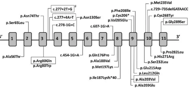
To date, a total of 29 mutations in the HSD17B3
gene have been identiied (Figure 1), including 21 missenses, one nonsense mutation, two frameshifs leading to downstream premature stop codon, four located at splice junctions leading to aberrant tran-scripts, and one duplication of exons 3-10 (4,15,16).
Here, we report the clinical and molecular pic-tures of four cases of 17-b-HSD3 deiciency.
MATERIALS AND METHODS
Genomic DNA was obtained from peripheral blood by proteinase K/phenol extraction method (17). The 11 exons and exon/intron junction sequences of
HSD17B3 gene were ampliied by polymerase chain
reaction (PCR). Primers used for PCR were chosen with Prime 3 primer designing tool; primer sequences are available upon request. Puriication of PCR prod-ucts was carried out using the Wizard® SV Gel and PCR clean-up system (Promega, Madison, WI, USA). Direct PCR fragment sequencing with sense and an-tisense primers was performed using Big Dye® Ter-minator Cycle Sequencing Kit V3.1 Ready Reaction (ABI PRISM/PE Biosystems, Foster City, CA, USA). Sequences were obtained in an automatic sequencer ABI 3130 DNA Analyzer (ABI PRISM/PE Biosys-tems), and were compared with the HSD17B3
nor-mal sequence (ENSEMBL – ENSG00000130948) using Chromas (reduced version-free software) and CLC Sequence Viewer v.6.2 (free software).
Figure 1. HSD17B3 gene is represented with its 11 exons. Mutations found in 17-b-HSD3 deiciency are indicated. Black boxes highlight recurrent mutations also identiied in this paper, and the # shows the mutation identiied here for the irst time.
Cop
yright
© ABE&M t
odos os dir
eit
os r
eser
vados
.
CASE REPORTS
Case 1
A 16-year-old girl from Pariconha (Alagoas – North-eastern region of Brazil) was referred to us due to genital ambiguity and virilization at puberty. She was born at term by cesarean section to a 48-year-old mother, and weighted over 4.5 kg. Her parents were consanguineous (second cousins), and family history showed a sister with primary amenorrhea and absence of uterus, who had both gonadectomy and genitoplasty done during childhood. On physical ex-amination, her weight and height were 68 kg and 168.4 cm, respectively; genitalia presented a 5.7-cm phallus and palpable gonads in labioscrotal folds with volumes of 15 cm3 and 10 cm3 for the right and the left gonad, respectively. A single perineal urethral opening with a short vagina were also observed. In addition, she presented facial hair and pubertal de-velopment Tanner 3 for breasts, and Tanner 5 for pubic hair (Figures 2A and 2B). The karyotype was 46,XY and laboratory data are shown in table 1.
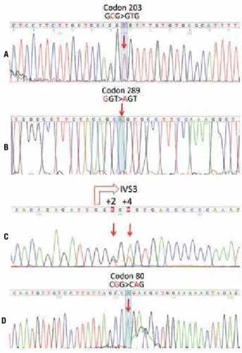
HSD17B3 gene sequencing revealed the
homozy-gous p.Ala203Val missense mutation within exon 9 (Figure 3A). After psychological evaluation of the patient and the family, they decided to maintain the female gender. Bilateral gonadectomy and feminiz-ing genitoplasty were performed, and hormonal replacement with estrogens was administrated. The analysis of both gonads indicated pubertal testes with discrete peritubular ibrosis. As she maintained the female gender, vaginal dilation will be performed as soon as she decides to initiate sexual activity.
Case 2
A 4‐year‐old girl, from Oliveira dos Brejinhos (Ba-hia – Northeastern region of Brazil), was referred to us to investigate genital ambiguity with palpable gonads. She was the second child of non-consangui-neous parents, and was born at term after an unevent-ful pregnancy. At physical examination, her weight was 16.2 kg and height 102 cm. A 3.7-cm phallus, single perineal opening, labioscrotal fusion, and bi-lateral palpable gonads within inguinal region were observed. Cytogenetic analysis indicated a 46,XY karyotype. Laboratory data are shown in table 1.
HSD17B3 gene sequencing showed homozygosis
for the p.Gly289Ser substitution in exon 11 (Figu-re 3B). Her family decided to maintain the female
gender. Therefore, gonadectomy, clitoroplasty, and introitoplasty were performed. Histological analysis of the gonads showed normal pre‐pubertal testes.
Case 3
A 13‐year‐old girl from São Luís (Maranhão –North-eastern region of Brazil) was referred to us due to signs of virilization such as acne, hirsutism, voice deepening, and phallus enlargement since the age of 11. She was born at term after an uneventful preg-nancy. Her parents were healthy, non-consanguin-eous, and from small cities Maranhão State (For-tuna and Buriti Bravo). She had two sisters, both with telarche, and without signs of virilization, and one brother without sex ambiguity. On physical ex-amination, the patient was 164 cm tall and weighed 59.8 kg. Her breasts were Tanner stage 1. She had increased hair on the abdomen and face, phallus en-largement (3.7 cm), and the skin of labioscrotal folds were pigmented and rugged. Pubic hair was Tanner stage 4. Her vagina ended in a 3-cm pouch, and no gonads were palpable (Figures 2C and 2D). Karyo-type was 46,XY. Results of hormonal evaluation are shown in table 1. MRI of the abdomen and pelvis did not show an uterus or a prostate. However, sem-inal vesicles were identiied, and both gonads were found at the inguinal region. HSD17B3 gene
se-quencing showed heterozygosis for two nucleotide changes within intron 3 splice donnor consensus sequence, c.277+4A>T, and the novel c.277+2T>G (Figure 3C). The analysis of the family indicated that the irst variant was inherited from her father, and the second from her mother (data not shown). After female gender identity was conirmed by long-term psychological evaluation, video laparoscopy was per-formed to remove the gonads, and estrogen replace-ment therapy was initiated, followed by vaginal dila-tion. Histologic examination of both gonads showed bilateral testes with slight peritubular ibrosis, germ-cell aplasia, and Leydig germ-cell hyperplasia, without evi-dence of malignancy.
Case 4
Cop
yright
© ABE&M t
odos os dir
eit
os r
eser
vados
.
showed a 2-year-old female maternal cou sin who presented clitoromegaly, and a 12-year-old female paternal cousin with hirsutism. Upon physical ex-amination, the patient was 160 cm tall and weighed 41.4 kg. She had a 4.5-cm phallus, a single perine-al opening, partiperine-al fusion of labioscrotperine-al folds with pigmentation, and bilateral palpable gonads in the lower third of inguinal canal. Facial, axillary, ab-dominal, and pubic hair (Tanner 3) were observed, as well as Tanner 3 breast development. Karyotype was 46,XY. Hormonal evaluation data are shown in table 1. Pelvic ultrasonography did not show a ute-rus or a prostate. Genitography showed a urogenital sinus with a short vagina. HSD17B3 gene
sequenc-ing showed homozygosis for p.Arg80Gln mutation in exon 3 (Figure 3D). The patient decided to main-tain the female gender; therefore, gonadectomy and feminizing genitoplasty were performed. Histologi-cal gonadal analysis indicated normal pubertal testes. Estrogen replacement therapy was initiated, and the patient decided to postpone vaginal dilation.
Figure 2. External genitalia of patients with HSD17B3 deiciency. Case 1
– igures A and B, and case 3 – igures C and D.
A
A
C
C
B
B
D
D
Figure 3. Parts of the eletropherograms obtained for HSD17B3 gene
sequencing: A) Homozygous GCG>GTG nucleotide change within exon 9
identiied in case 1; this substitution causes the replacement in residue
203, usually alanine, by valine (p.Ala203Val). B) Homozygous GGT>AGT
nucleotide change within exon 11 identiied in case 2; this substitution causes the replacement in residue 289, usually glycine by serine
p.Gly289Ser. C) Compound heterozygosis for c.277+2T>G and
c.277+4A>T nucleotide changes in case 3, which were inherited from the mother and the father, respectively (data not shown); the two mutations are within the donor splice site consensus sequence of intron 3; the empty
red arrow indicates the beginning of IVS3 at exon 3/intron 3 boundary. D)
Homozygous CGG>CAG nucleotide change within exon 3 identiied in case 4; this substitution causes the replacement in residue 80, usually arginine by glutamine (p.Arg80Gln).
DISCUSSION
Cop
yright
© ABE&M t
odos os dir
eit
os r
eser
vados
.
for HSD17B3 mutations. On the other hand, case 2,
who has non-consanguineous parents, also showed a homozygous mutation in the HSD17B3 gene. In
this case, the patient and her parents were born at a small city of the countryside of the State of Bahia.
Different HSD17B3 gene mutations confer a wide
range of phenotypic characteristics to 46,XY affected individuals. These may vary from predominantly fe-male genitalia, as in case 3; to mild virilized fefe-male genitalia, as in case 2; to evident genital ambiguity with palpable gonads, as in cases 1 and 4; to pre-dominantly male genitalia with microphallus and hy-pospadias (4-8). This variability in phenotypes may correlate with partial activity of mutated 17-b-HSD3 in the testes, or to an extragonadal conversion of Δ4 to T by other 17-b-HSD isoenzymes (11).
As the most common clinical presentations of 17-b-HSD3 deiciency are female or mild virilized female genitalia, most patients do not have the diag-nosis of 46,XY DSD at birth, and are registered and raised as females (4,5,8). Therefore, diagnosis is only established at puberty for most patients (4-8). Con-versely, some cases may be diagnosed early, because they seek medical care during childhood due to some degree of virilization with palpable gonads (4,5,7).
If 17-b-HSD3 deiciency is not diagnosed in childhood, and gonadectomy is not performed, pa-tients may present virilization at puberty (4,5,7).
Main signs of virilization are increased hair growth all over the body and face, deepening of the voice, and android fat distribution, in addition to phallus elongation reaching 5-8 cm in length, which may also be observed in response to peripheral T con-version. However, the phallus will always be shorter than a normal-sized penis (4,5,7).
The diagnosis of 17-b-HSD3 deiciency may be suspected upon laboratory investigation, and may be conirmed with molecular analysis. As veriied in all cases reported here, patients have, respectively, high and low to normal Δ4 and T serum concen-trations (4,5,7). When T/Δ4 ratio is less than 0.8, 17-b-HSD3 deiciency is suggested (7,14). The lit-erature also refers to increased serum concentrations of DHEA and DHEA sulfate (4,5,7). However, these elevated values are not always observed.
The mutation p.Arg80Gln identiied in case 4 is the most common one in Mediterranean (21) and Bra-zilian patients (6). McKeever and cols. (22)
demon-strated that p.Arg80Gln causes a signiicant decrease in the rate of enzymatic reaction leading to approxi-mately 5% of residual enzyme activity, and a 1/60 re-duction in the binding afinity of the mutant protein to NADPH cofactor. Therefore, it is suggested that the R in residue 80 of the protein structure should be critical in maintaining appropriate levels of enzymatic activity, and to promote normal human male
devel-Table 1. Hormonal proile of four cases with HSD17B3 deiciency
Case 1 Case 2 Case 3 Case 4
Male reference range (12 – 19 yr)
Age (years) 16 4 13 14
Puberty + - + +
LH (IU/L) 12.1 0.4 16.0 10.5 0.5 - 5.3
#0.1 - 1.2
FSH (IU/L) 7.4 2.0 6.7 8.7 1.4 - 18.1
#0.8 - 2.3
T (ng/mL) 4.2 0.8* 3.4 3.1 0.1 - 5.0
Δ4 (ng/mL) 6.8 1.3* 6.5 4.8 0.7 - 1.9
DHT (ng/mL) 0.49 0.21* 0.42 0.58 0.25 - 1.20
E1 (pg/mL) 32.1 NP 41.0 42.7 17.0 - 44.0
E2 (pg/mL) 20.9 NP 25.8 22.2 < 50.0
DHEA sulfate (μg/mL) 3.8 1.9 2.5 3.9 1.0 - 4.2
T/DHT ratio 8.6 3.8* 8.1 5.3 < 10.0
T/Δ4 ratio 0.62 0.61 0.50 0.65 > 0.8
E2/E1 ratio 0.65 NP 0.63 0.52
-* After 3 consecutive days of 1,500 IU of intramuscular of human chorionic gonadotropin;
# pre-pubertal male reference range; Δ4: androstenedione; E1: estrone; E2: estradiol;
Cop
yright
© ABE&M t
odos os dir
eit
os r
eser
vados
.
opment. This mutation was irst described in a Pales-tinian family from the Gaza Strip (20). Further, it was also identiied in Brazilian families with no Palestin-ian ancestry, and of probable Portuguese origin (6). However, its recurrence, not only in patients of Arab origin, but also in Dutch patients, was reported for individuals who were homo- or heterozygous for this mutation (5). Therefore, p.Arg80Gln may represent a founder effect, common among Arabs from differ-ent regions of Israel, Lebanon, and Syria (23). This fact led to the hypothesis that it has been introduced in Portugal and Spain by Phoenicians who migrated from Syria, Lebanon, and Israel around 750 B.C. (24,25). Its introduction in the Netherlands could have occurred during the Spanish domination in the sixteenth and seventeenth centuries and, in Brazil, by Portuguese colonizers and during the Dutch invasion of the Northeastern region of the country.
The p.Ala203Val mutation identiied in case 1 was irst described by Geissler and cols. (26), in a patient from São Paulo – Brazil. This mutation was assayed for the ability to convert Δ4 into T, and it complete-ly inactivated the enzyme 17-b-HSD3, indicating a good correlation with the phenotype of case 1.
Moghrabi and cols. (27) reported the missense substitution p.Gly289Ser (SNP – CM023631). This mutation has been considered a polymorphism since it is frequent in all populations reported in the screening of 1,000 genomes (28). It also did not al-ter the in vitro enzymatic activity. However, it was
identiied in a compound heterozygous patient who also carried the well-known deleterious p.Asn130Ser mutation. It was supposed that this patient carried alterations in regulatory regions of the HSD17B3
gene that may explain the phenotype (5). In addi-tion, p.Gly289Ser amino acid substitution has been associated with increased risk of developing prostate cancer, and to hypospadias in individuals carrying the S289 allele in homozygosis, once mRNA expres-sion levels were signiicantly lower for the mutant S289 than for the wild-type G289, indicating that it may not be as neutral as it had been initially con-sidered (29,30). Since we did not evaluate either 5’ or 3’ regulatory regions, we can speculate that the homozygous p.Gly289Ser in case 2 may be associ-ated with the phenotype, and some other nucleotide change in regulatory regions could act synergistically with the mutation.
Case 3 was found to be compound heterozy-gous for two nucleotide changes affecting intron 3
splice donor region. The c.277+4A>T mutation was reported in 1996 by Andersson and cols. (9), who identiied it in homozygosis in three families, and in heterozygosis in two other families. Those results indicated it as one of the most prevalent mutations found in subjects with 17-b-HSD3 deiciency. It is located within the intron 3 canonical splice donor site, and was shown to disrupt normal splicing by Boehmer and cols. (5), who analyzed the HSD17B3
cDNA prepared from testis mRNA of a homozygous patient. The result of cDNA sequencing showed a transcript where exon 3 had been skipped and, in minor amounts, a transcript with deletion of both exons 3 and 4 (5,31).
The mutation c.277+4A>T is found in popula-tions worldwide, including Dutch, Germans, white Australians and white Americans, who share the same marker genotype, and are likely to be identical by de-scent (5). The c.277+2T>G mutation was also identi-ied in case 3. It changes the almost invariant GT at the splice donor site to GG in intron 3. This mutation was identiied here for the irst time, and it probably suppresses the normal splicing so that it can either ac-tivate a cryptic splice site, or lead to exon 4 skipping.
The in silico search for splicing sites using the online
Splice Site Prediction NNSPLICE 0.9 version indi-cated that the mutation eliminated the normal donor splice at that position (data not shown). As testicu-lar samples are not accessible for in vitro studies, the
construction of mini-genes will be an alternative to test an aberrant splicing process in this case.
Cop
yright
© ABE&M t
odos os dir
eit
os r
eser
vados
.
In conclusion, we report mutations in HSD17B3
gene, including the novel c.277+2T>G mutation, in four cases of 17-b-HSD3 deiciency with different clinical laboratorial and presentations.
Disclosure: no potential conlict of interest relevant to this article was reported.
REFERENCES
1. Andersson S, Moghrabi N. Physiology and molecular genetics of 17beta-hydroxysteroid dehydrogenases. Steroids. 1997;62:143-47. 2. Lukacik P, Kavanagh KL, Oppermann U. Structure and function of human 17beta-hydroxysteroid dehydrogenases. Mol Cell Endo-crinol. 2006;248:61-71.
3. Labrie F, Luu-The V, Lin SX, Labrie C, Simard J, Breton R, et al. The key role of 17 beta-hydroxysteroid dehydrogenases in sex steroid biology. Steroids 1997;62:148-58.
4. George MM, New M I, Tem S, Sultan C, Bhangoo A. The clinical and molecular heterogeneity of 17bHSD3 enzyme deiciency. Horm Res Paediatr. 2010;74:229-40.
5. Boehmer AL, Brinkmann AO, Sandkuijl LA, Halley DJ, Niermeijer MF, Andersson S, et al. 17beta-hydroxysteroid dehydrogenase-3 deiciency: diagnosis, phenotypic variability, population genetics, and worldwide distribution of ancient and de novo mutations. J Clin Endocrinol Metab. 1999;84:4713-21.
6. Mendonça BB, Inacio M, Arnhold IJ, Costa EM, Bloise W, Martin RM, et al. Male pseudohermaphroditism due to 17beta-hydroxys-teroid dehydrogenase 3 deiciency. Diagnosis, psychological eval-uation, and management. Medicine (Baltimore). 2000;79:299-309. 7. Lee YS, Kirk JM, Stanhope RG, Johnston DI, Harland S, Auchus
RJ, et al. Phenotypic variability in 17beta-hydroxysteroid dehy-drogenase-3 deiciency and diagnostic pitfalls. Clin Endocrinol (Oxf). 2007;67:20-8.
8. Faienza MF, Giordani L, Delvecchio M, Cavallo L. Clinical, endo-crine, and molecular indings in 17beta-hydroxysteroid dehydro-genase type 3 deiciency. J Endocrinol Invest. 2008;31:85-91. 9. Andersson S, Geissler WM, Wu L, Davis DL, Grumbach MM, New
MI, et al. Molecular genetics and pathophysiology of 17 beta-hydroxysteroid dehydrogenase 3 deiciency. J Clin Endocrinol Metab. 1996;81:130-6.
10. Mendonca BB, Arnhold IJ, Bloise W, Andersson S, Russell DW, Wilson JD. 17Beta-hydroxysteroid dehydrogenase 3 deiciency in women. J Clin Endocrinol Metab. 1999;84:802-4.
11. Prehn C, Möller G, Adamski J. Recent advances in 17beta-hydroxysteroid dehydrogenases. J Steroid Biochem Mol Biol. 2009;114:72-7.
12. Hiort O, Reinecke S, Thyen U, Jurgensen M, Holterhus PM, Schon D, et al. Puberty in disorders of somatosexual differentiation. J Pediatr Endocrinol Metab. 2003;16(suppl 2):297-306.
13. Cohen-Kettenis PT. Gender change in 46,XY persons with 5alpha-reductase-2 deiciency and 17beta-hydroxysteroid dehydroge-nase-3 deiciency. Arch Sex Behav. 2005;34:399-410.
14. Faisal Ahmed S, Iqbal A, Hughes IA. The testosterone: andro-stenedione ratio in male undermasculinization. Clin Endocrinol (Oxf). 2000;53:697-702.
15. Ben Rhouma B, Belguith N, Mnif MF, Kamoun T, Chari N, Kamoun M, et al. A novel nonsense mutation in HSD17B3 gene in a Tunisian pa-tient with sexual ambiguity. J Sex Med. 2012 [Epub ahead of print].
16. Neocleous V, Sismani C, Shammas C, Efstathiou E, Alexandrou A, Ioannides M, et al. Duplication of exons 3-10 of the HSD17B3 gene: a novel type of genetic defect underlying 17b-HSD-3 dei-ciency. Gene. 2012;499:250-5.
17. Sambrook J, Fritsch EF, Maniatis TE. Molecular cloning, a labora-tory manual New York: Cold Spring Harbor; 1989.
18. Saez JM, De Peretti E, Morera AM, David M, Bertrand J. Familial male pseudohermaphroditism with gynecomastia due to a tes-ticular 17-ketosteroid reductase defect. I. Studies in vivo. J Clin Endocrinol Metab. 1971;32:604-10.
19. Saez JM, Morera AM, De Peretti E, Bertrand J. Further in vivo studies in male pseudohermaphroditism with gynecomastia due to a testicular 17-ketosteroid reductase defect (compared to a case of testicular feminization). J Clin Endocrinol Metab. 1972;34:598-600.
20. Rösler A, Silverstein S, Abeliovich D. A (R80Q) mutation in 17 beta-hydroxysteroid dehydrogenase type 3 gene among Arabs of Israel is associated with pseudohermaphroditism in males and normal asymptomatic females. J Clin Endocrinol Metab. 1996;81:1827-31.
21. Rösler A. 17 beta-hydroxysteroid dehydrogenase 3 deicien-cy in the Mediterranean population. Pediatr Endocrinol Rev. 2006;3(suppl 3):455-61.
22. Mckeever BM, Hawkins BK, Geissler WM, Wu L, Sheridan RP, Mosley RT, et al. Amino acid substitution of arginine 80 in 17b-hidroxysteroide dehydrogenase 3 and its effect on NADPH cofator binding and oxidation/reduction kinetics. Biochim Bio-phys Acta. 2002;1601:29-37.
23. Rosler A, Belanger A, Labrie F. Mechanisms of androgen produc-tion in male pseudohermaphroditism due to 17b-hydroxysteroid dehydrogenase deiciency. J Clin Endocrinol Metab. 1992;75:773-8. 24. Culigan W. Phoenicia and Phoenician colonization. In: Boardman
J, Edwards IE, Hammond NG, Sollberger E, Walker CB, eds. The Cambridge ancienty history, 2nd Ed. Cambridge University Press; 1991. p. 461-546.
25. Cavalli-Sforza LL, Menozzi P, Piazza A. The history and geography of human genes. Princeton: Princeton University Press; 1994. p. 217, 242-245, 260.
26. Geissler WM, Davis DL, Wu L, Bradshaw KD, Patel S, Mendonça BB, et al. Male pseudohermaphroditism caused by mutations of testicular 17b-hidroxysteroide dehydrogenase 3. Nat Genet. 1994;7:34-9.
27. Moghrabi N, Hughes IA, Dunaif A, Andersson S. Deleterious mis-sense mutations and silent polymorphism in the human 17b-hy-droxysteroid dehydrogenase 3 gene (hsd17b3). J Clin Endocrinol Metabol. 1998;83(8):2855-60.
28. http://www.ensembl.org/Homo_sapiens/Variation/Population?db=cor e;g=ENSG00000130948;r=9:98997588-99064434;t=ENST00000375263 ;v=rs2066479;vdb=variation;vf=16374979. Accessed on: Sept 30, 2012. 29. Margiotti K, Kim E, Pearce CL, Spera E, Novelli G, Reichardt JK. Association of the G289S single nucleotide polymorphism in the HSD17B3 gene with prostate cancer in Italian men. Prostate. 2002;53:65-8.
30. Sata F, Kurahashi N, Ban S, Moriya K, Tanaka KD, Ishizuka M, et al. Genetic polymorphisms of 17 b-hydroxysteroid dehydrogenase 3 and the risk of hypospadias. J Sex Med. 2010;7(8):2729-38. 31. Mains LM, Vakili MB, Lacassie Y, Andersson S, Lindqvistc A, Rock
JA. 17beta hydroxysteroid dehydrogenase 3 deiciency in a male Pseudohermaphrodite. Fertil Steril. 2008;89(1):228.e13-228.e17. 32. Lee PA, Houk CP, Faisal A, Hughes IA, International Consensus