Fracture of the acromion associated with arterial lesion: case
report
Fratura do acrômio associada a lesão arterial: relato de caso
Marcos Rassi Fernandes, Rui José Fernandes*
Introduction
he acromion (AC) is a strong bone that functions as a shield, protecting the structures of the subacromial region. It absorbs and dissipates the forces applied to the shoulder region;1 however, high energy force may produce fractures,
as well as other severe lesions. herefore, diagnosis may not be immediately performed due to the necessity of assessing associated lesions.2
Acromion fracture is rare and accounts for only 9% of scapular fractures, which correspond to 1% of all the frac-tures.3 It may result from a direct force applied to the upper
region of the AC, or a deviation or superior dislocation of the humeral head, which may also bring about a severe dis-ruption of the rotator cuf.4
Fractures frequently present a minimal deviation and do not require surgical treatment. It is important to distinguish them from the os acromiale,1-3 which is the failure of the AC’s
ossiication centers’ fusion. In the case of a deviated fracture, association with brachial plexus lesion should be considered.5
Most AC fractures are concomitant with fractures of the caracoid,6 clavicule,7 scapular neck or body,8
acro-mioclavicular dislocation9 or glenohumeral dislocation,3
but the neurovascular exam in high-magnitude trauma is always important.
his case report discusses a fracture of the AC’s basis associated with ipsilateral ulnar artery lesion, its treatment and evolution, drawing attention to possible associations between these two lesions. he study was approved by the Research Ethics Committee of Hospital Geral de Goiânia.
Case report
A 21-year-old male patient presented with pain in the let shoulder, superior and posterior region, ater a skate ac-cident. He was then admitted in another service, where he
Abstract
Fracture of the acromion and arterial injury are a rare association. he clinical picture is characterized by shoulder pain, functional disability and swelling of the afected limb with decreased distal pulse and temperature. Radiography of the shoulder and arteriography deine the diagnosis and assist in postoperative follow-up. he authors report a rare case of fracture of the acromion associated with injury of the ipsilateral ulnar artery and describe its treatment, as well as pre- and postoperative evaluations.
Keywords: Acromial lesions, bone fracture surgery, acromion radiography, acromion surgery, internal ixation of adult fractures.
Resumo
A fratura do acrômio com lesão arterial é uma associação rara. O quadro clínico caracteriza-se por dor no ombro, incapacidade funcional e edema do membro acometido, com pulso e temperatura distais diminuídos. A radiograia do ombro e a arteriograia deinem o diagnóstico e auxiliam na evolução pós-operatória. Os autores relatam um caso raro de fratura da base do acrômio associada a lesão da artéria ulnar ipsilateral, seu tratamento, bem como a avaliação pré e pós-operatória.
Palavras-chave: Lesões do acrômio, cirurgia de fraturas ósseas, acrômio, radiograia do acrômio, cirurgia do acrômio, ixação interna de fraturas de adultos.
* Médicos assistentes, Grupo de Ombro e Cotovelo, Hospital Ortopédico de Goiânia, Goiânia, GO, Brazil. No conlicts of interest declared concerning the publication of this article
was anesthetized to ‘put the shoulder right.’ He came to the session with simple shoulder radiographs. he mother also reported a decrease in temperature in the patient’s hand. At physical exam, he presented pain at palpation of the AC’s posterior region, a signiicant swelling in all the let upper limb (LUL) and hematoma in the medial surface of the arm. he LUL had no functional ability, associated with iliform radial pulse and hand temperature decreased. No neuro-logical deicit was observed. Simple shoulder radiographs showed fracture of the AC’s basis with inferior and medial deviation (Figure 1). he patient was referred to the angio-logy division for an emergency evaluation. A color Doppler exam, with linear transducer of LUL’s deep vases, was per-formed. No signs suggesting acute deep venous thrombosis were discovered. Radial artery was patent, with triphasic low, while distal ulnar artery had inverted and reduced low, with a short occluded segment in proximal forearm. Aterwards, a digital angiography was performed on the
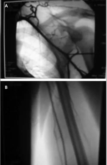
LUL, assessing the following aspects: patent subclavian ar-tery, patent brachial artery (Figure 2), patent radial arar-tery, ulnar artery occluded soon ater its origin with retrograde reilling (Figure 3), patent supericial palmar arch, and par-tially opaciied deep palmar arch.
Vascular team chose to treat the arterial occlusion with acetylsalicylic acid (100 mg) and observation. hree days ater irst consultation, an ultrasonography was performed on the let shoulder, and the diagnosis evidenced the pre-sence of difuse subcutaneous swelling of shoulder and pro-ximal arm; however, no lesion was observed in the rotator cuf. he patient was then discharged for surgical treatment of the AC fracture 10 days ater trauma.
With an incision of approximately 8 cm in the superior surface of the shoulder, following the Langer skin lines and fracture ater reduction, ixation was performed with two parallel Steinman wires, 2.5 mm, with threaded tips, poste-rior to anteposte-rior (Figure 4). he patient remained motionless
Figure 1– A) Acromion fracture. Scapular proile evidencing a fracture with inferior deviation; B) axillary proile with medial deviation
B A
Figure 2– Patent arteries A) subclavian and B) brachial A
B A
during 21 days. Ater that, passive and pendular exercises were resumed. he bolts were removed 6 weeks from pos-toperative, with the fracture consolidated (Figure 5). At3 month’s follow-up, the patient did not present shoulder pain, his movements had been recovered and the LUL had no edema, with normal pulse and temperature.
Discussion
he scapula plays an important role in the functioning of the arm. It is congruent with the rib cage and stabilizes the superior extremity against the thorax, in addition to joi-ning the appendicular to the axial skeleton through the gle-noid, clavicule and sternoclavicular and acromioclavicular articulations.1,2
he low incidence of scapular fractures is due to its gre-at mobility and position between muscle layers. A signii-cant trauma is necessary to fracture this bone.2,3 Associated
lesions are common, therefore fractures are usually diagno-sed late. Fischer et al.5 observed that 57% of patients with
scapular fracture presented lesion of the brachial plexus and arterial lesion of the ipsilateral extremity. Armstrong and Vanderspuy10 reported that associated lesions are
respon-sible for 10% of deaths in patients with scapular fracture. Studies on acromion fractures are rare, and most of them comprise case reports. Mcgahan & Rab11 reported a
case of acromion fracture associated with acromioclavicu-lar dislocation and axilacromioclavicu-lary nerve lesion. Weber et al.12 also
reported a deviated and isolated acromion fracture, which reduced the subacromial space. Surgical treatment was ne-cessary, evidencing that all fractures are combined, as de-monstrated by Goodrich et al.3 and Goss.6
Mcgahan & Rab13 evidenced that traumas of the
acromioclavicular articulation or acromion present a high incidence of peripheral nerves lesions, demanding
Figure 4– Fixed fracture with two Steinmann 2.5 mm wires
Figure 5– Consolidated acromion fracture
a great deal of attention at the performance of neurolo-gical exams in these patients. Baldwin et al.,14 in a recent
study, concluded that lesions of the upper limb, thorax and pelvic ring are frequently associated in patients with scapular fracture, due to the frequency of high-energy traumas in these cases.
Radiographic evaluation should always be the irst to be performed, and the axillary side is the best inciden-ce for the diagnosis. Computed tomography sometimes is necessary for assessing the fracture deviation and studying other associated bone lesions.2,4 Magnetic resonance and
ul-trasonography are helpful in assessing the sot parts of the shoulder region.2 In this case report, a simple radiograph
was suicient for fracture diagnosis. Ultrasonography did not evidence lesion of the rotator cuf.
Kuhn et al.8 have proposed a classiication of acromion
fractures to indicate those in which surgical intervention would be necessary. Minimally deviated type I fractures and deviated type II fractures with no reduction of subacromial space would require non-surgical treatment. In deviated type III lesions with space reduction surgery is indicated to prevent secondary impact.
Literature on the association of acromion fracture with arterial lesion of any topography is very scarce. Few studies show this concomitance, although some represent correla-tions with all the types of scapular fractures, such as Fischer et al.,5 who evidence an 11% incidence of arterial lesion,
and Stein et al.,15 who reported the case of a closed fracture
of the scapula neck with axillary artery lesion.
his case report showing a closed fracture of the acro-mion associated with a lesion of the ulnar artery is, as far as our knowledge is concerned, the irst to be described in literature showing exams, treatment and evolution. he most likely mechanism was an upper limb indirect trau-ma, with the elbow slightly lexed and the ist dorsilexed due to the presence of an arterial lesion of the distal fore-arm to the shoulder fracture (AC), despite the absence of rotator cuf lesion. herefore, arterial lesion did not occur due to a fracture deviation, which would happen if the le-sion was more proximal. High-energy trauma should be the irst parameter for considering the possibility of an as-sociation between these lesions. Surgical treatment, since it is a deviated fracture with subacromial space reduction, was correctly indicated, with good evolution, no pain and no movement restriction. hus, a possible pseudarthrosis or vicious consolidation was avoided. he option of per-forming an artery occlusion with clinical treatment was due to the good distal perfusion through radial artery,
with a good illing of the deep palmar arch and retrograde reilling of ulnar artery.
he presence of an acromion fracture should always alert the physician assisting the patient, because severe le-sions may be associated. A complete neurovascular exam is important. herefore, surgical treatment of the deviated acromion fracture seems to be the best option when suba-cromial space is reduced. Associated arterial lesions should always be remembered.
References
1. De Palma AF. Fractura del acromion. In: De Palma AF, editor. Cirurgia del ombro. Philadelphia: Lippincott; 1985. pp. 484-5.
2. Butters KP. Fractures and dislocations of the scapula. In: Rockwood CA, Green DP, Bucholz RW, editors. Fractures in adults. Philadelphia: Lippincott; 1984. pp. 990-1019.
3. Goodrich JA, Crosland E, Pye J. Acromion fracture associated with posterior shoulder dislocation. J Orthop Trauma. 1998;12:521-3.
4. Getz C, Deutsch A, Williams Junior GR. Scapular and glenoid frac-tures. In: Warner JJP, Iannotti JP, Flatow EL, editors. Complex and re-vision problems in shoulder surgery. Philadelphia: Lippincott; 2005. pp. 378-80.
5. Fischer RP, Flynn TC, Miller PW, hompson DA. Scapular frac-tures and associated major ipsilateral upper torso injuries. Curr Concepts Trauma Care. 1985;1:14-6.
6 Goss TP. he scapula: coracoid, acromial and avulsion fractures. Am J Orthop (Belle Mead NJ). 1996;25:106-15.
7. Ogawa K, Naniwa T. Fractures of the acromion and the lateral sca-pular spine. J Shoulder Elbow Surg. 1997;6:544-8.
8. Kuhn JE, Blasier RB, Carpenter JE. Fractures of the acromion process: a proposed classiication system. J Orthop Trauma. 1994;8:6-13.
9. Kurdy NM, Shah SV. Fracture of the acromion associated with acromioclavicular dislocation. Injury. 1995;26:636-7.
10. Armstrong CP, Van der Spuy J. he fractured scapula: importance in management based on a series of 62 patients. Injury. 1984;15: 324-9.
11. Mcgahan JP, Rab GT. Fracture of the acromion associated with an axillary nerve deicit: a case report and review of the literature. Clin Orthop Relat Res. 1980;147:216-8.
12. Weber D, Sadri H, Hofmeyer P. Isolated fracture of the posterior angle of the acromion: a case report. J Shoulder Elbow Surgery. 2000;9:534-5.
13. Mcgahan JP, Rab GT. Fractures of the scapula. J Trauma. 1980;20:880-3.
14. Baldwin KD, Ohman-Strickland P, Mehta S, Hume E. Scapula frac-tures: a marker for concomitant injury? A retrospective review of data in the National Trauma Database. J Trauma. 2008;65:430-5.
Correspondence: Marcos Rassi Fernandes Av. Azaléias, Qd. 10 Lt. 20, Residencial Jardins Viena CEP 74935-187 – Aparecida de Goiânia, GO Tel.: +55 (62) 3523.1247, +55 (61) 9602.7575 E-mail: [email protected]
Author contributions: RConception and design: MRF