Brazilian guidelines for community-acquired
pneumonia in immunocompetent adults - 2009*
Diretrizes brasileiras para pneumonia adquirida na comunidade em adultos imunocompetentes - 2009
Ricardo de Amorim Corrêa, Fernando Luiz Cavalcanti Lundgren, Jorge Luiz Pereira-Silva, Rodney Luiz Frare e Silva, Alexandre Pinto Cardoso,
Antônio Carlos Moreira Lemos, Flávia Rossi, Gustavo Michel, Liany Ribeiro, Manuela Araújo de Nóbrega Cavalcanti, Mara Rúbia Fernandes de Figueiredo, Marcelo Alcântara Holanda, Maria Inês Bueno de André Valery, Miguel Abidon Aidê,
Moema Nudilemon Chatkin, Octávio Messeder, Paulo José Zimermann Teixeira, Ricardo Luiz de Melo Martins e Rosali Teixeira da Rocha,
em nome da Comissão de Infecções Respiratórias e Micoses – Sociedade Brasileira de Pneumologia e Tisiologia
Abstract
Community-acquired pneumonia continues to be the acute infectious disease that has the greatest medical and social impact regarding morbidity and treatment costs. Children and the elderly are more susceptible to severe complications, thereby justifying the fact that the prevention measures adopted have focused on these age brackets. Despite the advances in the knowledge of etiology and physiopathology, as well as the improvement in preliminary clinical and therapeutic methods, various questions merit further investigation. This is due to the clinical, social, demographical and structural diversity, which cannot be fully predicted. Consequently, guidelines are published in order to compile the most recent knowledge in a systematic way and to promote the rational use of that knowledge in medical practice. Therefore, guidelines are not a rigid set of rules that must be followed, but first and foremost a tool to be used in a critical way, bearing in mind the variability of biological and human responses within their individual and social contexts. This document represents the conclusion of a detailed discussion among the members of the Scientific Board and Respiratory Infection Committee of the Brazilian Thoracic Association. The objective of the work group was to present relevant topics in order to update the previous guidelines. We attempted to avoid the repetition of consensual concepts. The principal objective of creating this document was to present a compilation of the recent advances published in the literature and, consequently, to contribute to improving the quality of the medical care provided to immunocompetent adult patients with community-acquired pneumonia.
Keywords: Pneumonia; Diagnosis; Epidemiology; Practice guideline; Primary prevention.
Resumo
A pneumonia adquirida na comunidade mantém-se como a doença infecciosa aguda de maior impacto social quanto à morbidade e a custos relacionados ao tratamento. Os grupos etários mais suscetíveis de complicações graves situam-se entre os extremos de idade, fato que tem justificado a adoção de medidas de prevenção dirigidas a esses estratos populacionais. Apesar do avanço no conhecimento no campo da etiologia e da fisiopatologia, assim como no aperfeiçoamento dos métodos propedêuticos e terapêuticos, inúmeros pontos merecem ainda investi-gação adicional. Isto se deve à diversidade clínica, social, demográfica e estrutural, que são tópicos que não podem ser previstos em sua totalidade. Dessa forma, a publicação de diretrizes visa agrupar de maneira sistematizada o conhecimento atualizado e propor sua aplicação racional na prática médica. Não se trata, portanto, de uma regra rígida a ser seguida, mas, antes, de uma ferramenta para ser utilizada de forma crítica, tendo em vista a variabili-dade da resposta biológica e do ser humano, no seu contexto individual e social. Esta diretriz constitui o resultado de uma discussão ampla entre os membros do Conselho Científico e da Comissão de Infecções Respiratórias da Sociedade Brasileira de Pneumologia e Tisiologia. O grupo de trabalho propôs-se a apresentar tópicos considerados relevantes, visando a uma atualização da diretriz anterior. Evitou-se, tanto quanto possível, uma repetição dos conceitos considerados consensuais. O objetivo principal do documento é a apresentação organizada dos avanços proporcionados pela literatura recente e, desta forma, contribuir para a melhora da assistência ao paciente adulto imunocompetente portador de pneumonia adquirida na comunidade.
Descritores: Pneumonia; Diagnóstico; Epidemiologia; Guia de prática clínica; Prevenção primária.
* Study carried out by the Respiratory Infections and Mycosis Committee of the Brazilian Thoracic Association, Brasília, Brazil. Correspondence to: Comissão de Infecções Respiratórias e Micoses - Sociedade Brasileira de Pneumologia e Tisiologia, SEPS 714 / 914 - Bloco E - Sala 220/223, Asa Sul, CEP 70390-145, Brasília, DF, Brasil.
Tel 55 61 3245-1030; 0800 61 6218. E-mail: [email protected] Financial support: None.
the 30 days preceding the disease; and those undergoing treatment in dialysis clinics) that are more appropriately classified as having hospital-acquired pneumonia.(2,3)
The diagnosis is based on the following: symptoms of acute lower respiratory tract infec-tion (cough and one or more of the following symptoms: expectoration, shortness of breath and chest pain); focal findings on physical examination of the chest; and systemic mani-festations (confusion, headache, sweating, chills, myalgia and temperatures higher than 37.8°C). These indications can be corroborated by a chest X-ray finding of new pulmonary opacity. Other clinical conditions can manifest in a similar manner, which can pose difficulties for primary and emergency care physicians in making a diagnosis of CAP. The clinical findings are only moderately accurate and do not allow a diag-nosis of CAP to be confirmed or excluded with any degree of certainty. In addition, the hetero-geneity of the physical examination conducted by primary and emergency care physicians, as well as the relative lack of experience of such health professionals in comparison with those who specialize in detecting radiological altera-tions, contributes to the difficulty in diagnosing CAP.(4,5)
Incidence and mortality
Most of the studies of CAP conducted in Brazil have focused on the etiology and treat-ment of the disease, and official statistics are a valuable source of information regarding the occurrence of CAP.
According to the Hospital Information Service of the Unified Health Care System, pneu-monia was the leading non-obstetric cause of disease-related hospitalization in Brazil in 2007, accounting for 733,209 hospitalizations. Among such hospitalizations, there was a predominance of males, as well as greater occurrence between the months of March and July.(6)
The rate of hospitalization for pneumonia has decreased over the last decade,(7) whereas the
rate of in-hospital pneumonia-related mortality has increased, which might be due to various factors, such as hospitalization of patients with pneumonia that was more severe and the aging of the population. The highest rates of hospi-talization for pneumonia are observed among
Methodology of the guidelines
This is a review and update of the previous guidelines published in 2004 by the Brazilian Thoracic Association. The present document presents certain topics that had not been discussed previously, as well as recently published data, and focuses exclusively on community- acquired pneumonia (CAP) in immunocompetent patients.
At the end of each section of this update are the principal recommendations and respec-tive levels of evidence, according to the current guidelines of the Brazilian Medical Association.
The participants of this edition of 2008 were divided into four work groups. In each group, an editor was responsible for distributing the topics among the members of the group.
• Group I: Definition, incidence, mortality,
etiology, diagnostic criteria and radiolo-gical diagnosis
• Group II: Diagnostic and complementary
tests, etiologic investigation, severity and locale of treatment
• Group III: Treatment, treatment failure
and prevention
• Group IV: Severe CAP: adjuvant treatment
Levels of evidence
The present guidelines were based on up-to-date evidence in the literature, classi-fied according to the recommendations of the Brazilian Medical Association (Chart 1). The final work of each group was extensively discussed among the editors and participants of the work groups.
Definition and clinical manifestations
Pneumonia is an acute infectious disease that is inflammatory, affects the air spaces and is caused by viruses, bacteria or fungi. The term CAP refers to pneumonia acquired outside the hospital environment or special health care units, or to pneumonia that manifests within the first 48 h after admission to a health care facility.(1)
Relevant points
• Hospitalizations for pneumonia showed a
predominance of males and greater occur-rence between the months of March and July (level II evidence).
• The rate of hospitalizations for pneumonia
has been decreasing since the last decade (level II evidence).
• The rate of in-hospital mortality has
increased, which might be due to various factors, such as hospitalization of patients with pneumonia that was more severe
and the aging of the population (level IV
evidence).
• The pneumonia-related mortality rate
differs among age groups and, as in other Latin American countries, has increased over the last decade among patients over 70 years of age (level II evidence).
Diagnostic and complementary tests
Radiological diagnosis
The present guidelines reiterate the previous recommendation that anteroposterior and lateral chest X-rays be taken, because, in addition to being central to the diagnosis of CAP, this aids in individuals less than 5 years of age and among
those over 80 years of age, showing opposite temporal trends: a downward trend among the former and an upward trend among the latter.
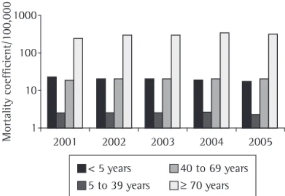
Respiratory diseases constitute the fifth leading cause of death in Brazil. Pneumonia is the second most common respiratory disease, causing 35,903 deaths in 2005, 8.4% of which occurred among patients less than 5 years of age and 61% of which occurred among patients over 70 years of age. The pneumonia-related mortality rate increased in the period between 2001 and 2004. In 2005, however, it fell to levels below 20/100,000 population, according to the latest mortality statistics provided by the National Ministry of Health.
The pneumonia-related mortality rate differs among age groups. Over the last 5 years, the rate has increased significantly among patients over 70 years of age, reaching levels above 500/100,000 population among those over 80 years of age. The lowest rates are found in the 5-49 year age bracket (less than 10/100,000 population); among those less than 5 years of age, the mortality rate remains at approximately 17/100,000 population, showing a slight downward trend (Figure 1). These data are similar to those obtained in other Latin American countries such as Chile.(8)
Chart 1 - Levels of evidence of the recommendations according to the Brazilian Medical Association.
Level of evidence
Evidence source Definition
I Randomized
controlled trials Rich database
Level I evidence is based on well-designed randomized controlled trials, which provide a consistent model of findings regarding the population to which the recommendation is addressed. Level I evidence requires a substantial number of studies, involving an adequate number of participants.
II Randomized
controlled trials Limited database
Level II evidence is based on intervention studies, which include a limited number of patients, post hoc analyses or analyses of subgroups of randomized controlled trials, or meta-analyses of randomized controlled trials. In general, level II evidence is pertinent when there are few randomized trials, when the trials are limited in scope, when the trials are conducted among a population that differs from the recommended target population or when the results are somehow inconsistent. III Nonrandomized trials
Observational studies
Level III evidence is based on nonrandomized, uncontrolled trials or on observational studies.
IV Consensus among the participants of the panel
Evidence level IV is attributed only to cases in which some type of support
when the clinical profile is unfavorable but chest X-ray is normal; when the objective is to detect complications such as loculated pleural effusion and encapsulated abscess in the airways; and when it is necessary to differentiate inflamma-tory infiltrate from lung masses.(17,18)
In cases of pleural effusion greater than 5 cm in the posterior mediastinum revealed by lateral chest X-ray in the orthostatic position, or in cases of loculated pleural effusion, thoracentesis should be performed in order to exclude the diagnoses of empyema and complicated para-pneumonic effusion. Thoracentesis is strongly recommended in cases of pleural effusion that occupy more than 20% of the hemithorax.(19)
Ultrasound is useful in cases of small pleural effusion or in suspected cases of loculated pleural effusion because it can determine the exact location of the effusion for subsequent drainage.(9,20)
Radiological progression after hospital admission might occur regardless of the etiology, and the therapeutic regimen should not be changed unless the clinical profile has shown no improvement.(10) Radiological resolution is
relatively slow and occurs after clinical recovery. Complete resolution of radiological alterations occurs two weeks after the initial presentation in half of all cases and six weeks after the initial presentation in two thirds.(11) Advanced age,
COPD, immunosuppression, alcoholism, diabetes and multilobar pneumonia are independently associated with slower resolution. Pneumonia caused by Mycoplasma sp. resolves more rapidly. Pneumonia caused by Legionella sp. resolves in a particularly slow manner. Residual lesions are found in 25% of the cases of Legionella sp. and bacteremic pneumococcal pneumonia.(10) Chest
X-ray should be repeated six weeks after the onset of symptoms in smokers over 50 years of age (risk of bronchial carcinoma) and in cases in which the symptoms persist or physical exami-nation reveals abnormal findings.(11,21)
Recommendations
• Anteroposterior and lateral chest X-rays
should be taken for the initial approach to patients suspected of having CAP (level III evidence).
• Chest X-ray is the only complementary test
to which low-risk CAP outpatients should be submitted (level I evidence).
assessing severity, identifies multilobar involve-ment and might suggest alternative etiologies, such as abscess and TB. Chest X-rays can reveal concomitant conditions, e.g., bronchial obstruc-tion or pleural effusion, as well as being useful in monitoring treatment response. However, the classification of CAP according to radiological patterns (lobar pneumonia, bronchopneumonia and interstitial pneumonia) is of limited use in predicting the causal agent: it cannot distin-guish between the groups of agents (bacterial and nonbacterial).(9-15) Specific agents can cause
different manifestations that can change or become more intense throughout the course of the disease, being frequently influenced by the immunological status.(13)
Chest X-ray is the imaging method of choice for the initial approach to CAP because its cost-effectiveness is excellent, the doses of radiation employed are low and the method is widely available.
Half of the cases diagnosed as CAP in Brazil in reality are not.(16) The greatest diagnostic
difficulty lies in the interpretation of the chest X-rays by non-specialists.
Cavitation is suggestive of an anaerobic etiologic agent, Staphylococcus aureus and, occasionally, gram-negative bacilli. In such cases, screening for TB should always be performed. Swollen lobes causing bulging fissures is a nonspecific finding that reflects intense inflam-matory reaction.(10)
A CT scan of the chest is useful in a number of situations: when a chest X-ray alone does not clearly show whether or not there is infiltrate;
1000
100
10
1
Mortality coefficient/100,000 2001 2002 2003 2004 2005
< 5 years 5 to 39 years
40 to 69 years ≥ 70 years
• Arterial blood gas analysis should be
conducted in cases of SpO2 ≤ 90% on room air or in cases of severe pneumonia (level I evidence).
• Hypoxemia is an indication for supple -mental oxygen and hospital admission (level I evidence).
Complementary tests
Urea levels higher than 65 mg/dL (corre-sponding to a value ≥ 11 mmol/L) constitute a strong marker of disease severity.(25-27) The blood
workup shows low sensitivity and specificity, being useful as a criterion for severity and therapeutic response. Leukopenia (< 4,000 leukocytes/mm3)
denotes poor prognosis.(28,29) Glycemia,
electro-lyte and transaminase levels have no diagnostic value; they can, however, influence the decision of hospitalizing a patient by allowing the identi-fication of concomitant diseases.(30,31)
C-reactive protein
As a marker of inflammatory activity, C-reactive protein has prognostic value in follow-up treatment. High C-reactive protein levels after 3-4 days of treatment and a < 50% reduction in initial C-reactive protein levels suggest worse prognosis or complications. The impact of C-reactive protein levels on the diag-nosis requires further investigation and definition of cut-off values before it can be used routinely in clinical practice. There is a lack of consistent data as to whether or not C-reactive protein can aid in the decision of using antibiotics.(32-35)
Procalcitonin
Procalcitonin is a marker of inflamma-tory activity that can be determined in various manners: using a commercially available
mono-• The radiological patterns cannot be used
to predict the causal agent or to distin-guish between the groups of agents (level III evidence).
• A CT scan of the chest should be performed
when there is doubt regarding the presence of inflammatory infiltrate; in order to detect complications; and in suspected cases of neoplasia (level III evidence).
• Significant pleural effusion (≥ 5 cm, seen on lateral chest X-ray in the orthostatic position from the posterior ridge) should be drained.
• Ultrasound is useful in cases of small pleural
effusion or in suspected cases of loculated pleural effusion (level III evidence).
• Chest X-ray should be repeated six weeks
after the onset of symptoms in smokers over 50 years of age and in cases in which the symptoms persist or physical examina-tion reveals abnormal findings.
• Persistence of radiological findings after
six weeks requires additional investigation
(level IV evidence).
Peripheral oxygen saturation and
arterial blood gas analysis
Peripheral oxygen saturation (SpO2) should be monitored routinely before recommending oxygen therapy. Arterial blood gas analysis should be conducted in cases of SpO2 ≤ 90% on room air or in cases of severe pneumonia. Hypoxemia is an indication for supplemental oxygen and hospital admission.(22-24)
Recommendations
• The SpO2 should be monitored routinely before the initiation of oxygen therapy (level I evidence).
Chart 2 - Most common pathogens in community-acquired pneumonia, in decreasing order.
Outpatients with CAP (mild) Inpatients with CAP (not in ICU) ICU patients with CAP (severe)
• S. pneumoniae • S. pneumoniae • S. pneumoniae • M. pneumoniae • M. pneumoniae • Gram-negative bacilli • C. pneumoniae • C. pneumoniae • H. influenzae • Respiratory viruses • Respiratory viruses • Legionella sp.
• Haemophilus influenzae • Haemophilus influenzae • S. aureus • Legionella sp.
to the high efficacy of empirical treatment and the low mortality among such patients (< 1%). The elucidation of the etiology of CAP does not result in lower mortality when compared with early, appropriate empirical antibiotic therapy. (41)
When empirical treatment fails in severe CAP patients, etiologic diagnosis and specific treat-ment correlate with lower mortality. Treattreat-ment initiation should not be delayed in order to perform tests to determine the etiology of CAP.(41,42) The most common etiologic agents
according to CAP severity and treatment locale are shown in Chart 2.
Sputum examination
Although sputum examination is commonly used for establishing the etiologic diagnosis of CAP, the beneficial role of this examina-tion in the initial management of CAP is still controversial.(43,44) Some of the obstacles to the
performance of sputum examination include the need for collecting the samples in an appropriate manner, the lack of standardization of the tech-niques used for sample preparation, the variable examiner ability to interpret the results and the lack of a gold standard for the microbiological diagnosis of CAP.(45) Valid sputum samples are
defined as those that contain < 10 epithelial cells and > 25 polymorphonuclear cells per low-power microscopic field.
clonal immunoluminometric assay, considered a less sensitive method; using a polyclonal immunoassay system (Kryptor; B·R·A·H·M·S Aktiengesellschaft, Hennigsdorf, Germany), considered a more sensitive method but not widely available in practice; and more recently,
using the VIDAS system of ELISA (bioMérieux,
Marcy l’Étoile, France), which is almost as sensi-tive as the Kryptor system and more readily available, although the assay kit is expensive. Studies involving patients at different levels of risk of death have demonstrated that procal-citonin levels tend to be higher in bacterial pneumonia patients with pneumonia severity index (PSI) scores of I or II than in nonbacterial pneumonia patients with the same PSI scores.
(36-38) No etiology-related differences were found
among patients with pneumonia that was more severe; however, higher procalcitonin levels correlated with complications and death. (37)
Procalcitonin is a better marker of severity than are C-reactive protein, IL-6 and lactate. Elevated serum procalcitonin levels are also found in other lung diseases such as chemical pneu-monitis and smoke inhalation injury in burn patients.(34,36,39,40)
Etiologic investigation
The methods used for determining the etiology of CAP have low immediate yield and are unnecessary in outpatients, which is due
Chart 3 - Complementary tests indicated for etiologic investigation of community-acquired pneumonia.
Evidence Blood
culture
Sputum smear microscopy and sputum culture
Urinary antigen test for pneumococcus and Legionella sp.
Bronchoalveolar lavage or transtracheal
aspiration
Other
ICU admission Severe CAP
Yes Yes Yes Yes Aspirate
if tracheal intubation is
performed
Alcohol abuse Yes Yes
Clinical treatment failure
Yes Yes Yes Yes*
Structural disease
No Yes No No
Cavitation Yes Yes No No AFB
Pleural effusion Yes Yes Yes No Thoracentesis
who do not respond to empirical treatment. Percutaneous puncture lung biopsy is contrain-dicated in patients on invasive mechanical ventilation.(49-51)
When tracheal intubation and initiation of mechanical ventilation are indicated, material should be collected from the lower airways using tracheal aspirate or bronchoscopic techniques, in order to perform quantitative culture.
Secretion collection through bronchoscopy poses lower risks to patients than does transtra-cheal aspiration and lung puncture.(52-57)
Serologic tests
Serologic tests should not be routinely requested. Serologic tests allow the establishment of retrospective diagnosis of infection caused by certain microorganisms that are difficult to culture (Mycoplasma, Coxiella, Chlamydophila and Legionella, as well as viruses). Serologic tests are considered positive when the titer obtained in the convalescent phase, i.e., four to six weeks after defervescence, is four times greater than that obtained in the acute phase. Because of this technical characteristic, serologic tests are not useful in treating patients individually; they are, however, useful in establishing the epidemio-logical profile of a given region or an epidemic outbreak.(1,58)
Urinary antigen tests
Urinary antigen tests are simple, rapid and not influenced by the use of antibiotics. The Due to the high prevalence of
pulmo-nary TB and mycoses in Brazil, screening for acid-fast bacilli in the sputum using the Ziehl-Neelsen technique, as well as screening for fungi, should be performed in suspected cases of CAP, according to the Tuberculosis Control Guidelines.(46)
Blood culture
Because blood culture commonly shows a low yield, it should be reserved for severe CAP patients and for inpatients who do not respond to the therapeutic approach used. False-positive results are common, especially if antibiotics have previously been used, and rarely result in a change of approach. Sputum samples should be collected before initiation or change of treat-ment and should not delay the administration of the first dose of antibiotics.(28,29,47,48)
Other techniques for the collection of
samples for microbiological examination
Other available techniques for obtaining samples from the lower airways include tracheal aspirate, mini-bronchoalveolar lavage and bronchoscopic protected specimen brush or bronchoalveolar lavage, as well as transthoracic needle aspiration biopsy.
These procedures should not be routinely recommended for most CAP patients. However, such procedures are useful in patients admitted to the intensive care unit (ICU) and in those
Chart 4 - Scores according to demographic, clinical and laboratory factors, as proposed by Fine et al.a
Demographic factors Radiological and laboratory findings
Age pH < 7.35 +30
Men 1 point/year of age Urea > 65 mg/dL +20
Women age -10 Sodium < 130 mEq/L +20
Nursing home residents age +10 Glucose > 250 mg/dL +10
Hematocrit < 30% +10
PaO2 < 60 mmHg +10
Pleural Effusion +10
Comorbidities Physical examination
Neoplasia +30 Altered mental status +20
Liver disease +10 Respiratory rate > 30 breaths/min +20
CHF +10 Systolic BP < 90 mmHg +20
Cerebrovascular disease +10 Temperature < 35°C or > 40°C +15
Kidney disease +10 Heart rate ≥ 125 bpm +10
inpatients who do not respond to the initial treatment (level III evidence).
• In cases of severe CAP, microbiological
investigation should be performed through blood culture, sputum culture, culture of tracheal aspirate or culture of samples obtained by bronchoscopy in patients on mechanical ventilation (level II evidence).
• Screening for the S. pneumoniae urinary antigen should be performed in severe CAP patients, and screening for the L. pneumo-phila urinary antigen should be performed specifically in all patients who do not respond to the initial treatment (level II evidence).
Classification of severity and choice
of treatment locale
Patients diagnosed with CAP should be eval-uated for disease severity. This evaluation will aid in the choice of treatment locale, in the extent of etiologic investigation and in the choice of antibiotic therapy. Socioeconomic factors should also be taken into consideration when making this decision.
Disease severity scores(72) or prognostic
models(73) evaluate the 30-day mortality risk and
are used to identify low-risk patients, who are therefore candidates for outpatient treatment.
Pneumonia severity index
The PSI score comprises 20 variables that include demographic characteristics, concomi-tant diseases, abnormal laboratory test results, radiological alterations and physical examination findings. The points attributed to each vari-able allow the severity to be stratified into five test for Legionella pneumophila becomes
posi-tive from day 1 of disease and remains so for weeks. Its sensitivity ranges from 70 to 90%, and its specificity is approximately 100%. Since the test detects the L. pneumophila serogroup 1 antigen (the most prevalent serogroup), infec-tions caused by other serogroups, although less common, might not be identified.(59-62)
The test for pneumococci has a sensitivity ranging from 50% to 80% (greater than that of sputum examination and blood culture) and a specificity of 90%.(63,64) Previous use of
antibi-otics does not affect the results. False-positive results can occur in cases of colonization of the oropharynx, especially among children with chronic lung diseases. The test is rapid and effec-tive, with good sensitivity and specificity.(65-68)
PCR
The greatest potential of PCR lies in the identification of L. pneumophila, Mycoplasma pneumoniae and Chlamydophila pneumoniae, as well as of other habitually noncolonizing pathogens.
The use of PCR, for the detection of one causative agent, or multiplex PCR, for the detec-tion of M. pneumoniae, C. pneumoniae and Legionella spp., shows good sensitivity and specificity; however, these tests are not available in most clinical laboratories.(68-71)
The etiologic tests recommended for use in specific situations are shown in Chart 3.
Recommendations
• Although glycemia, electrolyte and tran -saminase levels have no diagnostic value, they can influence the decision to hospita-lize a patient by allowing the identification of concomitant diseases (level II evidence).
• Because blood culture commonly has a low
yield, it should be reserved for use in severe CAP patients and for inpatients who do not respond to the therapeutic approach used (level III evidence).
• Serologic tests are not useful in treating
patients individually; they are, however, useful in establishing the epidemiolo-gical profile of a given region or epidemic outbreak (level III evidence).
• Screening for the etiologic agent should
be performed in severe CAP patients and
Chart 5 - Classification of patients with
community-acquired pneumonia based on risk, according to the Pneumonia Severity Index score.(73)
Score Points Mortality, % Recommended treatment locale
I 0.1 Outpatient clinic
II ≤ 70 0.6 Outpatient clinic
III 71-90 2.8 Outpatient clinic
or day hospital
IV 91-130 8.2 Hospitalization
The present guidelines reiterate the recom-mendation of the previous guidelines regarding the need to evaluate concomitant diseases, the extent of radiological involvement, the degree of oxygenation, psychosocial factors, socioeco-nomic factors and the viability of oral medication use in terms of their influence on the choice of treatment locale. Due to the simplicity of the CURB-65 and CRB-65 scores, as well as the fact that they are immediately applicable and easy to use, we recommend their use as appropriate criteria for the stratification of CAP severity at the primary and emergency care levels (Chart 6).
If not contraindicated by socioeconomic complications, decompensated concomitant diseases, hypoxemia or inability to take oral medication, hospital admission is indicated for patients with a score of at least two points on the CURB-65 or at least one point on the CRB-65. When such contraindications exist, the attending physician should prescribe outpatient treatment.
Severe community-acquired
pneumonia
From a practical standpoint, severe CAP is defined as CAP in which the clinical profile is more likely to worsen or the risk of death is higher. Admission to the ICU is mandatory for the appropriate management of severe CAP patients. Septic shock and the need for mechanical venti-lation are absolute criteria for ICU admission.(78)
classes, based on the risk of death (Charts 4 and 5). However, the primary objective of the original study was to identify low-risk patients.(30,73,74)
The PSI score might underestimate CAP severity among young patients without concomitant diseases. In addition, because the PSI is complex and requires extensive laboratory evaluation, it is not considered ideal for routine use in clinical practice.
The British Thoracic Society disease
severity score
The disease severity score proposed by the British Thoracic Society is based on variables indic-ative of severe CAP, as follows: mental confusion (defined as an abbreviated mental test score ≤ 8); urea > 50 mg/dL; respiratory rate ≥ 30 breaths/ min; systolic blood pressure < 90 mmHg or diastolic blood pressure ≤ 60 mmHg; and age ≥ 65 years. The name of this score (CURB-65) is an acronym based on the key term of each risk factor assessed (confusion, urea, respiratory and blood) and can be presented in its simplified form, CRB-65 (without urea levels). In this score, each variable represents 1 point, and the total possible score comprises 4 points (CRB-65) or 5 points (CURB-65), as shown in Figures 2 and 3.(25,72,75-77)
The greatest limitation of the CURB-65 and CRB-65 is the fact that they do not include concomitant diseases that can increase the risk of CAP, e.g., alcoholism, heart failure, liver failure and neoplasia.
CURB-65 Score
Low mortality, 1.5%
Likely candidate for
outpatient treatment Consider hospitalization
High mortality, 22%
Inpatient with severe CAP scores 4-5: Evaluate the possibility
of ICU admission Intermediate mortality, 9.2%
0-1 2 3 or +
• The possibility of treatment reevaluation
should be guaranteed for CAP patients recei-ving home treatment (level III evidence).
• CAP patients who meet major or minor
criteria for severe CAP should receive ICU treatment (level I evidence).
Treatment
Empirical treatment vs. specific
treatment
In the vast majority of CAP cases, it is not possible to determine the etiologic agent prior to the therapeutic moment of decision. Empirical antibiotic therapy is habitually targeted at the most prevalent microorganisms. More often than not, more than one pathogen is present and atyp-ical pathogens are included. This requires wider empirical coverage, particularly in cases that are more severe. Specific therapy has the potential to minimize adverse effects, decrease the induc-tion of resistance to antimicrobial agents and reduce costs.(42,81,83,84) Specific therapy can replace
empirical treatment of hospitalized patients when the specific pathogen is identified within the first 48-72 h after treatment initiation. In this context, the identification of the agent can narrow the empirical regimen or influence the choice of anti-microbial agent to be administered orally in the subsequent therapeutic approach.(81)
Recommendations
• The selection of the initial therapeutic
regimen for CAP patients takes into The currently used criteria were defined
by Ewig et al. and have a sensitivity of 78%, a specificity of 94%, a negative predictive value of 95% and a positive predictive value of 75% in selecting patients who require ICU admission . (78-80)
The present guidelines corroborate the adop-tion of such criteria in order to define severe CAP and to recommend ICU admission (Chart 7). If a patient meets two of the minor criteria or one of the major criteria for CAP severity, ICU admission is indicated.(81,82)
Recommendations
• The decision of whether to hospitalize a
CAP patient is the prerogative of the atten-ding physician, and the currently available evaluation scores are tools that inform that decision (level III evidence).
• The use of the CURB-65 or CRB-65 scores
is recommended to aid in the choice of treatment locale (level III evidence).
• Psychosocial and socioeconomic factors
should be taken into consideration when selecting the treatment locale (level III evidence).
• CAP patients presenting with septic shock,
requiring vasopressors or with acute respiratory failure (requiring mechanical ventilation) should be admitted to the ICU, as should patients who meet at least two of the minor criteria for CAP severity (level III evidence).
Figure 3 - CRB-65 score. CRB-65: C: mental confusion; R: respiratory rate ≥ 30 breaths/min; B: blood pressure
(systolic < 90 mmHg or diastolic ≤ 60 mmHg); and 65: age ≥ 65 years. CRB-65 Score
Low mortality, 1.2%
Likely outpatient treatment
Evaluate possibility of hospitalization
High mortality, 31%
Urgent hospitalization Intermediate mortality, 8.15%
caused by atypical pathogens showed that values were similar in North America, Europe, Latin America and Asia (22%, 28%, 21% and 20%, respectively).(86) However, in that study, the
proportion of patients who received empirical antibiotic therapy with coverage of atypical pathogens was 91%, 74%, 53% and 10%, respec-tively. Comparing those patients with those not receiving such therapy, the authors observed the following: less time to achieve clinical stability (3.2 vs. 3.7 days; p < 0.001); shorter hospital stay (6.1 vs. 7.1 days; p < 0.01); lower overall mortality rate (7.0% vs. 11.1%; p < 0.01); and lower CAP-related mortality (3.8% vs. 6.4%; p < 0.05).
In general, the empirical antibiotic therapy for CAP covers atypical pathogens.(87) Although
most guidelines recommend empirical antibiotic coverage of atypical pathogens, there is contro-versy regarding the level of scientific evidence on which this practice is based. A recently published observational study involving hospitalized patients (n = 201) revealed that previous outpatient treat-ment with a beta-lactam antibiotic correlated with an increased possibility (approximately three times greater) of an atypical pathogen being present, whereas the possibility of pneumococci being present was reduced to one third.(88) The need for
and effectiveness of empirical antibiotic coverage of atypical pathogens in hospitalized patients with mild CAP have been recently reevaluated in three studies (systematic review or meta-analysis), the outcome measures of which were the effi-cacy of the practice and on the related mortality rates.(83,89,90) These reviews principally compared
quinolone monotherapy with beta-lactam mono-therapy. In the most recent of the three, which included 5,244 patients from 25 randomized studies, no significant difference in mortality was observed between patients treated with antibi-otics that cover atypical pathogens (quinolones) and those treated with a beta-lactam (relative risk = 1.15; 95% CI: 0.85-1.56). No significant difference was observed regarding adverse effects or the need to discontinue treatment. In these reviews, the overall frequency of adverse effects was similar; however, patients treated with beta-lactam antibiotics presented more adverse effects in the gastrointestinal tract than did those treated with quinolones. (90) The two
system-atic reviews presented similar results.(83,89) In the
three reviews previously cited, the use of empir-consideration the most prevalent
microor-ganisms (level III evidence).
• Specific treatment targeted at a previously
identified pathogen or pathogens is to be preferred over empirical treatment. However, specific treatment is typically unfeasible at the therapeutic moment of decision (level III evidence).
• The identification of the agent(s) allows the
selection of specific therapy targeted at the pathogen(s) and the selection of antimi-crobial agent to be used in the subsequent therapeutic approach, potentially reducing treatment cost, adverse effects and resis-tance induction (level II evidence).
Empirical antibiotic coverage of atypical
pathogens
When CAP patients are submitted to specific diagnostic tests, atypical pathogens are frequently identified. Depending on the meth-odology employed, these agents can appear in isolation or as an integral part of a polymicro-bial etiology.(85) In a large study based on the
secondary analysis of international registries of hospitalized patients, the incidence of CAP Chart 6 - Steps for evaluating treatment locale of
patients with community-acquired pneumonia. 1 - Evaluate the presence of concomitant diseases 2 - Evaluate CRB-65
3 - Evaluate the degree of oxygenation and radiological involvement
• SpO2 < 90% - indication for hospitalization
• Chest X-ray
– Radiological extent
– Pleural effusion suspected of empyema 4 - Evaluate social and cognitive factors
• Absence of a family member or caregiver in
the residence
– need for observing treatment response
• Capacity of understanding the treatment
prescribed
5 - Evaluate economic factors
• Access to medication • Return for evaluation
6 - Evaluate acceptability of oral medication 7 - Clinical judgment
Chart 7 - Criteria for defining severe
community-acquired pneumonia.
• Major criteria: the presence of one criterion is an
indication for ICU treatment
- Septic shock requiring vasopressors
- Acute respiratory failure requiring mechanical ventilation
• Minor criteria: the presence of two criteria is an
indication for ICU treatment - Arterial hypotension - PaO2/FiO2 ratio < 250
- Presence of multilobar infiltrate ICU: intensive care unit.
with coverage of atypical pathogens (quinolone or macrolide). In a retrospective study involving 515 patients (of which 261 received combined therapy) with severe CAP (cases of severe CAP
were defined as those with a PSI score of V),
lower 14-day and 30-day mortality rates were found among the patients who received combi-nation antibiotic therapy, a result that was not observed among patients with CAP that was less severe.(91) In another observational study,
survival rates were higher among patients with severe CAP and septic shock who received combination therapy than they were among patients who received monotherapy, even when the latter was considered appropriate for the etiologic agent. Combination therapy showed no advantage over monotherapy in patients without septic shock.(92) Combination therapy
also seems to be more beneficial than mono-therapy in cases of pneumococcal CAP with bacteremia.(93) Possible explanations include
a possibly unapparent coinfection caused by atypical pathogens (found in 18-38% of the cases in some studies) or the immunomodula-tory effects of macrolides, or a combination of the two.(94)
Recommendations
• Combination therapy is not superior to
monotherapy among low-risk patients (level II evidence).
• Combination therapy should be recom -mended for patients with severe CAP, particularly for those with accompanying bacteremia, respiratory failure or septic shock (level II evidence).
• In comparison with monotherapy, combina -tion therapy with two effective antibiotics reduces mortality among bacteremic pneu-mococcal pneumonia (level II evidence).
Predictors of specific pathogens
The principal predictive factors of CAP caused by penicillin-resistant pneumococci are age ≤ 4 years (OR = 5.3; 95% CI: 2.2-12.6), immu-nosuppression (OR = 3.0; 95% CI: 1.5-6.0) and recent use of a beta-lactam (OR = 2.1; 95% CI: 1.0-4.5).(95) The principal independent predictors
of CAP caused by gram-negative bacilli include likely aspiration (OR = 2.3; 95% CI: 1.02-5.20; p = 0.04), hospitalization within the past 30 days ical antibiotic coverage of atypical pathogens
showed better results only in the subgroup of patients in which Legionella sp. was subsequently detected. Quinolone coverage did not result in worse disease progression in patients with CAP caused by pneumococci.(83,90) However, the
validity of such observational studies is ques-tionable.(87) Further prospective studies of such
patients, comparing beta-lactam mono therapy with beta-lactam-macrolide combination therapy and electing the mortality rate as the primary outcome measure, are needed.
Recommendation
• Although there is no conclusive evidence
that empirical antibiotic coverage of atypical pathogens is superior, this thera-peutic approach to hospitalized patients might result in lower mortality rates among patients with pneumonia caused by Legionella sp. It might also result in reduced hospital stay, overall mortality and mortality from pneumonia caused by atypical pathogens (level II evidence).
Combination therapy vs. monotherapy
Potential benefits of early initiation of
antibiotic therapy
At least two retrospective studies involving CAP patients aged ≥ 65 years who were hospi-talized in the USA demonstrated lower mortality among patients who received earlier antibiotic therapy.(97,98) One of the studies(98) proposed an
interval of 8 h between patient admission and the administration of the first dose, whereas the other(97) established a 4-h limit for the
admin-istration of the first dose. Another group of authors reported that early antibiotic therapy can reduce the length of hospital stays.(99)
Among patients with bacteremic pneumococcal pneumonia, initiation of antibiotic therapy within the first 4 h after hospital admission reduces mortality and length of hospital stay.(100)
However, in a prospective observational study involving 409 patients with moderate to severe CAP, no direct correlation was found between early antibiotic therapy and the time required (OR = 3.5; 95% CI: 1.7-7.1; p < 0.001),
anti-biotic use for more than 48 h within the past 30 days (OR = 1.9; 95 CI%: 1.01-3.70; p = 0.049) and parenchymal lung diseases (OR = 2.8; 95% CI: 1.5-5.5; p = 0.02).(96) A specific analysis
of a subgroup of patients with CAP caused by Pseudomonas aeruginosa showed that a history of lung disease (OR = 5.8; 95% CI: 2.2-15.3; p < 0.001) and recent hospitalization (OR = 3.8; 95% CI: 1.8-8.3; p = 0.02) were independent predictors of CAP caused by P. aeruginosa. In that study, infection caused by gram-negative bacilli was an independent variable for risk of death (OR = 3.4; 95% CI: 1.6-7.4; p = 0.002).(96)
Recommendation
• The predictors of risk for specific pathogens
should be taken into consideration when selecting the empirical antibiotic therapy for CAP patients (level II evidence).
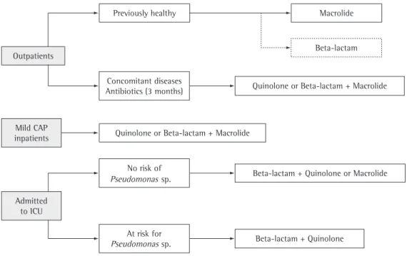
Outpatients
Previously healthy Macrolide
Beta-lactam
Quinolone or Beta-lactam + Macrolide
Beta-lactam + Quinolone or Macrolide
Beta-lactam + Quinolone
Quinolone or Beta-lactam + Macrolide Concomitant diseases
Antibiotics (3 months)
No risk of
At risk for Mild CAP
inpatients
Admitted to ICU
Pseudomonas sp.
Pseudomonas sp.
Figure 4 - Initial empirical antibiotic therapy according to degree of severity.
of penicillin and its derivatives (amoxicillin and ampicillin) in respiratory tract infections.(104)
The original definition of penicillin resistance (MIC ≥ 2 mg/L) was based on populations of patients with meningitis. This criterion was—and is—indistinctly applied to pneumonia isolates. In various regions, this has given rise to policies regarding the use of non-beta-lactam antibi-otics that are based on questionable resistance rate data. These resistance rates need to be definitively reevaluated. For pneumonia-related S. pneumoniae strains (i.e., those unrelated to meningitis) isolated from the respiratory tract, the blood, or both, separate categories of i.v. to achieve clinical stability, considering periods
of less than 4 h, of 4-6 h and of more than 6 h.(101) In a recent review of the topic, it was
suggested that early antibiotic therapy is poten-tially more beneficial for elderly patients who are antimicrobial treatment-naïve . (102) However,
the pressure to administer the first dose in less than 4 h rather than in 8 h might lead to errors in the diagnosis of CAP.(103) Therefore, the ideal
time frame between admission and the first dose of antibiotics remains controversial. It is recommended, however, that the first dose be administered to hospitalized patients “as early as possible”, preferably at admission, particularly in the emergency room.(81)
Recommendation
• Antibiotic therapy for CAP patients should
be initiated as early as possible, since it can reduce mortality rates, shorten hospital stays and control costs (level III evidence).
Antibiotic resistance in S. pneumoniae:
major changes in the Clinical Laboratory
Standards Institute criteria
The selection of an antimicrobial agent to treat infections caused by S. pneumoniae is based on various factors, such as the site of infection, resistance to penicillin (and other agents), degree of severity, pharmacokinetics / pharmacodynamic s of the drug and patient age. In community-acquired infections, empirical treatment can be guided by local epidemiological surveillance studies. In severe infections, it is important that culture and antibiogram be performed when treatment adjustment is needed. The interpreta-tion of the minimum inhibitory concentrainterpreta-tion (MIC) of penicillin for S. pneumoniae should always consider the clinical sample in which the agent was isolated, as well as clinical suspi-cion of the type of infection. The disk method routinely used in laboratories is not definitive in detecting penicillin resistance, and the results obtained should always be confirmed by deter-mining the MIC (mg/L).
The definition of penicillin resistance based on the MIC for S. pneumoniae strains was changed in 2008 by the Clinical Laboratory Standards Institute (CLSI) as a result of evidence of the pharmacokinetics and pharmacodynamics
Chart 8 - Principal causes and patterns of treatment failure.
Progressive (respiratory failure, shock) < 72 h
Severity of disease presentation Microorganismo não tratado Atypical pathogens
(mycobacteria, viruses, Nocardia sp., fungi) Antimicrobial resistance
Infectious complication Pulmonary
(empyema, parapneumonic effusion) Metastatic (endocarditis, meningitis, arteritis) Noninfectious cause
Incorrect diagnosis (PET, ARDS, vasculitis) > 72 h
Infectious complication Nosocomial superinfection Exacerbation of underlying disease Noninfectious cause (PET, AMI) Nonresponsive (persistent symptoms)
> 72 h
Nonresponsive microorganism Not covered
Resistant
Local complication
(empyema, parapneumonic effusion) Nosocomial superinfection
Noninfectious causes
Pneumonia-related complication (COP) Incorrect diagnosis
(PET, CHF, vasculitis, II, neoplasia) Antibiotic-related fever
ratory tests can screen for penicillin resistance using an oxacillin disk, which indicates decreased sensitivity when it reveals halos < 20 mm, this interpretation should be confirmed by a quanti-tative method for the determination of penicillin (MIC), because sensitive strains might be present even if halos of 20 mm are shown. Only the MIC value can definitively classify a S. pneu-moniae strain as being resistant.(105) Knowledge
of the distinct local mechanisms of resistance and their clinical and laboratory interpretation are of utmost importance in order to select the appropriate treatment and contribute to the penicillin sensitivity, based on the MIC, have
been established: sensitive, ≤ 2 mg/L; interme-diate, ≥ 4 mg/L < 8 mg/L; and resistant, ≥ 8 mg/L. It should be borne in mind that these criteria
do not apply to penicillin V (oral). According
to the new CLSI guidelines, meningitis-related S. pneumoniae strains (isolated from the cerebro-spinal fluid, blood, or both) are classified (based on the MIC) as penicillin-sensitive (< 0.06 mg/L) or penicillin-resistant (≥ 0.12 mg/L).
Local rules for the use of antibiotic therapy in CAP patients need to be revised, since they were based on earlier studies. Although routine
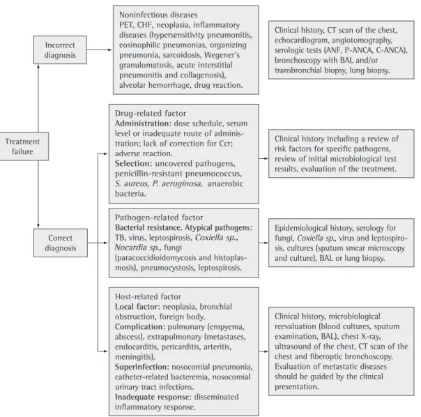
labo-Figure 5 - Principal causes of treatment failure and specific diagnostic approach. PTE: pulmonary thromboembolism; CHF: congestive heart failure; BAL: bronchoalveolar lavage; Ccr: creatinine clearance; ANF: antinuclear factor; and ANCA: antineutrophil cytoplasmic antibody.
Drug-related factor
Administration: dose schedule, serum level or inadequate route of adminis-tration; lack of correction for Ccr; adverse reaction.
Selection: uncovered pathogens, penicillin-resistant pneumococcus,
S. aureus, P. aeruginosa, anaerobic
bacteria.
S. aureus, P. aeruginosa,
Host-related factor
Local factor: neoplasia, bronchial obstruction, foreign body.
Complication: pulmonary (empyema, abscess), extrapulmonary (metastases, endocarditis, pericarditis, arteritis, meningitis).
Superinfection: nosocomial pneumonia, catheter-related bacteremia, nosocomial urinary tract infections.
Inadequate response: disseminated inflammatory response.
Noninfectious diseases
PET, CHF, neoplasia, inflammatory diseases (hypersensitivity pneumonitis, eosinophilic pneumonias, organizing pneumonia, sarcoidosis, Wegener’s granulomatosis, acute interstitial pneumonitis and collagenosis), alveolar hemorrhage, drug reaction.
Clinical history, CT scan of the chest, echocardiogram, angiotomography, serologic tests (ANF, P-ANCA, C-ANCA), bronchoscopy with BAL and/or transbronchial biopsy, lung biopsy.
Clinical history including a review of risk factors for specific pathogens, review of initial microbiological test results, evaluation of the treatment. Treatment
failure
Correct diagnosis Incorrect diagnosis
Pathogen-related factor
Bacterial resistance. Atypical pathogens: TB, virus, leptospirosis, Coxiella sp.,
Nocardia sp., fungi
(paracoccidioidomycosis and histoplas-mosis), pneumocystosis, leptospirosis.
Coxiella sp., Epidemiological history, serology for fungi, Coxiella sp., virus and leptospiro-sis, cultures (sputum smear microscopy and culture), BAL or lung biopsy.
Coxiella sp.
Clinical history, microbiological reevaluation (blood cultures, sputum examination, BAL), chest X-ray, ultrasound of the chest, CT scan of the chest and fiberoptic bronchoscopy. Evaluation of metastatic diseases should be guided by the clinical presentation.
two studies.(107,108) One of the studies, an open
randomized study, compared azithromycin with cefuroxime and erythromycin, as well as with the cefuroxime-erythromycin combination, in terms of efficacy and safety. In that study, 80% of the patients were low-risk patients (Fine score I-III). In the other study, which involved two groups of patients, azithromycin and the cefuroxime-erythromycin combination were both effective in 91% of the patients (61/67 and 71/78, respec-tively). Despite the similar effectiveness of the two treatments (p = 0.95), the incidence of adverse effects was higher in the latter group (p < 0.001). The degree of CAP severity was not reported in that study.
Recommendation
• In hospitalized patients, empirical treat -ment with azithromycin as monotherapy should be limited to patients with mild CAP (level II evidence).
Ertapenem for hospitalized patients
Ertapenem is a beta-lactam that is structurally similar to meropenem (possessing a 1-beta-me-thyl group) and is indicated for patients with moderate to severe infection caused by gram-positive pathogens, as well as infection caused by aerobic or anaerobic gram-negative bacteria. It is administered i.m. or i.v. in a single daily dose of 1 g. It can be used to treat infections (soft tissue infections, abdominal infections, acute pelvic infections and complicated urinary tract infections), CAP and sepsis. It is particularly useful in cases of infection caused by bacteria producing extended spectrum beta-lactamases. It is not indicated in cases of infection caused by penicillin-resistant S. pneumoniae, methicil-lin-resistant S. aureus, Pseudomonas spp. and Acinetobacter spp.(109)
Two studies compared ertapenem with ceftri-axone in terms of their efficacy and safety when used in the treatment of CAP patients. One of the studies (n = 658), a double-blind, rand-omized, prospective multicenter study, showed no significant difference between the two regi-mens regarding their efficacy (91.9% vs. 92.0%, respectively; 95% CI: 4.5-4.4). The incidence of adverse effects was similar for the two treat-ments.(110)
development of strategies for the use of the different classes of antimicrobial agents. The Brazilian Epidemiological Surveillance Program evaluated 6,470 invasive S. pneumoniae strains (isolated from respiratory samples, cerebros-pinal fluid samples and blood samples) between 1993 and 2004.(106) The proportion of penicillin-
resistant strains was found to range from 10.2% to 27.9%. In 1993 and 2004, respectively, 9.1% and 22.0% of the strains were classified as inter-mediate, compared with 1.1% and 5.9% that were classified as resistant, the rates for both categories presenting a temporal increase. These rates were cross-referenced with the CLSI criteria of the year of publication and will need to be recalculated according to the 2008 classifica-tion of resistance. It is of note, however, that none of the strains showed a MIC > 4 mg/L. Therefore, according to the new CLSI criteria, none of the invasive pneumonia-related strains evaluated in that study were resistant to peni-cillin (MIC ≥ 8 mg/L). Most of those isolates were from patients less than 5 years of age and belonged to serotype 14, which is part of the heptavalent pneumococcal conjugate vaccine. Other resistance rates shown in that study were as follows: trimethoprim- sulfamethoxazole (65%), tetracycline (14.6%), erythromycin (6.2%), chloramphenicol (1.3%) and rifampicin (0.7%). None of the strains showed resistance to levofloxacin. Based on the new CLSI criteria, pneumonia-related strains isolated in Brazil show higher rates of resistance to macrolides than they do to penicillin.
Recommendations
• Surveillance studies show that, according
to the 2008 CLSI criteria, invasive strains of S. pneumoniae isolated in Brazil are uniformly sensitive to penicillin (level III evidence).
• Penicillin resistance can only characterized
by determination of the MIC, interpreted in view of the new cut-off values establi-shed in the 2008 CLSI criteria.
Monotherapy with intravenous
azithromycin for hospitalized patients
of induction of resistance and reduces costs, as well as improving treatment adherence and tolerability. The bactericidal activity of fluo-roquinolones is concentration-dependent. The pharmacodynamic properties of fluoroqui-nolones (the ratio between the area under the curve and the MIC required to inhibit 90% of the colonies [MIC90] and the ratio between maximum concentration [Cmax] and MIC90) allow a reduc-tion in treatment durareduc-tion without jeopardizing treatment efficacy. A double-blind, randomized, multicenter, phase III study demonstrated that gemifloxacin mesylate, when used at a dose of 320 mg/day for 7 days, shows the same clin-ical, bacteriological and radiological efficacy as the amoxicillin-clavulanate combination (1 g/125 mg three times a day for 10 days).(112)
Another study compared the efficacy of gemi-floxacin (320 mg/day) for 5 days is not inferior to 7 days with respect to clinical, bacteriological and radiological efficacy in the CAP population studied.(113) Yet another study demonstrated that
the use of moxifloxacin (400 mg/day) and levo-floxacin (500 mg/day), for 7-14 days, produced comparable results in a population of elderly CAP inpatients, except for the fact that treat-ment duration was shorter when moxifloxacin was used.(114) Because the C
max/MIC90 of
levo-floxacin is inferior to that of moxilevo-floxacin, the use of higher doses (750 mg / day) makes it possible to reduce treatment duration without jeopardizing treatment efficacy. A double-blind, randomized, multicenter study compared the efficacy of levofloxacin at 750 mg/day for 5 days with that of levofloxacin at 500 mg / day for 10 days in patients with moderate to severe CAP and obtained similar results (92.4% vs. 91.1%, respectively).(115) A meta-analysis performed
to evaluate the efficacy of regimens of short duration (< 7 days) in adult patients with mild to moderate CAP, totaling 2,796 patients in 15 selected studies, showed that the efficacy of such regimens is similar to that of traditional regimens.(116)
Two recent randomized comparative studies demonstrated the efficacy and safety of a new microsphere formulation of azithromycin (not available in Brazil), used orally in a single dose, in patients with low-risk CAP.(117,118) A
double-blind, randomized, multicenter, phase III study (n = 499) compared the efficacy (bacteriological and clinical cure) of the treatment of patients The overall analysis of two studies involving
a total of 857 severe CAP patients (defined as
those with PSI scores of IV or V), 31% of whom
had accompanying COPD and were treated with ertapenem or ceftriaxone, revealed that clinical responses were similar between patients with COPD and those without (90% vs. 93%; OR = 0.7; 95% CI: 0.4-1.2).(111)
The initial antibiotic therapy for CAP outpa-tients recommended in the present guidelines takes into consideration three important aspects: the high proportion of CAP agents that are sensitive to beta-lactams in Brazil; the lack of definitive data with regard to empirical antibiotic coverage of atypical pathogens in cases of mild CAP; and the predominance of studies involving inpatients, rather than outpatients. However, it is of note that the coverage of macrolides has been broader than has that of amoxicillin (Figure 4). In all situations, patients undergoing treatment should be reevaluated at 48-72 h after treat-ment initiation. This reevaluation should be based almost exclusively on clinical data, and the repetition, in this period, of radiological examinations is not justified in stable patients showing satisfactory clinical evolution.
Recommendations
• Ertapenem constitutes an acceptable
alternative for CAP patients presenting risk factors for gram-negative patho-gens, except Pseudomonas spp. and Acinetobacter spp. (level III evidence).
• Ertapenem might be useful in CAP patients
who have recently used antibiotics and in those with polymicrobial infection (level III evidence).
• The initial antibiotic therapy for CAP
outpatients recommended in the present guidelines takes into consideration the high proportion of CAP agents that are sensitive to beta-lactams in Brazil and the lack of definitive data with regard to empirical antibiotic coverage of atypical pathogens in cases of mild CAP; in addition, the cove-rage of macrolides is broader than is that of amoxicillin (level II evidence).
Treatment duration
shown that 10-24% of inpatients(119-122) and 7%
of outpatients(123,124) do not present an adequate
clinical response. Treatment failure is an impor-tant prognostic factor for mortality among CAP patients, CAP-related mortality rates being approximately 40%.(119) The relevance of
treatment failure can also be measured by the morbidity associated with it, which translates to longer hospital stays, need for ICU admis-sion, complications and, indirectly, increased treatment costs.(121) There is no consensus
regarding the definition of treatment failure. Among outpatients, treatment failure is defined as the need for hospitalization or for a change in the antibiotic therapy.(123,124) Among
inpa-tients, treatment failure is classified as early or late. (120-122) Early treatment failure is
character-ized by respiratory failure (requiring mechanical ventilation) or septic shock, or both, within the first 72 h after admission. Late treatment failure occurs after 72 h of treatment and is character-ized by persistent or recurrent fever associated with respiratory symptoms, by respiratory failure (requiring mechanical ventilation), by septic shock or by a combination of respiratory failure and septic shock. The principal causes of early and late treatment failure are summarized in Chart 8.
Few prospective studies have been conducted with the specific objective of evaluating the risk factors related to treatment failure by means of multivariate analysis.(120-122) The independent
risk factors for early treatment failure include age > 65, PSI score > 90, inappropriate treat-ment, infection caused by Legionella sp. or by gram-negative bacteria, presence of multilobar infiltrate, pleural effusion and cavitation. (121,122)
Late treatment failure has been linked to neoplasm, high PSI score, neurological disease, aspiration, liver cirrhosis, multilobar infiltrate and pleural effusion.(120,122) Influenza
vaccina-tion, initial treatment with fluoroquinolones and the presence of COPD have been considered protective factors against treatment failure.(120)
Biological markers have been shown to be useful in identifying patients at increased risk of treatment failure. In a recent study, C-reactive protein levels higher than 21.9 mg/dL on the first day of treatment were an independent predictor of treatment failure (OR = 2.6).(125) In
the same study, C-reactive protein levels higher than 21.9 mg/dL and procalcitonin levels higher with low-risk CAP (Fine score I or II) using 2 g
of azithromycin microspheres taken orally in a single dose (efficacy of 92.6%) with the effi-cacy of slow-release clarithromycin at 1 g/day for 7 days (efficacy of 94.7%). No significant difference was found between the treatments regarding the incidence of adverse effects. (117)
Another study, which involved 427 CAP patients (Fine scores I-III), compared the efficacy of azithromycin microspheres taken orally in a single dose (n = 213, azithromycin group) with that of levofloxacin at 500 mg/day for 7 days (n = 214, levofloxacin group). The rate of clin-ical cure was similar in the two groups (89.7% and 93.7%, respectively). The two groups behaved similarly with regard to bacterial eradi-cation (90.7% vs. 92.3%; 95% CI: 5.5-8.8). No significant difference was observed between the two groups regarding the incidence of adverse effects (19.9% vs. 12.3%).(118)
The Cmax and the area under the curve 24 h after a 2-g dose of azithromycin microspheres have been found to be twice and three times greater, respectively, than those observed when the traditional regimens of 1.5 g of azithro-mycin (500 mg/day for 3 days or 500 mg on day 1 and 250 mg/day for 4 more days) are used.(117)
Therefore, higher serum and tissue concentration, reduced treatment duration and greater treat-ment adherence can be achieved, maintaining efficacy without jeopardizing tolerability.
Recommendations
• Adult patients with mild to moderate CAP
can be effectively treated with antibiotics administered for a period ≤ 7 days (level I evidence).
• This recommendation is consistent with
the classes of antibiotics habitually recom-mended (level I evidence).
• Although the results have been promising,
there is still no solid clinical evidence for the empirical use of azithromycin microspheres, taken in a single dose, in the treatment of patients with low - to - moderate-risk CAP (level II evidence).
Treatment failure: definition, predictors of risk, markers and practice