Nasalance Changes Following Various Endonasal
Surgeries
Hazem Saeed Amer
1Ahmed Shaker Elaassar
1Ahmad Mohammad Anany
1Amal Saeed Quriba
21Department of Otorhinolaryngology - Head and Neck Surgery, School
of Medicine, Zagazig University, Zagazig, Egypt
2Department of Otorhinolaryngology - Phoniatric Unit, Zagazig
University, School of Human Medicine, Ringgold Standard Institution, Zagazig, Egypt
Int Arch Otorhinolaryngol 2017;21:110–114.
Address for correspondenceHazem Saeed Amer, MD, Department of Otorhinolaryngology-Head and Neck Surgery, Faculty of Medicine, Zagazig University, Zagazig 0020552322665, Egypt
(e-mail: [email protected]).
Introduction
The resonance of voice is determined by two factors: voice source and vocal tract. The supraglottic larynx, tongue, lips, palate, pharynx, nasal cavity, and possibly the sinuses, act as resonators. Any alterations in the configuration of these structures may produce substantial changes in voice resonance.1
One of the most important factors of speech quality is nasal resonance. Vocal amplification in the oral and nasal cavities is responsible for resonance and is classified into hypernasality,
hyponasality, and mixed resonance. Hyponasal speech may be present in patients with nasal obstruction such as choanal atresia, nasal polyps, and septal deviation, since narrow nasal airways and hypernasal speech usually exist in patients with a cleft palate and velopharyngeal incompetence.2
The nasometer is a computer-based instrument that con-sists of a headset that has directional microphones for the nose and mouth. These microphones are separated by a baffle that rests against the upper lip. Use of the nasometer in the evaluation of resonance is done by picking up acoustic energy from the nasal and oral cavities, then computing the ratio of Keywords
►
speech
►
turbinate
►
endoscopy
►
sinusitis
Abstract
Introduction
There is change in nasalance post endonasal surgery which is not
permanent.
Objectives
The objective of this study is to evaluate the long-term nasalance changes
following different types of endonasal surgeries.
Methods
We included in this study patients who underwent sinonasal surgery at the
Otorhinolaryngology Department in Zagazig University Hospitals from February 2015
until March 2016. We divided the patients into two groups according to the surgeries
they underwent: Group (A) was the FESS group and group (B), the septoturbinoplasty
group. We checked nasalance using a nasometer before and after the sinonasal surgery.
Results
Nasalance increased at one month after the operation in both groups.
However, it returned to nearly original levels within three months postoperatively.
Conclusion
FESS, septoplasty, and turbinate surgery may lead to hypernasal speech.
This hypernasal speech can be a result of change in the shape and diameter of the
resonating vocal tract. Hypernasal speech in these circumstances may be a temporary
fi
nding that can decrease with time. Surgeons should inform their patients about the
possibility of hypernasality after such types of surgery, especially if they are professional
voice users.
received August 6, 2016 accepted November 6, 2016 published online February 10, 2017
DOI http://dx.doi.org/ 10.1055/s-0037-1598035. ISSN 1809-9777.
Copyright © 2017 by Thieme-Revinter Publicações Ltda, Rio de Janeiro, Brazil Original Research
nasal acoustic energy to total (nasal plus oral) acoustic energy, and displaying this in real time. In this way, an average“nasalance”score can be computed for a given speech segment. This instrument gives objective information regard-ing resonance and nasality.3,4However, the examiner must
interpret the scores based on knowledge regarding resonance and articulation.5
Chronic rhinosinusitis is an inflammatory disease of sino-nasal mucosa. The swollen sinosino-nasal mucosa and closed sinus ostia decrease the nasality. Recently, functional endoscopic sinus surgery (FESS) has been introduced as a successful management for chronic rhinosinusitis. Studies have re-ported that nasal volumes change after FESS.2
Although many previous studies6–8 have reported that patients who have undergone endoscopic sinus surgery for chronic rhinosinusitis complained from perceptual changes in voice sound, the relationship between nasality and func-tional endoscopic sinus surgery (FESS) has not received enough attention by researchers.
The current study aimed at evaluating nasalance changes after different types of endonasal surgeries including FESS, septoplasty, and turbinate surgery.
Patient and Methods
We conducted this study at the Otorhinolaryngology Depart-ment over the period from February 2015 until March 2016. The study received approval from the Institutional Review Board of our institution. All participating subjects gave in-formed consent. The study included 48 patients (Arabic speakers), which we divided into two groups: group (A) included patients who had chronic rhinosinusitis and under-went FESS through the Messerklinger technique; and group (B) included patients who had chronic nasal obstruction due to deviated nasal septum and hypertrophied inferior turbi-nate and underwent septoplasty with or without turbiturbi-nate surgery.
We excluded from the study patients with extensive sinonasal polyposis, patients with history of cleft lip, cleft palate, or submucosal cleft palate. We also excluded patients with history of previous nasal surgery, tonsillect-omy, or uvulopharyngopalatoplasty, or a combination of these.
The participants included were preoperatively subject to full history taking, full nasal examination, including anterior rhinoscopy, and endoscopic endonasal examination CT scan for nose and paranasal sinuses for all cases.
Participants also underwent postoperative endoscopic endonasal examination at one week, one month, and three months postoperatively to assess postoperative surgical results.
We conducted a speech assessment on all participants one to two days preoperatively then repeated it at one month and three months postoperatively.
Every patient went through the speech assessment pro-tocol that is applied in the Phoniatric Unit of our depart-ment.9,10It included:
(A) Video-nasoendoscopy:
We examined all the patients preoperatively using aflexible fiberoptic nasopharyngeal endoscope (Xion Medical, Berlin, Germany). We recorded and graded the velopharyngeal valve movement and closure from grade 0 to grade 4 as follows: (0¼the resting (breathing) position or no movement; 2¼
half the distance to the corresponding wall; 4¼the max-imum movement reaching and touching the opposite wall). Pattern of VPI closure was specified, whether circular, cor-onal, sagittal, or others. While the patient was repeating the speech samples following a recommendation given by an International Working Group,11 we also documented the
pattern of velopharyngeal valve closure as circular, coronal, or sagittal. This velopharyngeal endoscopy was done to exclude the presence of associated velopharyngeal incompe-tence. We excluded from the study any patient that had even a mild degree of velopharyngeal incompetence.
(B) Auditory Perceptual Assessment (APA) of speech: The subjective evaluation of patients’speech in a free con-versation included type (hyponasality or hypernasality) and degree of nasality, consonant precision, the compensatory articulatory mechanisms (glottal and pharyngeal articula-tion), facial grimace, audible nasal emission of air, and the overall intelligibility of speech. All these elements are graded along a four-point scale in which 0¼normal and 4¼severe affection.
(C) Nasometry:
We assessed nasalance using Nasometer II 6400 (Kay Elemetric Corporation, Lincoln Park, NJ), which was used for the analysis of speech samples of all patients.
We asked all subjects to repeat two sentences in Arabic:
(I) Nasal sentence (/mama betnayem manal/) (This means: Mother helps a girl named Manal to sleep).
(II) Oral sentence (/Ali raħyelab korah/) (This means: A boy named Ali went to play football).
We applied speech assessment one to two days preopera-tively, then one month and three months after the operation, and speech evaluators were blind to the technique used.
Surgical Technique
Group (A) patients: underwent FESS by Messerklinger technique.
Group (B) patients: underwent septoplasty with or with-out turbinate surgery.
Statistics
Results
We included forty-eight Egyptian patients in this study. They were 31 men (64.6%) and 17 women (35.4%) and their ages ranged from 18 to 58 years (mean age¼38.5 years).
Group (A) included 23 (16 men and 7 women) patients who underwent FESS by Messerklinger technique; whereas group (B) included 25 patients (15 men and 10 women) who underwent septoplasty with or without turbinoplasty.
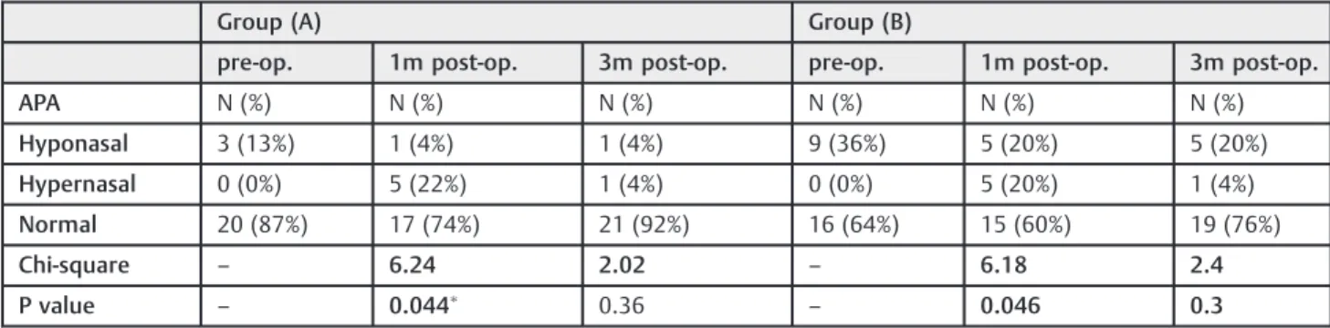
Velopharyngeal nasoendoscopy showed competent (nor-mal) closure of velopharyngeal valve in all study participants. Upon reviewing the APA results from group (A), they revealed the presence of hyponasal speech in 3 out of the 23 patients (13%), while the rest of the group showed normal speech. One month postoperative assessment revealed that only one patient (4%) had hyponasality while 5 (22%) had hypernasality. These results showed a highly significant dif-ference when compared with the preoperative results. Finally, three months postoperative assessment showed that only one patient (4%) had hyponasality and another one (4%) had hypernasality. These results, when compared with the pre-operative ones, showed non-significant difference (►Table 1). With regards to the results of the nasometry for the same group (group A), preoperative nasalance scores were 113 for oral sentence and 495 for nasal sentence. These scores increased one month postoperatively to 194 for oral sentence and 594 for nasal sentence and this increase is
statistically highly significant. Then, these scores return to be near the preoperative scores 3 months postoperatively as they were 135 for oral sentence and 523 for nasal sentence with non-significant increase when compared with the preoperative scores (►Table 2).
Upon reviewing the results of group (B), preoperative APA results revealed 9 out of 25 patients (36%) with hyponasal speech, while the rest of the group showed normal speech. One month postoperative APA assessment revealed that only 5 out of 25 patients (20%) still had hyponasality, while 5 (20%) had developed hypernasality with highly significant differ-ence when compared with the preoperative results. At three months postoperatively, the APA assessment showed that only 5 out of 25 patients (20%) still had hyponasality and only 1 out of 25 (4%) still had hypernasality, with non-significant difference when compared with the preoperative results (►Table 1).
With regards to the results of the nasometry for the same group (group B), preoperative nasalance scores were 84 for oral sentence and 445 for nasal sentence. At one month postoperatively they were 175 for oral sentence and 556 for nasal sentence with highly statistically significant difference, when compared with the preoperative scores. Then, three months postoperatively the scores were 93 for oral sentence and 463 for nasal sentence with non-significant difference when compared with the preoperative scores (►Table 2).
Table 2 Preoperative and postoperative results of nasometry of both groups
Group (A) Group (B)
pre-op. 1m post-op. 3m post-op. pre-op. 1m post-op. 3m post-op.
Nasometry MeanSD MeanSD MeanSD MeanSD MeanSD MeanSD
Oral sentence 113 194 135 84 175 93
T – 7.67 1.65 – 7.03 4
P – 0.001 0.1 – 0.001 0.3
Nasal sentence 495 594 503 445 556 463
T – 7.49 0.82 – 6.75 1.71
P – 0.001 0.41 – 0.001 0.09
Abbreviations: pre-op., preoperative; post-op., postoperative; SD, standard deviation.
Table 1 Preoperative and postoperative results of APA for both groups
Group (A) Group (B)
pre-op. 1m post-op. 3m post-op. pre-op. 1m post-op. 3m post-op.
APA N (%) N (%) N (%) N (%) N (%) N (%)
Hyponasal 3 (13%) 1 (4%) 1 (4%) 9 (36%) 5 (20%) 5 (20%)
Hypernasal 0 (0%) 5 (22%) 1 (4%) 0 (0%) 5 (20%) 1 (4%)
Normal 20 (87%) 17 (74%) 21 (92%) 16 (64%) 15 (60%) 19 (76%)
Chi-square – 6.24 2.02 – 6.18 2.4
P value – 0.044 0.36 – 0.046 0.3
Thus, nasometry for both oral and nasal sentences also showed significant increase in nasalance scores during the one-month postoperative assessment and then decreased again to near preoperative results at three months post-operative in both groups.
Discussion
Although the importance of certain supraglottic airspace resonators on the primary laryngeal sound is generally ac-knowledged, there is controversy concerning the real contri-bution of the nasal cavity and paranasal sinuses on speech.12 Nasality is one of the resonance disorders. Subjective judgments of nasality are made based on the perceptions of speech pathologists. However, subjective judgments often are incorrect. Therefore, several attempts have been made to objectively evaluate nasality.2
The development of computerized acoustic analysis sys-tems as an objective measure of voice has become readily available using a simple noninvasive technique.13
Many studies have been conducted worldwide to assess nasality in patients with cleft palate, motor speech disorders, hearing impairment, and functional nasality problems, but researchers must pay more attention to the relationship between nasality and functional endoscopic sinus surgery (FESS).6–8
The current study aimed at studying the relation between nasality and various types of endonasal surgeries including, FESS, septoplasty, and turbinoplasty. We also tried to estimate whether the effect of these endonasal surgeries on nasalance scores is permanent or temporary in terms of regaining preoperative values after a considerable period of time.
Results of both APA and nasometry in group (A) denoted that CRS may lead to mild degree of hyponasality (preopera-tive results). When CRS was managed by FESS, there was a highly significant degree of hypernasality, which appeared in the results of one month postoperative APA and nasometry. However, incidence of hypernasality decreased in the three months postoperative assessment (►Tables 1and2). These results agreed with those of Soneghet et al,12which found that FESS, despite being a minimally invasive technique, may lead to hypernasal speech.
Hong et al6also reported that the mean value of nasalance in patients with nasal polyposis was significantly lower than that of the healthy controls before FESS, but three weeks after surgery the patients’mean values had improved and were equal to those of the healthy controls.
Results of group (B) (septoplasty and turbinate surgery group) showed some similarity to those of group (A), but the incidence of preoperative hyponasality was higher than in group (B) (►Tables 1and2). This may be due to the presence of actual nasal obstruction in this group.
The results of the nasometry in both groups were nearly similar to other researchers who used different types of naso-metric languages for evaluation and scoring of nasalance.14,15 These results were also similar to several researches,16–18which demonstrated significant increases in nasalance scores, suggest-ing an increase in nasal acoustic energy.
This also, matched the opinions which reported that the anterior nasal obstruction due to septal deviation or hyper-trophy of the inferior turbinate increases the resistance to nasal airflow and sound transmission by reducing nasal airway patency. This may create enough impedance to reduce or even prevent sound from entering the nasopharynx, even when the velopharyngeal port is open during speech. On the other hand, obstruction of the anterior nasal cavity may add acoustic aspects to the speech signal that result in“ cul-de-sac”resonance.17,18
Although septoplasty does not affect the larynx or changes the structure of the vocal tract it decreases the pitch of the voice and the resonance so, it can consequently decreases the nasal resonance values which improve speech quality.15–18
The results of both groups are matched with those of Behrman et al,19who stated that the decreased tissue surface area and widened nasal passages after surgery will decrease the acoustic damping and increase the acoustic coupling with the paranasal sinuses, thereby increasing the amplitude or energy of the voice.
The increase of the nasalance in thefirst month after the operation is due the fact that the nasal cavity is usually covered with a mucosal crusting, resulting in decreased vibration. Decreased energy dampening, in turn, leads to more energy transfer to the nasal cavity.20
Normalization of nasality within an average of six weeks postoperatively may be due to the decrease of crusts and healing of the nasal and sinus cavity mucosa; subsequently, mucosal vibration and dampening function may normalize. In addition, if the middle turbinates properly cover the ethmoid and maxillary ostium, resonance might occur separately in the sinus and nasal cavities. As the patients have normal velopharyngeal function, the airflow regulation according to the changed nasal/oral impedance may also contribute to normalization of nasality.21
Endonasal operations cause widening of the nasal reso-nating cavities. This widening effect is not obvious during the early days and weeks after surgery due to the effect of edema and congestion which subside gradually. The widening of the nasal resonating cavities reaches its maximum after one month, which enhances nasal resonance and leads to in-creased nasalance. With time, the size of these cavities may decrease slightly again, leading to decreased nasalance again. To the best of our knowledge, this was the first study dealing with the effect of endonasal surgeries on nasalance to be conducted on Arabic speakers. We also used the naso-metric Arabic nasal and oral sentences.
Future studies are still needed for further evaluation of postoperative changes in nasalance scores in patients with severe disease and extensive nasal polyposis.
Conclusion
FESS, septoplasty, and turbinate surgery may lead to hyper-nasal speech, which may be due to changes in the shape and diameter of the resonating vocal tract.
Surgeons should inform their patients about the possibi-lity of temporary hypernasapossibi-lity after these types of surgery, especially if they are professional voice users.
References
1 Sataloff RT. Clinical anatomy and physiology of the voice. In: Professional Voice: The Science and Art of Clinical Care. New York, NY: Raven Press; 1991:7–18
2 Jiang RS, Huang HT. Changes in nasal resonance after functional endoscopic sinus surgery. Am J Rhinol 2006;20(04):432–437 3 Fletcher SG, Adams LE, McCutcheon MJ. Nasalance shaping
rou-tines. In: Instruction Manual for the Nasometer Model 6200. Pine Brook, NJ: Kay Elemetrics Corp; 1988
4 Dalston RM, Warren DW, Dalston ET. Use of nasometry as a diagnostic tool for identifying patients with velopharyngeal im-pairment. Cleft Palate Craniofac J 1991;28(02):184–188, discus-sion 188–189
5 Dalston RM, Warren DW, Dalston ET. A preliminary investigation concerning the use of nasometry in identifying patients with hyponasality and/or nasal airway impairment. J Speech Hear Res1991a;34(01):11–18
6 Hong KH, Kwon SH, Jung SS. The assessment of nasality with a nasometer and sound spectrography in patients with nasal poly-posis. Otolaryngol Head Neck Surg 1997;117(04):343–348 7 Hosemann W, Göde U, Dunker JE, Eysholdt U. Influence of
endo-scopic sinus surgery on voice quality. Eur Arch Otorhinolaryngol 1998;255(10):499–503
8 Chen MY, Metson R. Effects of sinus surgery on speech. Arch Otolaryngol Head Neck Surg 1997;123(08):845–852
9 Kotby N, Abdel Haleem EK, Hegazi M, Safe I, Zaki M. Aspects of assessment and management of velopharyngeal dysfunction in developing countries. Folia Phoniatr Logop 1997;49(03/04): 139–146
10 Emara TA, Quriba AS. Posterior pharyngealflap for velopharyngeal insufficiency patients: a new technique forflap inset. Laryngo-scope 2012;122(02):260–265
11 Golding-Kushner KJ, Argamaso RV, Cotton RT, et al. Standardiza-tion for the reporting of nasopharyngoscopy and multiview videofluoroscopy: a report from an International Working Group. Cleft Palate J 1990;27(04):337–347, discussion 347–348 12 Soneghet R, Santos RP, Behlau M, Habermann W, Friedrich G,
Stammberger H. Nasalance changes after functional endoscopic sinus surgery. J Voice 2002;16(03):392–397
13 Dang J, Honda K, Suzuki H. Morphological and acoustical analysis of the nasal and the paranasal cavities. J Acoust Soc Am 1994; 96(04):2088–2100
14 Kim YH, Lee SH, Park CW, Cho JH. Nasalance change after sinonasal surgery: analysis of voice after septoturbinoplasty and endoscopic sinus surgery. Am J Rhinol Allergy 2013;27(01):67–70
15 Mora R, Jankowska B, Dellepiane M, Mora F, Crippa B, Salami A. Acoustic features of voice after septoplasty. Med Sci Monit 2009; 15(06):CR269–CR273
16 Van Lierde KM, Wuyts FL, De Bodt M, Van Cauwenberge P. Age-related patterns of nasal resonance in normal Flemish children and young adults. Scand J Plast Reconstr Surg Hand Surg 2003; 37(06):344–350
17 Pegoraro-Krook MI, Dutka-Souza JCR, Williams WN, Teles Magal-hães LC, Rossetto PC, Riski JE. Effect of nasal decongestion on nasalance measures. Cleft Palate Craniofac J 2006;43(03):289–294 18 Watterson T, Lewis KE, Deutsch C. Nasalance and nasality in low pressure and high pressure speech. Cleft Palate Craniofac J 1998; 35(04):293–298
19 Behrman A, Shikowitz MJ, Dailey S. The effect of upper airway surgery on voice. Otolaryngol Head Neck Surg 2002;127(01):36–42 20 Borden GJ, Harris KS, Raphael LJ. Acoustics. In Physiology, Acous-tics, and Perception of Speech Raphael LJ (Eds). Philadelphia: Lippincott Williams & Wilkins; 2007:31–53