Adenoid Cystic Carcinoma Mimicking an
Oroantral Fistula: A Case Report
Bárbara Vanessa de Brito Monteiro
1Rafael Grotta Grempel
2Daliana Queiroga de Castro Gomes
3Gustavo Pina Godoy
3Márcia Cristina da Costa Miguel
11Oral Pathology Postgraduate Program, Federal University of Rio
Grande do Norte, Natal/RN, Brazil
2Dental School, State University of Paraiba, Araruna/PB, Brazil 3Dentistry Postgraduate Program, State University of Paraiba,
Campina Grande/PB, Brazil
Int Arch Otorhinolaryngol 2014;18:221–225.
Address for correspondence Márcia Cristina da Costa Miguel, PhD, Departamento de Odontologia, Programa de Pós Graduação em Patologia Oral, Universidade Federal do Rio Grande do Norte, Av. Senador Salgado Filho, 1787, Lagoa Nova, Natal/RN, CEP 59056-000, Brasil (e-mail: [email protected]).
Introduction
Adenoid cystic carcinoma (ACC) is one of the most frequent malignant salivary tumors, being more common in the minor salivary glands of the mouth, uncommon in the parotid glands, and rare in the nose and paranasal sinuses.1ACC is more prevalent in the sixth decade of life,2,3shows slow and indolent growth, and is frequently associated with distant metastases and high rates of recurrence of the primary tumor.4,5As a consequence, treatment is difficult2and the long-term survival rates are low.5 Histologically, ACC is characterized by three distinct growth patterns: cribriform, tubular, and solid; tumors presenting mainly solid features have a poor prognosis.6
Treatment modalities for ACC vary according to the stage of the tumor and include surgical resection with or without neck dissection depending on the existence of a clinical or radiologic suspicion of lymph node metastases.7According
to da Cruz Perez et al,3 the combination of surgery and radiotherapy is associated with better overall survival. In general, combined therapies are indicated more than surgery alone.2
The objective of the present study was to describe the case of a 50-year-old man with ACC of the maxillary sinus exteri-orized to the oral cavity. In addition, the literature was reviewed regarding the clinical and histopathologic features of ACCs occurring in the maxillary sinus glands.
Literature Review and Differential Diagnosis
ACC is a malignant tumor that can arise from a variety of anatomical sites, including the major and minor salivary glands, lacrimal glands, skin, and breasts. Most cases of head and neck ACC arise from the minor salivary glands (60%), occur more frequently in the palate, and can also involve the mucous glands of the upper respiratory tract.2,6Malignant sinonasal
Keywords
►
carcinoma
►
adenoid cystic
►
maxillary sinus
neoplasms
►
maxillary sinus
Abstract
Introduction
Adenoid cystic carcinoma (ACC) is one of the most frequent malignant
salivary gland tumors, which commonly affects the minor salivary glands of the mouth
and is rare in the nose and paranasal sinuses. In the maxillary sinus, ACC can mimic
in
fl
ammatory diseases and has a poor prognosis.
Objective
To report a case of a 50-year-old man with ACC of the maxillary sinus whose
clinical
fi
ndings in the alveolar ridge mimicked an oroantral
fi
stula.
Resumed Report
An excisional biopsy was performed and histopathologic analysis
revealed ACC. Lung metastases and residual tumor in the maxillary sinus were detected
by imaging methods. In view of the poor general health of the patient, no new surgical
intervention was performed and he was only treated by radiotherapy and follow-up.
Conclusion
Although rare in the maxillary sinus, ACC should be included in the
differential diagnosis of lesions affecting this site.
received May 2, 2013 accepted May 26, 2013
DOI http://dx.doi.org/ 10.1055/s-0033-1352507. ISSN 1809-9777.
Copyright © 2014 by Thieme Publicações Ltda, Rio de Janeiro, Brazil
tumors are relatively rare, accounting for only 3% of all cancers of the upper respiratory tract.8ACC of the maxillary sinus corresponds to 10% of all cases of sinonasal tract malignancies,9 and it is the second most common sinonasal tumor.8ACC is one of the most common malignant salivary gland tumors and is characterized by different histologic patterns, a variable clini-cal behavior,10and a prolonged clinical course.11
A slight preference of salivary gland tumors for men has been reported in the literature, and this preference has been specifically observed in the case of ACC.3 However, other studies have shown a preference of ACC for women.2Most patients with ACC have an asymptomatic mass that may have been present for months or even years before diagnosis. The tumor is characterized by a prolonged natural history and slow growth even in cases that develop local recurrence and distant metastases.12
ACC arising from the minor salivary glands has usually reached an advanced stage at the time of diagnosis, and its complete excision is limited by the large size of the tumor and proximity to important neural and vascular structures.6ACC of the maxillary sinus can be asymptomatic or can produce symptoms such as nasal obstruction, epistaxis, facial pain, nasal discharge, loss of smell, swelling, headache, and pares-thesia. These highly variable symptoms can mimic infl am-matory diseases such as chronic sinusitis, leading to a delay in diagnosis.8,13This fact was observed in the present case, in which the patient reported to have chronic sinusitis and the
initial diagnosis for the clinically observable lesion in the alveolar ridge was an oroantralfistula.
Case Report
A 50-year-old white man was seen at the Interdisciplinary League Against Oral Cancer, Paraiba State University, for examination of a painful lesion in the alveolar ridge. The patient reported slow growth of the lesion. He had a medical history for chronic sinusitis, in addition to a stroke 4 years earlier. Physical evaluation showed a slightly white and soft lesion in the right maxillary alveolar ridge, and closer exami-nation revealed the presence of an oroantral communication. On the basis of the hypothesis of an oroantralfistula estab-lished by the clinician, an excisional biopsy was performed.
The material was sent for anatomopathological analysis, and the histopathologic diagnosis was ACC. Analysis revealed the proliferation of round or cuboidal epithelial cells with scarce cytoplasm and large hyperchromatic oval nuclei. The epithelial cells were arranged in pseudocystic structures, corresponding to the cribriform pattern (►Fig. 1A), or in
tubular structures (►Fig. 1B), corresponding to the tubular
subtype of ACC. Few small islands of the solid pattern were also observed. The stroma consisted of densefibrovascular connective tissue, with the observation of extensive areas of hyalinization. In addition, the ciliated cylindrical
pseudostratified epithelium lining the maxillary sinus muco-sa showed continuation with the tumor (►Fig. 1C,D).
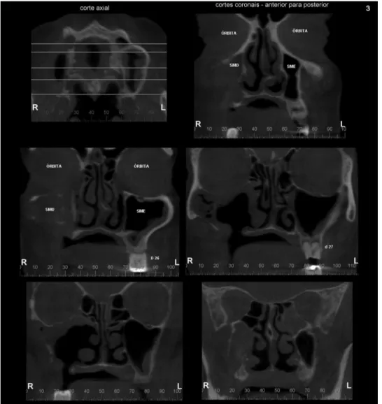
In view of the established diagnosis of ACC, control imag-ing exams were requested, which showed a radiodense image in the right maxillary sinus corresponding to the loss of definition of perisinus bone contours (►Figs. 2and3). Normal
transparency was noted in the other anterior paranasal cavities. The patient reported episodes of epistaxis.
The patient was referred to an oncologist and radiotherapy was recommended. The patient underwent 24 sessions con-sisting of daily irradiations of 4000 cGy in the right maxillary sinus in an attempt to reduce the size of the tumor, which was achieved in part. New imaging exams were performed after radiotherapy. Computed tomography revealed opacity in the superior lobes of both lungs, suggestive of secondary dissem-ination. Material of soft tissue density occupying part of the right maxillary sinus causing nasal septum deviation to the left was compatible with residual tumor. Clinical examination showed expansion and the absence of coalescence of the borders of the tumor in the maxillary ridge (►Fig. 4).
The patient underwent no further surgical intervention due to his poor general health and the fact that ACC is
associated with long survival even without any intervention. Ten months after the first computed tomography scan, a control exam was requested. Analysis showed an irregular bone contour accompanied by osteolytic foci in the right orbital floor, but no significant changes in the size of the tumor were observed. The patient is followed carefully.
Discussion
ACCs of the nose and paranasal sinuses are rare and show a peculiar clinical history. These tumors commonly arise from a mass or epistaxis and have propensity for perineural invasion and early hematogenous spread.1This could be observed in part in the present case, with the patients showing recurrent episodes of nosebleed. According to Lloyd et al,14ACCs grow unnoticed and are therefore diagnosed late, a fact contribut-ing to the poor prognosis of these tumors and the difficulty in achieving complete surgical resection.
The computed tomography images and panoramic radiog-raphy showed the presence of infiltrative growth toward the bone limits of the walls of the right maxillary sinus, and the tumor was only clinically noticed after rupture of the cortical
bone of the hard palate and exteriorization. Opacification of the affected maxillary sinus, osteolysis, extension to the orbitalfloor, and destruction of the sinus walls are common
findings in the cases reported in the literature.1
ACC is histologically characterized by the proliferation of round or cuboidal cells containing scarce cytoplasm and large, oval and hyperchromatic nuclei. These cells form sheets or islands and are usually surrounded by an abundant hyaline
stroma. ACC exhibits pseudocystic structures formed by neoplastic cells of epithelial or myoepithelial phenotype.4
The distinct histopathologic patterns of ACC are related to the prognosis of the disease. In the present case, the cribri-form and tubular patterns were the predominant types, but small islands of the solid pattern were also identified. The solid subtype of ACC has shown a poorer prognosis.6
Sinonasal tumors are uncommon, therefore difficulties exist in defining the characteristics and treatment options for the different histologic entities that can arise in this area. ACC continues to be difficult to treat and no standard therapy has been established for this tumor despite the large number of studies. Broad surgical excision is the treatment of choice; however, complete resection is often not achieved because of vascular invasion and perineural infiltration of the tumor.5
Postoperative radiotherapy is necessary, although ACC is considered to be radiosensitive but not radiocurable.5 Lupinetti et al observed significant improvement of overall survival in patients who had combination therapy when compared with those who had other treatment modalities such as surgery or radiotherapy alone.13In addition, postop-erative radiotherapy seems to increase local and regional control of the tumor and improves overall outcome.5,7In the present case, after the diagnosis of ACC, radiotherapy was chosen in view of the poor prognosis factors, including tumor
Fig. 3 Computed tomography showing material of soft tissue density occupying part of the right maxillary sinus causing nasal septum deviation to the left and loss of definition of perisinus bone contours.
location, identification of areas (although small) of the solid pattern, and the relatively large size of the tumor.
In addition, Gil et al demonstrated that ACCs of the para-nasal sinuses have a high propensity for perineural inva-sion.15 The lungs are the most frequently affected sites of distant metastases, followed by bone, liver, and brain. In-volvement of the brain may occur by direct extension.
Chemotherapy has not yet been established as an effective treatment modality of ACC.13Despite the success obtained with the combination of therapies, controversy still exists regarding the best treatment for advanced cases of ACC of the maxillary sinus. Although the present patient had lung metastases, chemotherapy was not indicated because of his unfavorable health condition and because this treatment has not been established as an effective tool for cases like the present one. Recurrences indicate that the tumor is practical-ly incurable, but survival after recurrence might be long.1
ACC of the head and neck has been characterized as a malignancy whose survival curve drastically declines after 5 years.6However, the extremely slow growth of the tumor and the gradual occurrence of metastases permit the patient to live a normal life for many years.5Overall 5-, 10-, 15-, 20-, 25-, and 30-year survival rates of 77.3, 59.3, 44.9, 35.0, 25.5, and 20.5%, respectively, have been reported.14 The 5-year survival rate for ACC of the maxillary sinus is 62.9%.13
Conclusion
In this reported case, due to the fact that ACCs are rare in maxillary sinus along with the clinical presentation that mim-icked an inflammatory disease, there was a delay in diagnosis and, consequently, a poor prognosis. However, the best treat-ment option was chosen after the established diagnosis.
References
1 Kumar VP, Rao PN, Kumar GA. Adenoid cystic carcinoma of nasal cavity—a case report. Indian J Otolaryngol Head Neck Surg 2003; 55:43–46
2 Kokemueller H, Eckardt A, Brachvogel P, Hausamen JE. Adenoid cystic carcinoma of the head and neck—a 20 years experience. Int J Oral Maxillofac Surg 2004;33:25–31
3 da Cruz Perez DE, Pires FR, Lopes MA, de Almeida OP, Kowalski LP. Adenoid cystic carcinoma and mucoepidermoid carcinoma of the maxillary sinus: report of a 44-year experience of 25 cases from a single institution. J Oral Maxillofac Surg 2006;64:1592–1597
4 Freitas VM, Scheremeta B, Hoffman MP, Jaeger RG. Laminin-1 and SIKVAV a laminin-1-derived peptide, regulate the morphology and protease activity of a human salivary gland adenoid cystic carci-noma cell line. Oral Oncol 2004;40:483–489
5 Mano T, Wada N, Uchida K, Muraki Y, Nagatsuka H, Ueyama Y. Central adenoid cystic carcinoma of the mandible with multiple bone metastases: case report. J Oral Maxillofac Surg 2010;68:446–451
6 Bradley PJ. Adenoid cystic carcinoma of the head and neck: a review. Curr Opin Otolaryngol Head Neck Surg 2004;12: 127–132
7 Gomez DR, Hoppe BS, Wolden SL, et al. Outcomes and prognostic variables in adenoid cystic carcinoma of the head and neck: a recent experience. Int J Radiat Oncol Biol Phys 2008;70:1365–1372
8 Rhee CS, Won TB, Lee CH, et al. Adenoid cystic carcinoma of the sinonasal tract: treatment results. Laryngoscope 2006;116: 982–986
9 Lin WY, Hsu WH. Tumor-to-tumor metastasis: maxillary sinus adenoid cystic carcinoma metastasizing to double primary lung adenocarcinoma. Ann Thorac Surg 2010;90:e59–e61
10 Dodd RL, Slevin NJ. Salivary gland adenoid cystic carcinoma: a review of chemotherapy and molecular therapies. Oral Oncol 2006;42:759–769
11 Singh S, Gokkulakrishnan, Jain J, Pathak S, Singh KT. Adenoid cystic carcinoma of buccal mucosa. J Maxillofac Oral Surg 2010;9: 273–276
12 Cohen AN, Damrose EJ, Huang RY, Nelson SD, Blackwell KE, Calca-terra TC. Adenoid cystic carcinoma of the submandibular gland: a 35-year review. Otolaryngol Head Neck Surg 2004;131:994–1000
13 Lupinetti AD, Roberts DB, Williams MD, et al. Sinonasal adenoid cystic carcinoma: the M. D. Anderson Cancer Center experience. Cancer 2007;110:2726–2731
14 Lloyd S, Yu JB, Wilson LD, Decker RH. Determinants and patterns of survival in adenoid cystic carcinoma of the head and neck, including an analysis of adjuvant radiation therapy. Am J Clin Oncol 2011;34:76–81