Arq Bras Cardiol volume 75, (nº 5), 2000
Bertola et al Cardiac findings in Noonan syndrome
4 0 9
Unidade de Genética do Instituto da Criança Faculdade de Medicina da Universidade de São Paulo
Mailing address: Chong Ae Kim – Genética – InCor – Av. Dr. Enéas C. Aguiar, 647 – 05403-900 - São Paulo, SP
Objective - To evaluate cardiac findings in 31
Noo-nan syndrome patients.
Methods – Thirty-one (18 males and 13
females)pa-tients from 26 families affected with Noonan’s syndrome were evaluated from the cardiac point of view with electro-cardiography and echodopplerelectro-cardiography.
Results – Twenty patients had some type of cardiac
abnormality. The most frequent was pulmonary valve ste-nosis followed by hypertrophic myocardiopathy, common-ly associated with valve defects. Upper deviation of the QRS axis was observed in 80% of these patients.
Conclusion - In view of the high frequency and
diver-sity of cardiac abnormalities present in Noonan syndrome, cardiac evaluation with electrocardiography and echo-cardiography should be performed in all patients diag-nostically suspected of having this disease.
Key words: Noonan syndrome, pulmonary valve steno-sis, hypertrophic myocardiopathy .
Arq Bras Cardiol, volume 75 (nº 5), 409-412, 2000
Débora Romeo Bertola, Chong Ae Kim, Sofia M. M. Sugayama, Lilian Maria José Albano, Jaqueline Wagenführ, Regina Lúcia Moysés, Claudette Hajaj Gonzalez
São Paulo, SP - Brazil
Cardiac Findings in 31 Patients with Noonan’s
Syndrome
Original Article
Noonan syndrome is a genetic disease of autosomal dominant inheritance, characterized by low stature, cranio-facial dysmorphism, webbed neck, cardiac abnormalities, cryptorchism in male patients, skeletal anomalies, and he-morrhagic diathesis.
Noonan and Ehmke recognized the disease as a dis-tinct entity in 1963 1; since then many cases have been described in the literature. Its incidence has been estimated at 1/1000 to 1/2500 live births 2, making it one of the most frequent syndromes associated with cardiac defects.
The most frequent cardiac abnormality in Noonan’s syndrome is pulmonary valve stenosis, present in approxima-tely 50% of the patients 3, followed by hypertrophic myocar-diopathy occurring in 25% 4. Both anomalies have peculia-rities in Noonan’s syndrome; in pulmonary valve stenosis, valves are often dysplasic, differing from the dome format and without the commissural fusion observed in nonsyndromic forms; hypertrophic myocardiopathy is frequently asso-ciated with cardiac defects, in particular, pulmonary valve stenosis. Although these cardiac findings are the most com-mon ones 5, practically all other types of cardiac anomalies have been encountered in Noonan syndrome.
The electrocardiograms of affected patients com-monly show an upper deviation of the QRS axis and deep S waves in the precordial derivations; these findings are not associated with a specific cardiac anomaly 5.
The gene responsible for Noonan syndrome has been mapped on the long arm of chromosome 12 6, but in some families of affected patients, this connection was not ob-served, indicating the genetic heterogeneity of the disease. Our objective was to study cardiac findings in indivi-duals with Noonan’s syndrome.
Methods
4 1 0
Bertola et al
Cardiac findings in Noonan syndrome
Arq Bras Cardiol volume 75, (nº 5), 2000
Noonan Syndrome diagnoses of these individuals, were based on the clinical criteria established by van der Burgt et al. 7. Twenty-six proposed patients and their first-degree relatives (parents and siblings) were evaluated; re-currence of the disease was observed in five relatives. One of the chosen patients was an adopted child and another’s father was not available for evaluation. Of the 29 remaining cases, 8 (28%) were familial and 21 (72%) sporadic. Eighteen patients were males and 13 were females, with ages ranging from 3 months to 41years (average 12 years) . One of the patients had already passed away at the time of the study; his data were obtained from medical files; photographs were used to analyze some of his craniofacial characteristics. The impossibility of detecting some of the craniofacial dysmorphisms in this patient is reflected in the total number of these findings shown (Table I).
The cardiac evaluation was based on a special physi-cal examination, on the electrocardiogram, and on the echo-cardiogram.
Subjects affected with Noonan’s syndrome were only included in the study after signing an informed consent, for themselves when adults, or by legal guardians when unde-rage.
Results
The most commonly observed clinical findings in pa-tients with Noonan’s syndrome were: low stature, cranio-facial dysmorphism, short or webbed, cardiac anomalies, sternal deformity, and the presence of pads at the tip of the fingers and toes (Table I).
Chromosome studies showed normal results for all patients.
Twenty (65%) of the patients had some form of echo-cardiographic abnormality (Table II).
Signs of right or left ventricular hypertrophy were pre-sent in 13 and 5 patients, respectively (Table III).
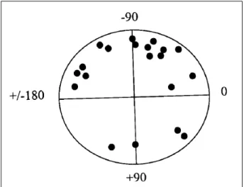
Upper deviation of the QRS axis was a rather common finding in 80% of these patients.
Discussion
Cardiac anomalies are common in Noonan syndrome and the major cause of morbidity and mortality in this disease.
In the present study, 65% of affected individuals had some form of cardiac abnormality. This frequency is higher than that estimated in other works (3,8), probably because the majority of our patients were sent by the Heart Institute and were known to have a cardiac anomaly, and also because a Genetic Unit of a tertiary hospital, in this case the Children’s Institute, tends to receive only the more serious cases.
A cardiac murmur was heard in 12 (60%) of the neona-tes with cardiac problems, allowing for a precocious, ade-quate follow-up.
In agreement with literature reports, the most frequent cardiac anomaly was pulmonary stenosis, found in 70% of
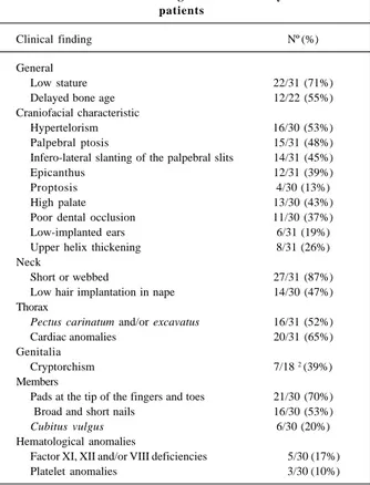
Table I – Clinical findings in 31 Noonan syndrome patients
Clinical finding Nº (%)
General
Low stature 22/31 (71%)
Delayed bone age 12/22 (55%)
Craniofacial characteristic
Hypertelorism 16/30 (53%)
Palpebral ptosis 15/31 (48%)
Infero-lateral slanting of the palpebral slits 14/31 (45%)
Epicanthus 12/31 (39%)
Proptosis 4/30 (13%)
High palate 13/30 (43%)
Poor dental occlusion 11/30 (37%) Low-implanted ears 6/31 (19%) Upper helix thickening 8/31 (26%) Neck
Short or webbed 27/31 (87%)
Low hair implantation in nape 14/30 (47%) Thorax
Pectus carinatum and/or excavatus 16/31 (52%) Cardiac anomalies 20/31 (65%) Genitalia
Cryptorchism 7/18 2 (39%)
Members
Pads at the tip of the fingers and toes 21/30 (70%) Broad and short nails 16/30 (53%)
Cubitus vulgus 6/30 (20%)
Hematological anomalies
Factor XI, XII and/or VIII deficiencies 5/30 (17%) Platelet anomalies 3/30 (10%)
(1)Two standard deviations below average; (2) total number of male patients.
Table II - Cardiac anomalies observed in Noonan syndrome patients
Cardiac anomalies Nº %
PVS and/or SVPS 10 50%
EPV/MVP 3 15%
ASD 1 5%
HM 4 20%
VSD 1 5%
Aortic valve thickening 1 5%
Total 20 100%
PVS– pulmonary valve stenosis; SVPS- supravalvar pulmonary steno-sis; MVP- mitral valve prolapse; HM- hypertrophic myocardiopathy; ASD- atrial septal defect; VSD- ventricular septal defect.
Table III – Electrocardiographic findings in Noonan syndrome patients
Findings Nº %
RVO 12 57%
LVO 5 24%
RAO 1 5%
RBB 2 9%
Sinus bradycardia 1 5%
Total 21 100%
Arq Bras Cardiol volume 75, (nº 5), 2000
Bertola et al Cardiac findings in Noonan syndrome
4 1 1
the patients with cardiac problems. The pressure gradient between the right ventricle and the pulmonary arterial trunk varied between 23 and 121mmHg in these patients. Three (21%) had a pressor gradient above 60mmHg characterizing important pulmonary stenosis, 4 (29%) slight stenosis (pressor gradient between 20 and 40mmHg), and 7 (50%) moderate pulmonary stenosis. The most common associated anomalies were interarterial communication and hypertro-phic myocardiopathy. Char et al. 9, studying 45 cases of Noonan’ syndrome, observed interarterial communication to be the most frequent cardiac defect associated with pulmonary stenosis.
Echocardiographic findings showed a dysplasic pul-monary valve in 4 (24%) of our cases of pulpul-monary valve stenosis. Burch et al. 4, in an echocardiographic study of 119 Noonan syndrome patients, diagnosed dysplasic valves in 8 (27%) of 30 of these patients who also had pulmonary stenosis.
Despite the presence of pulmonary valve dysplasia in one third of the patients, the majority of them had no evi-dence of dysplasia on echocardiography; this observation should be considered in the therapy of these individuals.
Percutaneous dilatation with a balloon catheter, the treatment of choice in cases of moderate and serious pulmo-nary stenosis, is rarely effective when the valve is dysplasic 5. Ishizawa et al. 10, who performed this procedure in 4 Noonan syndrome patients with dysplasic pulmonary valves, ob-tained good results in two of them. The authors postulated that valvuloplasty should be performed as an initial proce-dure in patients with Noonan syndrome and pulmonary ste-nosis, even when evidence of valve dysplasia is found. In our study, three patients, one with serious, two with mode-rate pulmonary stenosis, respectively, who underwent valvuloplasty, had a significant reduction in the pressor
gradient of those with moderate stenosis. It is important to point out that none of these three patients had signs of valve dysplasia on the echocardiogram. Four patients with Noonan syndrome underwent surgical correction of pul-monary stenosis (commissurotomy, pulpul-monary valvulo-plasty, myectomy, and amplification of the right ventricle’s outlet by insertion of a bovine pericardium graft), of these two had a pressor gradient above 110mmHg, one had mode-rate pulmonary stenosis, and in one, no reduction of in the pressor gradient was observed following valvuloplasty.
The second most common cardiac anomaly of Noo-nan’s syndrome is hypertrophic myocardiopathy, also ob-served in our study. Differently from the non-syndrome form, Noonan syndrome hypertrophic myocardiopathies are frequently associated with a valve anomaly, mainly pul-monary stenosis. Four of our patients with hypertrophic myocardiopathy also had involvement of pulmonary, aortic, or mitral valves.
Interventricular communication and aortic valve thi-ckening were other cardiac findings diagnosed with echo-cardiography in our study.
The electrocardiogram showed an upper deviation of the QRS axis in 16 (60%) patients with cardiac abnormalities. This was observed in cases of pulmonary valve stenosis, supravalvar pulmonary stenosis, hypertrophic myocardio-pathy, and interventricular communication. However, it was not found in patients affected by Noonan’s syndrome without cardiac anomalies. Neither was there a direct corre-lation between the degree of obstruction of the right ven-tricle’s outlet and axis deviation. The physiopathology of this symptom is not fully known, but appears to involve not only counterclockwise rotation of the heart, but also a dis-turbance in the conduction system of affected patients 11. This electrocardiographic finding may aid in the diagnosis of Noonan syndrome.
Cardiac involvement is rather frequent in Noonan syndrome patients, with peculiarities when compared with nonsyndromic cases. Affected individuals often have thoracic deformities, with pectus carinatum upwards, and excavatum downwards; both deformities are capable of in-terfering with cardiac auscultation. A detailed cardiac eva-luation, including electrocardiographic and echocardiogra-phic examinations, is recommended for every patient with a diagnostic suspicion of Noonan syndrome.
This is a relatively frequent autosomal dominant inhe-rited genic disease; the wide spectrum of its clinical mani-festations or phenotype variability calls for the services of various specialists including pediatricians, endocrinolo-gists, ophthalmoloendocrinolo-gists, cardioloendocrinolo-gists, and hematologists. A better understanding of this heterogeneous syndrome may lead to more adequate follow-up and treatment of affected patients.
4 1 2
Bertola et al
Cardiac findings in Noonan syndrome
Arq Bras Cardiol volume 75, (nº 5), 2000
1. Noonan JA, Ehmke DA. Associated noncardiac malformations in children with congenital heart disease. J Pediatr 1963; 63: 468-70.
2. Nora JJ, Nora AH, Sinha AK, Spangler RD, Lubs HA. The Ullrich-Noonan syn-drome (Turner phenotype). Am J Dis Child 1974; 127: 48-55.
3. Allanson JE - Noonan syndrome. J Med Genet 1987; 24: 9-13.
4. Burch M, Sharland M, Shinebourne E, Smith G, Patton M, McKenna W. Cardio-logic abnormalities in Noonan syndrome: phenotypic diagnosis and echocar-diographic assessment of 118 patients. J Am Coll Cardiol 1993; 22: 1189-92. 5. Noonan J, O’Connor W. Noonan syndrome: a clinical description emphasizing
the cardiac findings. Acta Pediatr Jpn 1996; 38: 76-83.
6. Jamieson CR, van der Burgt I, Brady AF, et al. Mapping a gene for Noonan syndro-me to the long arm of chromososyndro-me 12. Nature Genet 1994; 8: 357-60. 7. van der Burgt I, Berends E, Lommen E, et al. Clinical and molecular studies
References
in a large dutch family with Noonan syndrome. Am J Med Genet 1994; 53: 187-91.
8. Mendez HMM, Opitz JM. Noonan Syndrome: a review. Am J Med Genet 1985; 21: 493-506.
9. Char F, Rodriguez-Fernandez HL, Scott CI, Borgaonkar DS, Bell BB, Rowe RD. The Noonan syndrome – a clinical study of forty-five cases. BD: OAS 1972; VIII: 110-8.
10. Ishizawa A, Oho SI, Dodo H, Katori T, Homma SI. Cardiovascular abnormalities in Noonan syndrome: the clinical findings and treatments. Acta Pediatr Jpn 1996; 38: 84-90.