TECHNICAL NOTE
A new technique for the correction of hypoplastic
left heart syndrome
Roberto Rocha-e-Silva
Hospital das Clı´nicas, Instituto do Corac¸a˜o, Sa˜o Paulo, Brazil.
Email: [email protected] Tel.: 55 11 2661 5014
INTRODUCTION
Hypoplastic left heart syndrome (HLHS) inevitably results in death within a few weeks of birth, and the surgical treatment for this condition involves three complex surgeries that are associated with a high mortality and cost (1). The mortality rates for the first-stage procedure range from 25-80% at medical centers worldwide. Moreover, many children (possibly as many as 50%, according to hospitals where accurate records are kept) with this disease are not referred for surgery and progress to death within days or weeks (2-4). The two conventional techniques that are used for the Norwood procedure (the first-stage surgical correction) involve the construction of a systemic pulmon-ary shunt. This procedure performed using the Blalock-Taussig technique leads to a diastolic systemic pulmonary flow that mimics systemic valve insufficiency. When performed using the Sano modification technique, the Norwood procedure involves building a tube between the systemic ventricle and the pulmonary artery; this technique avoids the diastolic systemic pulmonary flow but allows the blood in the lungs to reflux to the single ventricle, which increases the volume overload. This overload associated with ventriculotomy may lead to dysfunction of the ventricle and arrhythmia. Moreover, both techniques necessitate the use of an artificial tube and carry a risk for thrombosis or stenosis (5-9). Additionally, fabrication of the neo-aorta is a laborious technique that requires the extended use of extracorporeal circulation (ECC).
Hybrid approaches involving the maintenance of the patent ductus arteriosus with the implantation of a stent (10,11) or the prolonged administration of prostaglandin E1 (12) associated with the banding of the pulmonary branches simplifies the surgical approach but have yielded unsatis-factory results (13,14).
PROPOSED PROCEDURE FOR THE CORRECTION OF HLHS, STAGE 1
1. Typical preparations for an intracardiac operation with ECC.
2. Median sternotomy.
3. Dissection of the brachiocephalic trunk and implanta-tion of a 3.5-mm Gore-Tex graft for arterial ECC access.
4. Dissection and cannulation of the femoral artery as a second route of arterial access to maintain systemic perfusion after aortic clamping.
5. Cannulation of both venae cavae.
6. Placement of tourniquets in the left carotid artery, left subclavian artery and descending aorta after the insertion of the ductus arteriosus.
7. Initiation of ECC and ligation of the ductus arteriosus. 8. Placement of a clamp at the aortic arch to isolate the ascending aorta and the brachiocephalic trunk from the systemic circulation, which serves to maintain the cerebral, right superior limb and coronary perfusion. 9. Closure of the left carotid artery, left subclavian artery
and descending aorta tourniquets with the continued maintenance of the descending aortic circulation via the femoral cannula.
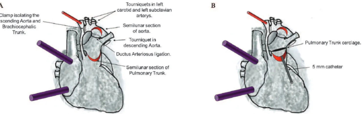
10. Autologous tissue is formed into a flap using a semilunar section of the initial portion of the anterior pulmonary trunk 3 cm above the pulmonary valve (Figure 1).
11. A second semilunar incision is made into the anterosuperior side of the aortic arch near the left carotid and left subclavian artery to create a second flap of autologous tissue (Figure 1A).
12. Introduction of a 5-mm catheter through the opening of the pulmonary artery towards the pulmonary branches. Around this catheter is placed the pulmon-ary trunk cerclage using cardiac tape sutured with 5-0 prolene. This procedure results in a systemic pulmon-ary shunt with autologous tissue (Figure 1B). 13. The free edges of the semilunar flaps of the aorta and
pulmonary trunk are rotated and sutured edge-to-edge, originating the autologous posterior neo-aortic wall (Figure 2A). A second line of stitches is placed to dorsally exclude the ductus arteriosus tissue.
14. A valved bovine pericardium patch is implanted from the anterior opening of the pulmonary trunk to the aortic arch and extends to the beginning of the descending aorta to give rise to the anterior wall of the neo-aorta (Figure 2B). The opening of the valve is placed near the lower edge of the aortic arch. This valve prevents diastolic systemic reflow to the pulmonary arteries and improves coronary perfusion pressure through the ascending aorta (retrograde
Copyrightß2012CLINICS– This is an Open Access article distributed under
the terms of the Creative Commons Attribution Non-Commercial License (http:// creativecommons.org/licenses/by-nc/3.0/) which permits unrestricted non-commercial use, distribution, and reproduction in any medium, provided the original work is properly cited.
No potential conflict of interest was reported.
CLINICS 2012;67(5):521-524 DOI:10.6061/clinics/2012(05)20
flow). The neo-aorta does not require mobilization of the ascending aorta or the pulmonary trunk.
15. A small right atriotomy is performed for atrial septum resection. After the appropriate maneuvers are per-formed to remove air that may be present, the tourniquets are released for subsequent finalization of the ECC.
16. The remainder of the procedure is performed accord-ing to the conventional protocol.
The procedure was simulated in an experimental mechanical model as described below.
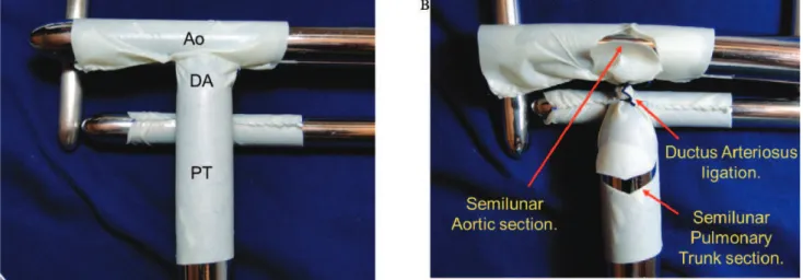
The model was constructed with bovine pericardium tissue (HP Bioprosthesis Ltda.). The pulmonary trunk was represented using an 18-mm pericardial tunnel. On its posterior superior face, a second 12-mm pericardial tunnel was placed to simulate the pulmonary branches. On the cranial face of the pulmonary trunk, an additional 18-mm
pericardium tube was placed to simulate the aortic arch. The pulmonary trunk segment between the pulmonary branches and the aorta represented the ductus arteriosus. All anastomoses were performed with prolene 6-0 (Figure 3A).
The ductus arteriosus was ligated. The semilunar open-ings of the aorta and pulmonary trunk used to form the autologous flaps (to construct the neo-aorta) were created as depicted in Figure 3B.
A 5-mm catheter was introduced through the opening of the pulmonary trunk towards the pulmonary branches. The pulmonary trunk cerclage was obtained by suturing a cardiac tape around the catheter with 5-0 prolene (Figure 4A). Following the removal of the catheter, a 5-mm systemic pulmonary shunt of autologous tissue was created.
The aortic flap was rotated caudally, and the pulmonary trunk flap was rotated cranially. These flaps were then sutured edge-to-edge with 6-0 prolene originating the Figure 1 -A) Cannulation of the brachiocephalic trunk (cannulation of femoral artery not showed in the figure) and double venae cavae for extracorporeal circulation (ECC). Placement of the tourniquet in the left carotid and left subclavian arteries and descending aorta. Subsequently, ECC is initiated, and the ductus arteriosus is ligated. A clamp is then placed at the aortic arch to isolate the ascending aorta and the brachiocephalic trunk from the systemic circulation. Next, the tourniquets are closed, and a semilunar section of the initial portion of the anterior pulmonary trunk (3 cm above the pulmonary valve) is used to create a flap. A second semilunar incision is made into the anterosuperior side of the aortic arch near the left carotid and left subclavian arteries to create a second flap. B) A 5-mm catheter is introduced through the opening of the pulmonary trunk towards the pulmonary branches. The pulmonary trunk cerclage is obtained by suturing a cardiac tape around the catheter with 5-0 prolene.
Figure 2 -A) The free edges of the semilunar flaps of the aorta and pulmonary trunk are rotated and sutured edge-to-edge originating the autologous posterior neo-aortic wall. B) A valved bovine pericardium patch is implanted from the anterior opening of the pulmonary trunk to the aortic arch and extends to the beginning of the descending aorta to give rise to the anterior wall of the neo-aorta.
New technique for HLHS correction
Rocha-e-Silva R CLINICS 2012;67(5):521-524
autologous posterior wall of the neo-aorta. There was no tension in this suture, and no mobilization of the aortic arch was required (Figure 4B).
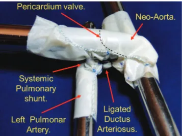
The anterior wall of the neo-aorta was created using a bovine pericardium patch sutured with prolene 6-0 (Figure 5). The valved patch was not used in this model, although the location of the valve position is shown in Figure 4B. The black dashed line indicates where the valve suture would be inserted into the pericardial patch that forms the anterior wall of the neo-aorta.
EXPECTED RESULTS
The construction of an experimental model used to demonstrate the novel technique for the correction of HLHS proved to be simple.
The proposed procedure requires fewer sutures, which excludes the need to implant a heterologous systemic
pulmonary shunt and decreases the surgical time. One patient who met the relevant criteria and required surgical correction was submitted to the procedure (15) and demonstrated that it could potentially reduce the perio-perative complications.
The technique described does not require any period of coronary ischemia or surgical manipulation of the ven-tricle, as described previously (15). The combined effect of these improvements should promote a higher quality of postoperative ventricular function. Moreover, mainte-nance of the anatomy of the ascending aorta simplifies the procedure and avoids distortions in the coronary arteries.
The in situ autologous systemic pulmonary shunt is simple to manufacture and should not generate thrombosis-related complications. In addition, the maintenance of the pulmonary trunk in situ (with the cerclage) should promote better development of the pulmonary tree.
Figure 3 -A) The pulmonary trunk (PT) was represented using an 18-mm pericardial tunnel. A second 12-mm pericardial tunnel is placed on its posterior superior face to simulate the pulmonary branches. An additional 18-mm pericardium tube, which simulated the aortic arch (Ao), is then placed on the cranial face of the pulmonary trunk. The pulmonary trunk segment between the pulmonary branches and the aorta represented the ductus arteriosus (DA). B) The DA is ligated, and the autologous flaps were obtained through the semilunar opening of the aorta and pulmonary trunk.
Figure 4 -A) Through the opening of the pulmonary trunk, a 5-mm catheter is introduced towards the pulmonary branches. The pulmonary trunk cerclage is obtained by suturing a cardiac tape with 5-0 prolene around the catheter. B) The aortic flap is rotated caudally, and the pulmonary trunk flap is rotated cranially. These flaps are then sutured edge-to-edge with 6-0 prolene originating the autologous posterior neo-aortic wall. The red marks indicate where the top of the valve of the pericardium should be placed.
CLINICS 2012;67(5):521-524 New technique for HLHS correction
Rocha-e-Silva R
The use of a valved pericardium for the creation of a neo-aorta should improve systemic perfusion (including the coronary circulation) and prevent diastolic systemic pul-monary reflow.
REFERENCES
1. Dean PN, Hillman DG, McHugh KE, Gutgesell HP. Inpatient Costs and Charges for Surgical Treatment of Hypoplastic Left Heart Syndrome. Pediatrics. 2011;128(5):e1181-6, http://dx.doi.org/10.1542/peds.2010-3742. 2. Karamlou T, Diggs BS, Ungerleider RM, Welke KF. Evolution of treatment options and outcomes for hypoplastic left heart syndrome over an 18-year period. J Thorac Cardiovasc Surg. 2010;139(1):119-26; discussion 126-7, http://dx.doi.org/10.1016/j.jtcvs.2009.04.061. 3. Rychik J, Szwast A, Natarajan S, Quartermain M, Donaghue DD, Combs
J, et al. Perinatal and early surgical outcome for the fetus with hypoplastic left heart syndrome: a 5-year single institutional experience. Ultrasound Obstet Gynecol. 2010;36(4):465-70, http://dx.doi.org/ 10.1002/uog.7674.
4. Hornik CP, He X, Jacobs JP, Li JS, Jaquiss RD, Jacobs ML, O’Brien SM, et al. Complications After the Norwood Operation: An Analysis of The Society of Thoracic Surgeons Congenital Heart Surgery Database. Ann Thorac Surg. 2011;92(5):1734-40, http://dx.doi.org/10.1016/j.athoracsur. 2011.05.100.
5. Raja SG, Atamanyuk I, Kostolny M, Tsang V. In hypoplastic left heart patients is Sano shunt compared with modified Blalock-Taussig shunt associated with deleterious effects on ventricular performance? Interact Cardiovasc Thorac Surg. 2010;10(4):620-3, http://dx.doi.org/10.1510/ icvts.2009.227322.
6. Ru¨ffer A, Danch A, Gottschalk U, Mir T, Lacour-Gayet F, Haun C, et al. The Norwood procedure - does the type of shunt determine outcome? Thorac Cardiovasc Surg. 2009;57(5):270-5, http://dx.doi.org/10.1055/s-0029-1185459.
7. Silva JP, Fonseca L, Baumgratz JF, Castro RM, Franchi SM, Lianza AC, et al. Hypoplastic left heart syndrome: the report of a surgical strategy and comparative results of Norwood x Norwood-Sano approach. Rev Bras Cir Cardiovasc. 2007;22(2):160-8.
8. Raja SG. Right ventricle to pulmonary artery shunt modification of Norwood procedure: Outcomes, concerns, and controversies. Ann Pediatr Cardiol. 2011;4(2):150-1, http://dx.doi.org/10.4103/0974-2069.84654.
9. Loomba RS, Shah PH, Chandrasekar S. Short-term outcome comparison of Norwood procedures with right ventricle to pulmonary artery conduit versus modified Blalock-Taussig shunt: A meta-analysis. Ann Pediatr Cardiol. 2011;4(2):145-9, http://dx.doi.org/10.4103/0974-2069.84653. 10. Bockeria L, Alekyan B, Berishvili D, Pursanov M, Krupianko SM,
Zarginava G, et al. A modified hybrid stage I procedure for treatment of hypoplastic left heart syndrome: an original surgical approach. Interact Cardiovasc Thorac Surg. 2010;11(2):142-5, http://dx.doi.org/10.1510/ icvts.2010.235374.
11. Galantowicz M, Cheatham JP, Phillips A, Cua CL, Hoffman TM, Hill SL, et al. Hybrid approach for hypoplastic left heart syndrome: intermediate results after the learning curve. Ann Thorac Surg. 2008;85(6):2063-70; discussion2070-1, http://dx.doi.org/10.1016/j.athoracsur.2008.02.009. 12. Sakurai T, Kado H, Nakano T, Hinokiyama K, Shiose A, Kajimoto M,
et al. Early results of bilateral pulmonary artery banding for hypoplastic left heart syndrome. Eur J Cardiothorac Surg. 2009;36(6):973-9, http:// dx.doi.org/10.1016/j.ejcts.2009.05.009.
13. Knirsch W, Liamlahi R, Hug MI, Hoop R, von Rhein M, Preˆtre R, et al. Mortality and neurodevelopmental outcome at 1 year of age comparing hybrid and Norwood procedures. Eur J Cardiothorac Surg. 2012 Jan 18. [Epub ahead of print].
14. Photiadis J, Sinzobahamvya N, Hrasˇka V, Asfour B. Does Bilateral Pulmonary Banding in Comparison to Norwood Procedure Improve Outcome in Neonates with Hypoplastic Left Heart Syndrome Beyond Second-Stage Palliation? A Review of the Current Literature. Thorac Cardiovasc Surg. 2012 Jan 3. [Epub ahead of print].
15. Rocha-e-Silva R, Mola R, Santos ES, Martins DM, Pesciotto VR, Hatori DM, et al. Surgical correction of hypoplastic left heart syndrome: a new approach. Clinics. 2012;67(5):535-9, http://dx.doi.org/10.6061/clinics/ 2012(05)24.
Figure 5 -The implantation of a bovine pericardium patch from the anterior opening of the pulmonary trunk to the aortic arch. This patch extends to the beginning of the descending aorta and gives rise to the anterior wall of the neo-aorta. The dashed line represents the valve suture on the pericardium.
New technique for HLHS correction
Rocha-e-Silva R CLINICS 2012;67(5):521-524