Faculdade de Ciências Médicas da Universidade Estadual de Campinas-UNICAMP Mailing address: Marco Antonio Volpe - Rua Baronesa do Japi, 129/13 – 13207-000 - Jundiaí, SP - Brazil
Objective – To describe a surgical procedure
utili-zing a malleable bovine pericardium ring in mitral valve repair and clinical and echodopplercadiographic results.
Methods – Thirty-two (25 female and 7 male)
patien-ts, aged between 9 and 66 (M=36.4±17.2) years, were stu-died over a 16-month period, with 100% follow-up. In 23 (72%) of the patients, the mitral approach was the only one applied; 9 patients underwent associated operations. The technique applied consisted of measuring the perime-ter of the anperime-terior leaflet and implanting, according to this measurement, a flexible bovine pericardium prosthesis for reinforcement and conformation of the posterior mitral annulus, reducing it to the perimeter of the anterior leaflet with adjustment of the valve apparatus.
Results – The patient survival ratio was 93.8%, with
2 (6.2%) fatal outcomes, one from unknown causes, the other due to left ventricular failure. Only one reoperation was performed. On echodopplercardiography, 88% of the patients had functional recovery of the mitral valve (50% without and 38% with mild insufficiency and no hemody-namic repercussions). Of four (12%) of the remaining patients, 6% had moderate and 6% had seigre insufficien-cy. Twenty-eight percent of class II patients and 72% of class III patients passed into classes I (65%), II (32%), and III (3%), according to NYHA classification criteria.
Conclusion – Being flexible, the bovine pericardium
ring fit perfectly into the valve annulus, taking into acco-unt its geometry and contractility. Valve repair was shown to be reproducible, demonstrating significant advantages during patient evolution, which did not require anticoa-gulation measures.
Key words: mitral valve surgery, annuloplasty, bovine pericardium
Arq Bras Cardiol, volume 75 (nº 5), 389-396, 2000
Marco Antônio Volpe, Domingo Marcolino Braile, Reinaldo W ilson Vieira, Dorotéia Rossi Silva Souza
Campinas, SP - Brazil
Mitral Valve Repair with a Malleable Bovine Pericardium Ring
To achieve perfect functioning of the mitral valve, it is necessary to maintain the functional integrity of its constituent elements, avoiding the effects of alteration consequent to congenital or acquired disease 1. These are
capable of causing valve annulus dilatation, commissure fu-sion, posterior cusp hypoplasia, shortening or lengthening of tendinous chords, and anterior or posterior chord rup-ture, leading to mitral insufficiency or stenosis 2.
Lillehey and collaborators in 1956 performed the first annuloplasty to correct mitral insufficiency, under direct vision 3. Subsequently, other surgeons attempted to
gene-ralize annuloplasty by using annulus plication 4. However,
this procedure led to a high rate of recurrence due to dehis-cence, thrombosis, loss of cusp complacency, and other problems 5.
The introduction of valve substitutes led to loss of interest in conservative procedures, and only a few groups continued to perform valve repair in selected cases 6,7.
Prosthesis diversity kept increasing, but patients with valve substituents started to experience complications, like fractures, wear and tear, thromboembolism, infection, hemolysis, rupture, and calcification, directly or indirectly associated with the prostheses 7-9.
Interest in mitral reconstruction returned in the seventies with the work of Carpentier et al., who proposed the utilization of a rigid prosthetic ring for mitral valve reconstruction 7,10,11. These authors also developed several
techniques for the correction of specific anatomical valve abnormalities among which were segmental resection of the posterior leaflet as well as chord shortening, lengthening, grafting, and division 11. These techniques started to be
used together with annuloplasty for recovery of valve function 7,12. Accumulated experience and new concepts
about the physiology of the mitral valve apparatus showed the limitations of this technique, such as fixation of the con-tractile annulus 13,14 and eventual obstruction of left
ventri-cle outflow 15-19.
Complete rings developed by Duran et al. 13,20 and by
Puig et al. 21,22 followed principles similar to these proposed
by Carpentier and colleagues, with the advantage of flexibi-lity respecting native annulus motiflexibi-lity 13,20-22, and reduction
the anterior valve annulus does not participate, to an expressive extent, in the dilatation of the mitral apparatus, and the placing of a complete annular prosthesis may distort it, harming the left ventricleoutflow tract. This observation led to the proposal of an exclusively posterior prosthesis annuloplasty 26 or of a merely continuous
suture, similar to a "De Vega" 25. Evolution confirmed the
superiority of rings for posterior conformation 26-30.
Valve repair techniques using rings are based on the anterior leaflet area, associated or not with intertrigonal or intercommissural distances for the selection of annular prosthesis size 10,13,27-30. In 1985, Braile et al. 26 developed
specific measuring devices aimed at the evaluation of the anterior cusp perimeter, thus introducing a new concept for the choice of annular prosthesis size. They utilized a fle-xible, radiopaque bovine pericardium ring for reinforcement and remodeling of the posterior mitral annulus, reducing it to the perimeter of the anterior leaflet.
The present study had as its objective the description as well as postoperative clinical and echodopplercardiogra-phic evaluation of a mitral valve repair technique using mal-leable bovine pericardium rings.
Methods
Thirty-two (25 female and 7 male) patients aged between 9 and 66 (M=36.4±17.2) years were studied. Pati-ents were operated upon with mitral valve repair with mal-leable bovine pericardium prostheses implanted in the pos-terior annulus, between August ’94 and December ’95 at the Cardiac Surgery Clinic of the Surgery Department of the School of Medical Sciences of the State University of Cam-pinas.
Rheumatic disease was the cause of valve lesions in 28 (87.5%) of the subjects, followed by myxomatous degenera-tion in 4 (12.5%). Only one patient had had previous surge-ry for replacement of the aortic valve by a mechanical pros-thesis. Table I presents the lesions that called for valve repair, emphasizing double mitral lesions, detected in 14 (44%) and pure mitral insufficiency detected in 12 (37%) of the patients.
The period of extracorporeal circulation varied from 51 to 132 (mean, 80.2±18.8) minutes, and aortic clamping varied between 29 and 100 (mean, 57.9±16.1) minutes. Cardiople-gics were administered by continuous infusion. Twenty-five, 27, 29, and 31-mm rings were used.
Table II shows the 23 (72%) procedures and the 9 (28%) operations associated with the plastic surgery. Mitral commissurotomy and tricuspid plastic were prominent pro-cedures and operationsperformed in 18 and 4 of the pati-ents, respectively.
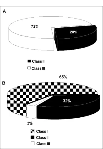
Clinical evaluation according to the New York Heart Association (NYHA) was done in the pre- and postopera-tive periods. Figure 1-A shows 23 (72%) functional class III and 9 (28%) functional class II preoperative patients.
Table I – Preoperation valve lesions of patients who underwent mitral valve repair with a malleable bovine pericardium ring
Lesions Patients
No %
Double mitral lesion 14 44
Pure mitral insufficiency 12 37
Pure mitral stenosis 5 16
Calcified mitral stenosis 1 3
Total 32 100
Table II – Procedures and operations associated with mitral valve repair with a malleable bovine pericardium ring
Associated procedures Patients
No %* %**
Anterior and posterior commissurotomy 17 74 53 Posterior papillarotomy 3 13 10
Anterior papillarotomy 2 9 6
Quadrangular resection of 1 4 3 the posterior cusp
Total 23 100 75
Associated operations No %* %**
Tricuspid valve repair 4 45 13
Aortic valve replacent 3 33 9
Aortic valve repair 2 22 6
Total 9 100 28
* Percentage of total number of procedures or associated operations; ** percentage of the total 32 patients.
A
B
28%
Fig. 1 - Clinical evaluation of patients who underwent preoperative postoperative with a malleable ring of bovine pericardium. (A) Preoperation; (B) postoperation.
Class II
Class III
Class I
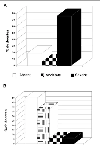
Degrees of mitral insufficiency were evaluated pre-and postoperatively by echodopplercardiography. Severe mitral insufficiency was detected in 75% of the patients, 12 (37.5%) with pure insufficiency and 12 (37.5%) with double lesions and a component of severe insufficiency. Of the remaining patients, two (6%) had double mitral lesions and a moderate insufficiency component, and 6 (19%) did not show insufficiency but had mitral stenosis (fig. 2-A).
In-hospital deaths occurred in the institution or within 30 days following surgery.
All patients underwent a medial sternotomy and systemic heparinization according to their activated coagu-lation time (≥480s). Following aortic cross clamping , infu-sion of blood cardioplegics was started from the aortic root, with a 12Fr cannula.
The right atrium was opened and a purce string aro-und the coronary sinus was made for introduction of a Fo-ley o 18 for continuous retrograde infusion of cardio-plegics. The left atrium was opened by a longitudinal inci-sion in the direction of the lower vena cava and below the upper caval vein.
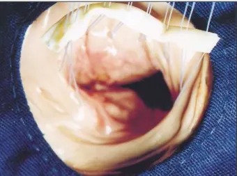
Adequate viewing of the mitral valve permitted the study of the valve’s anatomy and the evaluation of extant
damage to the subvalvar apparatus. Lengthening, retrac-tion, or rupture of the tendinous chords, as well as position and participation of the papillary musculature in the lesion were carefully evaluated. Two hooks with blunted, curved tips were utilized for gentle traction of the valve leaflets. When necessary, the tendinous chords, papillary muscles, and leaflets were handled. In the next step, measuring devices for the choice of the ring’s size were utilized. These instruments come in pairs and sustain in their extremes 21, 21/23, 25/27, and 29/31mm semicircles (fig. 3) for measure-ment of anterior leaflet perimeter (fig. 4).
In the following sequence, U-form stitches of 2.0 braided polyester thread were passed deep under the poste-rior mitral annulus, having as limits the two fibrous trigones and avoiding the coronary system. The same stitches were passed through the malleable ring and tied to retrieve the posterior mitral annulus, remodeling it to adequately fit the posterior to the anterior leaflet, guaranteeing valve compe-tence (figures 5 and 6). The mitral valve was tested by pas-sing a 12F probe through the aortic valve orifice to the left ventricle, with infusion of the blood supplied by the arterial line via a shunt. After the test, when necessary, an additio-nal repair was performed without difficulty, because the flexible rings allowed for a renewed approach to the valve, even after it was fixed. Testing was done until complete and adequate valve repair was achieved, and was followed by closing of the left atrium. After maneuvering to remove air from the cardiac cavities, cardioplegic infusion was
A
B
Fig. 3 - Anterior leaflet perimeter measuring devices.
Fig. 4 - Anterior leaflet perimeter measurements. Fig. 2 - Echodopplercardiographic evaluation of patients who underwent mitralplasty
with a malleable ring of bovine pericardium. (A) Preoperative; (B) Postoperative.
% de doentes
% de doentes
Absent Moderate Severe
stopped, and the aorta was unclamped; return of heartbeats occurred within a short time.
Clinical and echodopplercardiographic evaluations were performed based on the clinical history and pre- and postoperative examinations. The clinical classification was in agreement with the NYHA. Echodopplercardiographic evaluations by the transthoracic route used the ULTRA-MARK – 4 apparatus from ATL (Advanced Technology La-boratories) with 2.25, 23.3, and 5.0 MHZ transducers. The classification proposed by Colette et al. 31 and described by
Feigenbaum 32, which quantifies mitral insufficiency based
on the extension and magnitude of the regurgitation jet during left ventricular systole was used. Classes were iden-tified as absent – no detectable regurgitating jet; minimal – discrete, non-holosystolic jet regurgitation restricted to the vicinity of the mitral valve; moderate – considerable, holo-systolic regurgitation reaching the roof of the left atrium; severe – marked, holosystolic regurgitation reaching the roof of the left atrium, caught up to the level of the entrance of the pulmonary veins.
For a statistical analysis of the clinical and echodop-plercardiographic evaluations Wilcoxon’s test for paired data was used. Differences between values obtained
pre-and postoperatively were considered significant when the statistical probability of their casualty was <5%.
Results
During the evolution of the 32 patients, two death oc-curred, one immediate, the other delayed. Both patients had a rheumatic valve pathology and severe mitral and tricuspid valve insufficiency, and underwent valve repair and asso-ciated procedures in both valves. One of these patients, 19 years old, died on the third day following the operation from unknown causes; the other, nine years old, had to be reope-rated upon for mitral valve replacent nine months following valve repair. During evolution, he required prolonged ven-tilator support and experienced hemodynamic instability evolving into left ventricular failure, leading to death on the second day following the operation. All other patients were followed for 15 months, an equivalent of 214 months pa-tients, with an average evolution time of 6.7 months.
No fatal late complications directly related to the mal-leable ring occurred, which allows a survival index of 93.8%. One patient was re-operated upon nine months after mitral valve repair, with replacement of the valve due to the recur-rence of the rheumatic process and development of impor-tant delayed post-operation insufficiency. Thus, the reope-ration index was 3%, coinciding with the index of nonfatal delayed complications.
Clinical pre- and postoperation evaluations were made on the basis of the NYHA classification. Figure 1 shows that on a comparative evolution analysis of 31 patients, 20 (65%) passed on to functional class I; 10 (32%) to class II, and only one (3%) stayed in class III after reoperation.
Echodopplercardiographic studies shown on figure 2, allow comparison of pre- and postoperation findings and reveal that of the 32 patients; 16 (50%) had an evolution free of insufficiency, 12 (38%) had minimal insufficiency without hemodynamic repercussions, 2 (6%) had moderate, and 2 (6%) had severe insufficiency.
Wilcoxon’s test for paired data showed statistically significant differences for functional class and echodop-plercardiographic evolution between pre- and post-opera-tion.
Discussion
This work proposes the utilization of a biological ring restricted solely to the posterior part of the mitral annulus. It differs from the flexible synthetic ring developed by Duran and Ubago 13 of the second ring generation 21 by not
invol-ving the whole mitral valve circumference. During evolu-tion, it was shown to be satisfactory in patients who un-derwent annuloplasty, in agreement with previously pre-sented results 26,28,29.
In the decision for either valve repair or replacement, adequate patient selection and presence of associated risk factors should be considered 33. In a multivariate analysis,
Galloway and collaborators 33 by combining age, sex, and
Fig. 5 - Stitches passed through the posterior mitral annulus and the malleable bovine pericardium prosthesis.
various risk factors recorded four elements related to in-creased operation risk: age, functional class IV of the NYHA, previous cardiac surgery, and presence of previous cardiac interventions. According to the authors, the type of surgical valve conduct does not seem to influence opera-tion risk or late survival of patients who undergo valve re-pair or replacement. However, for Carpentier 11 valve
recons-truction is to be indicated for all lesions, even prior to consi-deration of age, cause of the disease, and patient condition. Furthermore, the younger the patient, the more urgent such an indication becomes.
In the present work, echodopplercardiography reaf-firmed the indication for reparative surgery in patients with functional class II (28%) before they developed greater lesions of the valve apparatus and evolution to ventricle de-terioration. This permitted the achievement of a better result, 65% of the patients passing to functional class I with re-covery of left ventricular geometry and mechanical function of the mitral valve, as is also reported in the literature 26,28,34,35.
A follow-up period of 16 months showed a survival ratio of 93.8%; other studies, with lower 29 or equal 36 follow-up
periods, reported 98% indexes. Studies with follow-up periods of five 26,28, eight 35 and 16 34 years, reported survival
rates of respectively, 90 to 98.8±1.2%, 88±4%, and 76.1±-3.1%. It is common to find valve repair leading to a higher survival rate in comparison with valve replacement 37.
Acquired experience, understanding of the role of the mitral valve apparatus in left ventricular function, and decreased postoperative morbidity and mortality led to the expansion of the number of valve repair interventions 30,38
known 39 to lead to a better postoperative performance of
the left ventricle as well. Long-term results showed ventri-cular function restored to normal levels, significantly imved survival ratios in particular following solely mitral pro-cedures 34, which in the present work were performed in
71.8% of the patients. Associated with this was the intro-duction of various valve repair techniques 13,21,40,41 as well as
recognition of the problems caused by valve replacement procedures like prosthesis degeneration, infection, trans-valvular gradients, paratrans-valvular leaking, hemolysis, and thromboembolic events 8,37.
Compared with valve replacement, repairs is asso-ciated with fewer complications 33,42. In this study,
postope-rative complications occurred in 3.1% of the cases, coinci-ding with reoperation in a patient with rheumatic valve pathology and severe mitral and tricuspid insufficiency, who underwent repair procedures in both valves. During later evolution, this patient required mitral valve replace-ment, which however, led to death. The literature shows a reoperation-free ratio of patients who undergo valve repair, fluctuating between 49 and 100% during a follow-up period of 4 to 17 years 26,30,33,43,44. The lower ratio was not always
related to a longer follow-up period.
Analysis of mitral valve repir reveals the importance of annulus remodeling using some type of prosthesis, bea-ring in mind that the absence of annuloplasty is associated with a higher incidence of precocious failure 30. A study by
Bernal et al. 34, utilizing a complete, malleable ring, revealed
that 89.9±3.2% of their rheumatic patients remained free of reoperation over a 16-year period. In this case, the risk of reoperation curve showed two critical periods with a high in-cidence, during the first two years, and 10 years after sur-gery. According to the authors, precocious reoperation is habitually associated with an inadequate indication or te-chnical, while delayed indications are in general, connected to the progress of the valve’s basic pathology. In the pre-sent study, however, such a progress precociously determi-ned reoperation.
In contrast, Braile and collaborators 26 revealed the
absence of reoperation after a follow-up period of five years of patients with predominantly rheumatic valve pathology who underwent posterior annuloplasty 26. Nevertheless,
rheumatic valve pathology is worrisome, with a high in-cidence of failure in the reparation procedures 11,33,43. These
are probably due to histopathological alterations occurring in the mitral valve during rheumatic disease, including among others, intercellular edema, collagen fiber fragmen-tation, and lymphocyte and plasma cell infiltration. Never-theless, Bernal et al. 34 and Antunes et al. 43 affirm that
val-valve repair is an excellent alternative for the treatment of patients with rheumatic mitral valve disease.
David et al. 35, using a complete, malleable ring in
pa-tients with degenerative valve pathology, found 95±2% free from reoperation for more than 8 years. They concluded that only an advanced degree of myxomatous alterations was related to a greater risk of reoperation. Cosgrove et al. 29, in a
study of patients with predominantly degenerative valve pathology treated with posterior annuloplasty, observed 97% of them to be free of reoperation for a year, and Camille-ri et al. 28 found 99% of patients to be free of this event for
over 5 years. Controversy exists over the reoperation ratio associated with valve repair ou replacement. Galloway et al. 33
and Cosgrove 44 report similar indexes for both procedures,
but some studies show higher values for plastic surgery 45.
Valve repair in rheumatic disease is associated with a higher index of thromboembolic complications, in compari-son to similar procedures in a degenerative or ischemic valve pathology 34. In the present study, although 87.5% of
the patients had a rheumatic valve pathology, no thrombo-embolic episodes were registered even in the absence of an-ticoagulants in patients who received only oral treatment with a dose of 200mg/day of acetylsalicylic acid, which is in agreement with Braile et al. 26 whose predominantly
rheu-matic valve pathology patients underwent the same tech-nique and a five-year follow-up period.
Bernal et al. 34 found 79.2±3.2% of carriers of rheumatic
the lesion, to preoperative risk factors, rather than to the surgical procedure proper 34,35.
David et al. 35 found after an eight-year follow-up
les-ser indexes of absence of thromboembolism, below 82±6%, in patients who underwent valve repair for degenerative mitral disease. An association between age (above 60 years) and a threefold higher risk of occurrence of this event was found. In the present study, this age group was repre-sented by only 12.5% of the patients. In contrast, consi-dering only predominantly degenerative valve pathology, Camilleri et al. 28 and Cosgrove et al. 29 observed 97 and 91%
of patients to be free of this complication after follow-up periods of, respectively, one and five years.
Endocarditis, not observed in this study also shows a low incidence in other literature reports. It was observed by Bernal et al. 34 in 0.8% of patients having rheumatic valve
pathology, followed for 16 years.
In a study of patients with a predominance of rheu-matic valve pathology, Braile et al. 26 found 95.7±4.5% of
them not to have this complication. The index for degenera-tive valve pathology varied between 98 and 100% during a follow-up period of one to eight years 28-30,35.
In the present work, mitralvalve repair, normally perfor-med in patients with a diagnosis of insufficiency or double lesion, was also indicated for carriers of mitral stenosis or calcification (19%) following commissurotomy. The tech-nique was effective in cases where, following this proce-dure, the valve had some insufficiency and was found to be of importance for postoperation evolution. The malleable bovine pericardium ring enabled a decrease in the area that the leaflets, with adequate coaptation, should close. This allowed for the recovery of the relationship between the valve annulus and the cusps, whose variation falls between 1:1.5 and 1:22 30,46.
Benefits were seen with echodopplercardiography, revealing an 88% recovery of valve function, regurgitation being either absent (50%) or minimal (39%) with a discrete, nonholosystolic jet restricted to the vicinity of the mitral valve, according to the classification proposed by Colette et al. 31. It is believed that one of the factors determining the
success of this technique lies in the measurement of the an-terior cusp’s perimeter in the choice of the size of the pros-thesis to be used in the posterior mitral annulus. Differing annuloplasty adopts as a basis for measurement in the choice of the different size rings, the intertrigonal or inter-commissural distance, associated or not with the measure-ment of the anterior leaflet’s area 10,13,27-30. Yet, the mitral
an-nulus does not maintain size and form constant during sys-tole and the annulus segment over the area of aortic-mitral continuity is significantly lengthened during ventricular systole 47. Thus, such measurements do not seem to favor
an adequate estimate for the selection of annular prosthe-sis’ size, justifying the development of devices to measure anterior cusp perimeters 26.
Nagastu et al. 48 experimentally demonstrated that
following mitral valve repair a fraction of the regurgitation of ≤30% is associated with full recovery of the contractile
function, alterations occurring at the cellular (myocyte) le-vel. Clinical evidence indicates pronounced improvement in diastolic functional response as well as of volume and di-mension of the left ventricle during a follow-up period of one year 14; this carries promise for the evolution of the
patients studied in this work.
Reconformation of the mitral annulus enables a direct im-provement of the cardiac muscle with a reduction in diastolic diameter of the left ventricle, as demonstrated in studies with longer follow-up periods 26,28,34,35. Braile et al. 26 reported a
significant difference in the p and postoperation re-lationship between aortic and left ventricle diameters, with reacquisition of normal values for this relation in the late postoperative period. These findings confirm the description of a dilatation of the mitral annulus occurring mainly in its pos-terior portion, with a symmetric or asymmetric pattern, depending on the type of disease 49. Thus, the anterior portion
of the annulus apparently does not require reconformation 26,30.
However, the success obtained in annuloplasty using rings did not prevent the use of rigid prostheses for mitral valve repair 10,27, a situation that has generated discussion
about the real performance of the various types of prosthe-ses in dilated valve annulus remodeling. Knowledge about the dynamic properties of the native mitral ring confirms the absence of its position fixed in a geometric plane, because its saddle format allows for a large flexion during systole (28,30). In normal situations, the valve annulus has a
sphin-cter-like function, with an approximately 26% reduction in
its area during ventriclar systole, acquiring an elliptical form, while in diastole it has a circular shape 30. Therefore,
insertion of a rigid ring could interfere with these properties in a negative manner 26,28-30,34. Due to its characteristics,
bo-vine pericardium adapts itself perfectly to the mitral ring, not interfering with its contractility 26, leading to the
satisfac-tory evolution of the patients verified in this study. In choosing the prosthesis for valve repair, it is impor-tant to consider the cause of the disease. Thus, for patients with mitral regurgitation due to a degenerative valve patho-logy and a contractile mitral annulus, flexible prostheses are indicated, but for those with regurgitation due to a rheuma-tic valve pathology and a valve annulus with impaired con-tractility, both types would be of benefit to left ventricular function. Avoidance of a drastic change from a regularly contractile annulus to a rigid one 14 is recommended.
Spence et al. 50, in an experimental study comparing
different types of annular prostheses, concluded that the rigid ones harm systolic function of the left ventricle. At a later date, the same group clinically confirmed this by repor-ting such rings to be a determinant for depressed ventricu-lar function 14, and supporting the use of malleable rings in
carriers of a rheumatic valve pathology with reduced annu-lar contractility, as occurred in 87.6% of the presently stu-died cases. Furthermore, the rigid ring may also cause the obstruction of the left ventricular outflow tract observed in 4.5 to 10% of the cases 15-18, a fact not observed in
annulo-plasty with completely flexible rings 14,31 or with flexible
In conclusion, the bovine pericardium ring was shown to be flexible, fitting itself perfectly to the valve an-nulus, respecting its geometry and contractility. It appears legitimate to affirm that mitral lesions due to different cau-ses, including insufficiency secondary to surgical treat-ment of mitral stenosis, may be adequately treated by the presently proposed technique, considering that 65% of the patients evolved to functional class I and 88%
achieved complete recovery of valve function without needing anticoagulation.
Acknowledgement
We thank Drs. Valentim Baccarin, Orlando Petrucci Jr. and Pedro Paulo Martins Oliveira for support received during this work.
1. Perloff JK, Roberts WC. The mitral apparatus. Functional anatomy of mitral regurgitation. Circulation 1972; 46: 227-39.
2. Cosgrove DM, Stewart WJ. Mitral valvuloplasty. Curr Probl Cardiol 1989; 4: 359-415.
3. Lillehei CW, Gott VL, Dewall RA. Surgical correction of pure mitral insuffici-ency by annuloplasty under direct vision. Lancet 1957; 77: 446-9. 4. Bigelow WG, Kuypers PJ, Heimbecker RO, Gunton RW. Clinical evaluation of
the efficiency of mitral annuloplasty. Ann Surg 1961; 154: 320-9.
5. Agathos EA, Starr A. Mitral valve replacement. Curr Probl Surg 1993; XXX: 481-600.
6. Reed GE, Pooley RW, Moggio RA. Durability of measured mitral annuloplasty: Seventeen-year study. J Thorac Cardiovasc Surg 1980; 79: 321-5.
7. Galloway AC, Colvin SB, Baumann FG, Harty S, Spencer FC. Current concepts of mitral valve reconstruction for mitral insufficiency. Circulation 1988; 78: 1087-98.
8. Reul GJ, Cooley DA, Duncan JM, et al. Valve failure with the Ionescu-Shiley bovine pericardial bioprosthesis: analysis of 2680 patients. J Vasc Surg 1985; 2: 192-204.
9. Braile DM, Volpe MA, Ramin SL, Souza DRS. Tratamento cirúrgico das valvopa-tias. In: Barretto ACP, Sousa AGMR, eds. Cardiologia: Atualização e Reciclagem. Rio de Janeiro: Atheneu, 1994: 476.
10. Carpentier A, Deloche A, Dauptain J, et al. A new reconstructive operation for correction of mitral and tricuspid insufficienty. J Thorac Cardiovasc Surg 1971; 61: 1- 13.
11. Carpentier A. Cardiac valve surgery - the “french correction”. J Thorac Cardiovasc Surg 1983; 86: 323-37.
12. Galloway AC, Colvin SB, Baumann FG. Long-term results of mitral valve reconstruction with Carpentier techniques in 148 patients with mitral insufficiency. Circulation 1988; 78(suppl 1): 97-105.
13. Duran CG, Ubago JLM. Clinical and hemodynamic performance of a totally flexible prosthetic ring for atrioventricular valve reconstruction. Ann Thorac Surg 1976; 22: 458-63.
14. David TE, Komeda M, Pollick C, Burns RJ. Mitral valve annuloplasty: the effect of the type on left ventricular function. Ann Thorac Surg 1989; 47: 524-8. 15. Kreindel MS, Schiavone WA, Lever HM, Cosgrove DM. Systolic anterior
motion of the mitral valve after Carpentier ring valvuloplasty for mitral valve prolapse. Am J Cardiol 1986; 57: 408-12.
16. Marwick TH, Stewart WJ, Currie P, Cosgrove DM. Mechanisms of failure of mitral repair: an echocardiographic study. Am Heart J 1991; 122: 149-56. 17. Krenz HK, Mindich BP, Guarino T, Goldman ME. Sudden development of
intraoperative left ventricular outflow obstruction: differential and mechanism: an intraoperative two-dimensional echocardiographic study. J Cardiovasc Surg 1990; 5: 93-101.
18. Stewart WJ, Currie PJ, Salcedo EE, et al. Intraoperative doppler color flow mapping for decision-making in valve repair for mitral regurgitation: technique and results in 100 patients. Circulation 1990; 81: 556-66.
19. Grossi EA, Galloway AC, Parish MA, et al. Experience with twenty-eight cases of systolic anterior motion after mitral valve reconstruction by the Carpentier technique. J Thorac Cardiovasc Surg 1992; 103: 466-70.
20. Duran CMG, Ubago JL. Conservative mitral valve surgery. Problems and developments in the techniques of prosthetic ring annuloplasty. In: Kalmanson D, ed. The Mitral Valve. A Pluridisciplinary Approach. Massachussets: Publishing Sciences Group Inc., 1975: 549.
21. Puig Messana M, Calbet JM, Castells E. Conservative surgery of the mitral valve. Annuloplasty on a new adjustable ring. In: Bircks W, Ostemeyer J, Schulte HD, eds. Cardiovascular Surgery. Berlim: Springer-Verlag, 1981: 30.
References
22. Murphy JP, Sweeney MS, Cooley DA. The Puig-Massana-Shiley annuloplasty ring for mitral valve repair: experience with 126 patients. Ann Thorac Surg 1987; 43: 52-8.
23. Rey MJ, Mercier LA, Castonguay Y. Echocardiographic diagnosis of left ventricular outflow tract obstruction after mitral valvuloplasty with flexible Duran ring. J Am Soc Echocardiogr 1992; 5: 89-92.
24. Cooley DA, Frazier OH, Norman JC. Mitral leaflet prolapse: surgical treatment using a posterior annular collar prosthesis. Bull Tex Heart Inst 1976; 3: 438-42. 25. Czer LSC, Maurer G, Trento A, et al. Comparative efficacy of ring and suture
annuloplasty for ischemic mitral regurgitation. Circulation 1992; 86(suppl II): 46-52.
26. Braile DM, Ardito RV, Pinto GH, et al. Plástica mitral. Rev Bras Cir Cardiovasc 1990; 5: 86-98.
27. Gregori F, Silva SS, Baba K, et al. Um novo modelo de anel protético para pacientes com insuficiência valvar mitral. Relato de dois casos. Arq Bras Cardiol 1988; 50: 417-20.
28. Camilleri L, Filaire M, Repossini A, et al. Mitral annuloplasty with a flexible linear reducer. J Card Surg 1995; 10: 99-103.
29. Cosgrove DM, Arcidi JM, Rodriguez L, Stewart WJ, Powell K, Thomas JD. Initial experience with the cosgrove-edwards annuloplasty system. Ann Thorac Surg 1995; 60: 499-504.
30. Camilleri LF, Miguel B, Bailly P, et al. Flexible posterior mitral annuloplasty: five-year clinical and doppler echocardiographic results. Ann Thorac Surg 1998; 66: 1692-7.
31. Colette V, Ameur A, Sylvie BAS, Lessana A, Abitbol G, Kalmanson D. Pulsed doppler echocardiographic indices for assessing mitral regurgitation. Br Heart J 1984; 51: 130-8.
32. Feigenbaum H. Acquired valvular heart disease. In: Feigenbaum H, ed. Echocardiography. Indianapolis: Lea & Febiger, 1993: 239.
33. Galloway AC, Colvin SB, Baumann FG, et al. A comparison of mitral valve reconstruction with mitral valve replacement: intermediate-term results. Ann Thorac Surg 1989; 47: 655-62.
34. Bernal JM, Rabasa JM, Vilchez FG, Cagigas JC, Revuelta JM. Mitral valve repair in rheumatic disease: the flexible solution. Circulation 1993; 88: 1746-53. 35. David TE, Armstrong S, Sun Z, Daniel L. Late results of mitral valve repair for
mi-tral regurgitation due to degenerative disease. Ann Thorac Surg 1993; 56: 7-14. 36. Carpentier AF, Lessana A, Relland JYM, et al. The “physio-ring”: an advanced
concept in mitral valve annuloplasty. Ann Thorac Surg 1995; 60: 1177-86. 37. Cohn LH, Kowalker W, Bhatia S, et al. Comparative morbidity of mitral valve
repair versus replacement for mitral regurgitation with and without coronary artery disease. Ann Thorac Surg 1988; 45: 284-90.
38. Lillehei CW, LevyMJ, Bonnabeau RC. Mitral valve replacement with preservation of papillary muscles and chordae tendineae. J Thorac Cardiovasc Surg 1964; 47: 532-43. 39. Goldaman ME, Mora F, Guarino T, Fuster V, Mindich BP. Mitral valvuloplasty is superior to valve replacement for preservation of left ventricular function: an intraoperative two-dimensional echocardiographic study. J Am Coll Cardiol 1987; 10: 568-75.
40. Frater RWM, Gabbay S, Shore D, Factor S, Strom J. Reproducible replacement of elongated or ruptured mitral valve chordae. Ann Thorac Surg 1983; 43: 14-28. 41. Gregori F, Takeda R, Silva S, Facanha L, Meier MA. A new technique for repair of mitral insufficiency caused by ruptured chordae of the anterior leaflet. J Thorac Cardiovasc Surg 1988; 96: 765-8.
42. Craver JM, Cohen C, Weintraub WS. Case-matched comparison of mitral valve replacement and repair. Ann Thorac Surg 1990; 49: 964-9.
44. Cosgrove DM. Mitral valve repair in patients with elongated chordae tendineae. J Cardiac Surg 1989; 4: 247-52.
45. Cohn LH. Mitral valve surgery: reconstruction versus replacement. In: International symposium advances in cardiology - controversial views. Berlim: Anais Berlim, 1984.
46. Brock RC. Surgical and pathological anatomy of the mitral valve. Brit Heart J 1952; 14: 489-513.
47. Glasson JR, Komeda M, Daughters GT, et al. Three-dimensional regional dynamics of the normal mitral anulus during left ventricular ejection. J Thorac Cardiovasc Surg 1996, 111: 574-85.
48. Nagatsu M, Ishihara K, Zile MR, et al. The effects of complete versus incomplete mitral valve repair in experimental mitral regurgitation. J Thorac Cardiovasc Surg 1994; 107: 416-23.
49. Galloway AC, Colvin SB, Baumann FG. Long-term results of mitral valve re-construction with Carpentier techniques in 148 patients with mitral insuffi-ciency. Circulation 1988; 78(suppl 1): 97-105.