São Paulo, SP - Rio de Janeiro, RJ - Instituto do Coração do Hospital das Clinicas FMUSP and Hospital Pró-Cardíaco, Rio de Janeiro.
Mailing address: Ricardo Vivacqua C. Costa - Av. Afrânio de Melo Franco, 365/101 Cep 22430-060 - Rio de Janeiro, RJ, Brazil - E-mail: [email protected] English version by Stela Maris C. e Gandour
Received: 11/04/02 Accepted: 03/31/03
Arq Bras Cardiol, volume 81 (nº 6), 581-5, 2003
Ricardo Vivacqua Cardoso Costa, Antonio Claudio Lucas da Nóbrega, Salvador Manoel Serra, Salete Rego, Mauricio Wajngarten
São Paulo, SP - Rio de Janeiro, RJ - Brazil
Influence of Skeletal Muscle Mass on Ventilatory and
Hemodynamic Variables During Exercise in Patients with
Chronic Heart Failure
Despite the marked reduction in mortality and morbidi-ty of cardiovascular diseases observed in recent decades, the incidence and prevalence of heart failure (HF) have sig-nificantly increased in both developed and developing countries 1.
Heart failure in its chronic form manifests with several signs and symptoms; its most significant clinical characte-ristic is the incapacity to tolerate progressively milder phy-sical effort, which is an indicator of the severity of heart fai-lure itself 2. The hemodynamic parameters at rest of patients with heart failure have been shown not to correlate with tho-se during effort 3, the latter having an independent prog-nostic power for overall and cardiovascular mortality 4. In addition, the symptoms usually reported by CHF patients underestimate their true physical capacity, which may be better assessed on cardiopulmonary exercise testing 5,6 that provides an assessment of the integrated cardiopulmonary function 7,8.
In the genesis of heart failure syndrome, central hemo-dynamic abnormalities are present, as are changes in skele-tal muscle function, which have been held responsible for fatigue and its consequent exercise limitation 9,10. These muscle changes reflexly activate the autonomous nervous system, playing an important role not only in the origin of the exercise limiting symptoms, but also in the progression of heart failure 11,12.
Despite the existence of some studies on the influence of the skeletal musculature on the functional capacity of pa-tients with heart failure, no report exists about the relation between skeletal muscle mass and ventilatory and hemody-namic variables, such as anaerobic threshold and oxygen pulse, which have been considered important functional and risk markers in that syndrome. Therefore, the present study aimed at assessing the influence of skeletal muscle mass on different ventilatory and hemodynamic variables in patients with chronic heart failure during exercise.
Objective - To assess the influence of skeletal muscle mass on ventilatory and hemodynamic variables during exercise in patients with chronic heart failure (CHF).
Methods - Twenty-five male patients underwent maxi-mum cardiopulmonary exercise testing on a treadmill with a ramp protocol and measurement of the skeletal muscle mass of their thighs by using magnetic resonance imaging. The clinically stable, noncachectic patients were assessed and compared with 14 healthy individuals (S) paired by age and body mass index, who underwent the sa-me examinations.
Results - Similar values of skeletal muscle mass were found in both groups (CHF group: 3863 ± 874 g; S group: 3743 ± 540 g; p = 0.32). Significant correlations of oxygen consumption in the anaerobic threshold (CHF: r = 0.39; P= 0.02 and S: r = 0.14; P = 0.31) and of oxygen pulse also in the anaerobic threshold (CHF: r = 0.49; P = 0.01 and S: r =0.12; P = 0.36) were found only in the group of patients with chronic heart failure.
Conclusion - The results obtained indicate that skele-tal muscle mass may influence the capacity of patients with CHF to withstand submaximal effort, due to limitations in their physical condition, even maintaining a value similar to that of healthy individuals. This suggests qualitative changes in the musculature.
Methods
This study selected 25 male patients with chronic heart failure in NYHA functional class III with the following etiologies: idiopathic (44%), arterial hypertension (32%), and alcoholism (24%). Patients with the following characte-ristics were excluded from the study: hospitalization or the-rapeutic change, or both, within less than 3 months; neuro-logic or locomotor afflictions, or another disease that could interfere with the tests to be performed; episodes of nonsus-tained ventricular tachycardia at rest; intracavitary throm-bus with emboligenic potential; and cachexia.
All patients were using angiotensin-converting enzy-me inhibitors, digoxin, and furosemide, in addition to the following medications: carvedilol (24%), spironolactone (40%), amiodarone (20%), and warfarin sodium (12%).
The resting electrocardiogram showed the following changes: left bundle-branch block (36%), left ventricular hy-pertrophy (32%), left bundle-branch anterior divisional block (12%), atrial fibrillation (12%), and left atrial hypertro-phy (8%).
A control group paired by age and body mass index was recruited for comparison. This group comprised 14 healthy individuals according to the results of a clinical exa-mination, electrocardiography, echocardiography, and car-diopulmonary exercise testing. They were using no medica-tion and were not engaged in a formal physical exercise pro-gram. Their characteristics are shown in table I.
This study was approved by the Committee on Ethics for the Analysis of Research Projects of the Hospital das Clínicas of the Medical School of USP and the Committee on Ethics in Research of the Hospital Procardíaco (Rio de Ja-neiro). All volunteers signed the formal written consent after receiving the necessary explanations.
All patients underwent treadmill testing (ATL10200, Inbramed, Brazil) with simultaneous analysis of pulmonary ventilation and expired gases with a metabolic analyzer (TEEM100, Aerosport, USA), 12-lead electrocardiographic recordings (Mason Likar system), and blood pressure mea-surement through sphygmomanometry. The ramp protocol was used adjusted to the clinical and biomechanical condi-tions of the patients, with a progressive increase in the in-tensity of effort and estimated duration between 8 and 12 mi-nutes, being interrupted by fatigue or dyspnea. Considering
the occurrence of natural adaptation to exercise testing, 2 tests were performed at an approximate interval of 1 week, the results of the second test being used for analysis.
The anaerobic threshold, obtained in all cases, was determined by 3 experienced examiners and defined when agreement, at least between 2 of them, occurred. The criteria used for its determination were as follows 13,14: a nonlinear increase in ventilation, ie, exponentiation of the ventilation curve; point of beginning of the consistent elevation of the curve of the oxygen ventilatory equivalent; and curve ele-vation referring to the expired oxygen fraction.
The muscle mass of the thigh was determined with magnetic resonance imaging with the patient lying in the dorsal decubitus position. The examinations were perfor-med in a Sigma 1.5 T Horizon LX 8.2 device with torso coil (General Electric, USA). The sequences processed were spin-echo T1 in 5.0-mm thick axial views at 8.0-mm intervals. Topogram was performed in the coronal plane. The skeletal muscle mass (MM) of the right and left thighs was measu-red in the spin-echo T1 sequence using an irregular cursor around the muscle groups, excluding the fat tissue and
bo-nes 15,16. The anatomic landmark for the first view of the
thigh was the hip joint, and the last view was that immedia-tely before the patella. All muscle mass values were added and multiplied by 13 mm and by muscle density (1.05); then, the mean muscle volume was calculated 17.
All variables had a normal distribution and homoge-neity of variances. Therefore, parametric procedures were used for all analyses. The Student t test for independent samples was used for comparing the results of the patients and controls. The correlation coefficient was used for mea-suring the association between 2 variables, 0.323 being the critical value for the number of patients studied, and 0.426 for the control group. Linear regression analysis through the least squares method was used to determine the first-degree equation that describes the relation between VE and VCO2. The correlation coefficient was > 0.95 in all cases. The slope calculated was used for analyzing the correlation with muscle mass. The significance level adopted was 5%.
Results
The results of the variables obtained in cardiopulmo-nary exercise testing and magnetic resonance imaging are shown in table II. As expected, the results indicate a lower functional capacity in the CHF patients as compared with that in healthy individuals, although no difference was found in regard to the skeletal muscle mass. Figure 1 depicts the behavior of pulmonary ventilation in relation to CO2 production in a CHF patient and in a healthy individual.
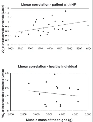
Table III shows the linear correlations between muscle mass and ventilatory and hemodynamic variables during exercise in patients and control individuals. Significant po-sitive and negative correlations were observed only in the group of patients. Figure 2 depicts the graphs with individu-al results and the line of linear regression between skeletindividu-al muscle mass and oxygen consumption at the anaerobic threshold in patients and control individuals.
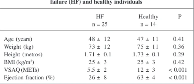
Table I - Characteristics (mean ± sd) of patients with heart failure (HF) and healthy individuals
HF Healthy P
n = 25 n = 14
Age (years) 48 ± 12 47 ± 11 0.410
Weight (kg) 73 ± 12 75 ± 11 0.360
Height (metros) 1.71 ± 0.1 1.73 ± 0.1 0.290
BMI (kg/m2) 25 ± 3 25 ± 3 0.420
VSAQ (METs) 5.5 ± 2 12 ± 3 < 0.001 Ejection fraction (%) 26 ± 8 63 ± 4 < 0.001
Discussion
Our study found a correlation between the skeletal muscle mass of the thighs and the variables obtained in the cardiopulmonary exercise test in patients with heart failure. A correlation between muscle mass and the variables was found not only at the peak of effort, but also at the anaero-bic threshold.
Quantitative similarity in the muscle mass obtained on magnetic resonance imaging was observed in patients with heart failure and in the control group. This may be explained by the fact that the patients were stable and not cachectic.
The anaerobic threshold was used for the functional assessment of patients, and more elevated values were served in the control group (P<0.001). This parameter is ob-tained in submaximal intensities of effort accompanying most of the individuals´ daily activities and is used for pro-gramming physical activity 18,19.
Fig. 1 - Examples of VE x VCO2 slope. CHF patients = 41; healthy individuals = 19. Relation between ventilation per minute and production of carbon dioxide in a pa-tient with heart failure and in a healthy individual. The ventilation necessary to eli-minate carbon dioxide increases earlier in the patient with heart failure as compared with that in the healthy individual.
Table II - Results (mean ± sd) of patients with heart failure (HF) and healthy individuals
HF Healthy P
n = 25 n = 14
Metabolic variables
VO2 peak (L/min) 1.3 ± 0.5 3.1 ± 0.9 < 0.001
VO2 anaerobic thresh (L/min) 0.8 ± 0.3 2.1 ± 0.7 < 0.001
Derived variables
VE/VCO2 peak 35 ± 8 24.1 ± 4.5 < 0.001
VE/VCO2 anaerobic thresh 33 ± 7 23.8 ± 4.1 < 0.001
VE x VCO2 slope 32 ± 9 21 ± 3 < 0.001
VE/VO2 peak 36 ± 7 25 ± 5 < 0.001
VE/VO2 anaerobic thresh 29 ± 6 21 ± 5 < 0.001
O2 pulse peak (mL/beat) 8.6 ± 2.7 18.4 ± 5 < 0.001
O2 pulse anaerobic thresh (mL/beat) 6.4 ± 2 15.2 ± 3.9 < 0.001
Muscle mass of the thighs (g) (RM) 3863 ± 874 3743 ± 540 0.320
VO2 anaerobic thresh (% peak) 62 ± 9 66 ± 10 0.100
Borg scale peak 18 ± 2 19 ± 3 0.090
Respiratory quocient 1 ± 0.09 1.1 ± 0.1 0.210
VO2 peak - oxygen consumption at peak effort; VO2 thresh - oxygen consumption at the anaerobic threshold; VE / VCO2 - ventilatory equivalent of carbon dioxide; VE / VO2
-ventilatory equivalent of oxygen; VE x VCO2 - linear relation between the volume expired and the ventilatory equivalent of carbon dioxide; O2 pulse- oxygen pulse.
Tale III - Correlations of the ventilatory hemodynamic variables with muscle mass of the thighs in patients with heart failure (HF) and
healthy individuals
HF Healthy
n = 25 n = 14
r P r P
Variables
VO2 peak x MM 0.36 0.04 0.14 0.32
VO2 anaerobic threshold x MM 0.39 0.02 -0.14 0.31 VE/VCO2 peak x MM -0.33 0.05 -0.15 0.30
VE/VCO2 anaerobic -0.36 0.04 -0.08 0.39 threshold x MM
VExVCO2 slope x MM -0.40 0.02 -0.07 0.40 VE/VO2 peak x MM -0.42 0.02 -0.07 0.30
VE/VO2 anaerobic threshold x MM -0.38 0.03 -0.11 0.36 O2 pulse peak x MM 0.47 0.01 0.12 0.36
O2 pulse anaerobic threshold x MM 0.49 0.01 0.12 0.68
In our study, the behavior of the VE x VCO2 slope rela-ted to ventilatory inefficiency and intolerance to exercise is worth noting, being an independent variable to estimate the prognosis in patients with heart failure 20. The mean values of the VE x VCO2 slope in the patients studied (32±9) are bellow 34, a value confirmed in the study by Chua et al 21 as characterizing severity and a poor prognosis.
Other studies have reported an inverse and significant
relation between muscle mass and the VE x VCO2 slope,
confirming the existence of a correlation with the accentua-tion of the ergoreflex 12,13 with values equivalent to those ob-tained in this study.
The participation of hemodynamic factors in this stu-dy was assessed with oxygen pulse 22,23, which correlates with systolic volume 24. In the group of patients with heart failure, a direct and significant relation was found between oxygen pulse and muscle mass at the peak of effort and anaerobic threshold. A report on the behavior of this hemo-dynamic variable related to skeletal muscle has not been found in the literature. One may suppose that the reduced values of oxygen pulse during exercise obtained in all pa-tients cause secondary hemodynamic changes that
interfe-re with muscle mass, with elevation in the arterial-venous oxygen content difference.
A direct and significant relation between skeletal mus-cle mass and oxygen consumption at the peak of effort was obtained in our study, and these values are similar to those reported by other authors 25,26.
According to the muscle hypothesis 27, the functional limitation of these patients could be attributed to the action of metabolic and structural factors in the skeletal muscula-ture, triggering the ergoreflex, with elevation in the sympa-thetic activity consequent to peripheral vasoconstriction, leading to ventilatory dysfunctions 28.
Another study on skeletal muscle mass by Toth et al 29 assessed 14 stable and noncachectic patients with NYHA functional class III heart failure and 52 healthy individuals. The values of muscle mass in the heart failure group and in the healthy individuals were, respectively, 3200±400 g and 3300±600 g (NS), and a significant relation to oxygen con-sumption was found at peak effort. Toth et al 29 used the same methodology used in our study with almost overlap-ping results. Those authors concluded that qualitative and not quantitative factors of skeletal musculature interfere with the functional condition of these patients; however, the factors were not studied at the anaerobic threshold.
The intrinsic changes in skeletal muscle influencing physical activity have been shown in several studies, such as that by Massie et al 30, who assessed 18 patients in NYHA functional class III and with an inverse and significant corre-lation between type II ab muscle fibers and peak VO2. Those authors found a metabolic change in skeletal muscle with a reduction in the oxidative activity of the enzymes.
The study by Okita et al 31 is worth noting. They as-sessed 12 patients with heart failure and 7 controls and cor-related the tolerance to exercise assessed at VO2 peak to the depletion in phosphocreatine and to the pH reduction in skeletal musculature. The strong relation between oxygen consumption at peak effort and cellular acidosis confirmed that these intrinsic changes are an important limiting factor to exercise.
The relation between muscle mass and oxygen con-sumption at the anaerobic threshold was a characteristic as-sessed in our study, but not in other studies published. This relation was direct and significant in patients with heart fai-lure, but nonsignificant in healthy individuals. This shows that the more altered the muscle mass, the lower the toleran-ce to lactacidemia during exercise, with a reduction in the functional capacity of these patients.
In conclusion, patients with heart failure have a corre-lation between the skeletal muscle mass of the thighs and the ventilatory and hemodynamic variables at the anaerobic threshold and at the peak effort that participate in the me-chanisms that reduce physical capacity.
VO
2
at the anaerobic threshold (L/min)
Fig. 2 - Linear correlation between VO2 at the anaerobic threshold and muscle mass of
the thighs. CHF patients: r = 0.39; P = 0.02. Healthy individuals: r = 0.14; P = 0.31.
Linear correlation - patient with HF
Linear correlation - healthy individual
VO
2
at the anaerobic threshold (L/min)
1. Eriksson H. Heart failure: a growing public health problem. J Intern Med 1995; 237: 135-41.
2. Sociedade Brasileira de Cardiologia. Revisão das II Diretrizes para o diagnóstico e tratamento da insuficiência cardíaca. Arq Bras Cardiol 2002; 79 (suppl.IV): 1-30. 3. Remme WJ, Swedberg K. Task force for the diagnosis and treatment of chronic heart failure, European Society of Cardiology. Eur Heart J 2001; 22:1527-60. 4. Weber KT, Kinasewitz GT, Janicki JS, Fishman AP. Oxygen utilization and
ven-tilation during exercise in patients with chronic cardiac failure. Circulation 1982; 65:1213-23.
5. Wright DJ, Tan L.B. The role of exercise testing in the evaluation and management of heart failure. Postgrad Med J 1999; 75:453-8.
6. Myers JN, Gullestad L, Vagelos R, et al. Cardiopulmonary exercise testing and prognosis in severe heart failure: 14mL/kg/min revisited. Am Heart J 2000; 139:78-84.
7. Itoh H, Taniguchi K, Koike A, Doi M. Evaluation of severity of heart failure using ventilatory gas analysis. Circulation 1990; 81(suppl.II): II 31-7. 8. Wajngarten M, Kalil LM, Negrão CE, et al. Cardiopulmonary exercise test in the
evaluation of healthy elderly men. Arq Bras Cardiol 1994; 63:27-33. 9. Wilson JR. Exercise intolerance in heart failure – importance of skeletal muscle.
Circulation 1995; 91:559-60.
10. Coats AJS, Clark AL, Peipoli M, Volterrani M, Poole-Wilson PA. Symptoms and quality of life in heart failure: the muscle hypotesis. Br Heart J 1994; 72 (suppl):S36-9.
11. Gitt AK, Wasserman K, Kilkowski C, et al. Exercise anaerobic threshold and ventilatory efficiency identify heart failure patients for high risk of early death. Circulation 2002; 106: 3079-84.
12. Ponikowski PP, Francis DP, Piepoli MF, et al. Enhanced ventilatory response to exercise in patients with chronic heart failure and preserved exercise tolerance. Circulation 2001;103:967-72.
13. Wasserman K, Mcilroy MB. Detecting the threshold of anaerobic metabolism in cardiac patients during exercise. Am J Cardiol 1964;14:844-52.
14. Shimizu M, Myers J, Buchanan N, et al. The ventilatory threshold: method, proto-col, and evaluator agreement. Am Heart J 1991;122:509-16.
15. Murphy WA, Totty WG, Caroll JE. MRI of normal and pathologic skeletal muscle. Am J Roentoenol 1986;146:565-74.
16. Shellock FG, Fukunaga T, Mink JH, Edgerton VR. Acute effects of exercise on MR imaging of skeletal muscle: concentric vs eccentric actions. Am J Roetoenol 1991;156:765-8.
References
17. Anderson MW. Muscle. In: Higgins CB, Hricak H, Helm CA. Magnetic Resonan-ce Imaging of the Body. Philadelphia: Lippincott-Raven, 3rd ed, 1997:1321-41. 18. Piña IL, Karalis DG. Comparison of four exercise protocols using anaerobic threshould measurement of funtional capacity in congestive heart failure. Am J Cardiol 1990; 65:1269-71.
19. Hambrecht R, Gielen S, Linke A, et al. Effects of exercise training on left ventricu-lar function and peripheral resistence in patients with chronic heart failure. JAMA 2000; 283: 3095-101.
20. Francis DP, Shmim W, Ceri DL. Cardiopulmonary exercise testing for prognosis in chronic heart failure: continuous and independent prognostic value from VE X VCO2 slope and peak VO2. Eur Heart J 2000; 21: 154-61.
21. Chua TP, Ponikowski P, Harrington D, et al. Clinical correlates and prognostic significance of the ventilatory response to exercise in chronic heart failure. J Am Coll Cardiol 1997;29:1585-90.
22. Agostini PG, Wasserman K, Perego GB, et al. Non-invasive measurement of stro-ke volume during exercise in heart failure patients. Clin Sci 2000; 98: 545-51. 23. Whipp BJ, Higgenbotham MB, Cobb FC. Estimating exercise stroke volume from
asymptotic oxygen pulse in humans. J Appl Physiol 1996; 81:2674- 9. 24. Metra M, Faggiano P, D’aloia A, et al. Use of cardiopulmonary exercise testing
with hemodynamic monitoring in the prognostic assessment of ambulatory pa-tients with chronic heart failure. J Am Coll Cardiol 1999; 4: 943-50. 25. Cicoira M, Zanolla L, Franceschini L, et al. Skeletal muscle mass independently
pre-dicts peak oxygen consumption and ventilatory response during exercise in nonca-chetic patients with chronic heart failure. J Am Coll Cardiol 2001;37: 2080-5. 26. Sunnerhagen KS, Cider A, Schaufelberger M, Hedberg M, Grimby G.Muscular
performance. J Card Fail 1998;4:97-104.
27. Clark AL, Poole-Wilson PA, Coats AJS. Exercise limitation in chronic heart fai-lure: central role of the periphery. J Am Coll Cardiol 1996;28:1092-102. 28. Scott AC, Wensel R, Davos CH, et al. Skeletal muscle reflex in heart failure
pa-tients: role of hydrogen. Circulation 2003;107:300-6.
29. Toth MJ, Gottlieb SS, Fisher ML, Poehlman ET. Skeletal muscle atrophy and peak oxygen consumption in heart failure. Am J Cardiol 1997; 79:1267-9. 30. Massie BM, Simonini A, Sahgal P, Wells L, Dudley GA. Relation of systemic and
local muscle exercise capacity to skeletal muscle characteristics in men with con-gestive heart failure. J Am Coll Cardiol 1996;27:140-5.