Alternative Endocardial Sites for Artificial Cardiac Stimulation
Otaviano da Silva Júnior, Celso Salgado de Melo, Marcelo Marra, Dalmo Correia
Hospital de Clínicas da Universidade Federal do Triângulo Mineiro, Uberaba, MG - BrazilKeywords
Pacemaker, artificial; septal stimulation; ventricular function, left.
Mailing address: Otaviano da Silva Júnior •
Rua Constituição, 730 - Abadia - 38081-300 - Uberaba, MG - Brazil E-mail: osilvajr@hotmail.com
Manuscript received October 21, 2009; revised manuscript received October 26, 2009; accepted November 30, 2009.
(RV)3-6. In the last 12 years, several studies have been carried out with the objective of finding alternative sites for the implant of electrodes in RV endocardial stimulation. The region of the bundle of His7-12, the right ventricular outflow tract (RVOT)13-23 and the mid-septal region24-26 have been assessed.
Most studies involved a small sample size, without randomization, and the criteria used to define the stimulation site might have led to the assessment of heterogeneous groups. Likewise, different methods have been used to evaluate the functional outcome of cardiac stimulation.
Therefore, in spite of the demonstration of the deleterious effects of the RV apical stimulation and potential benefits of the alternative sites, conflicting results have been reported and the site of choice for the implant of the right ventricular electrode is yet to be defined.
The objective of this study is to analyze the alternative sites for artificial cardiac stimulation in the context of evidence-based cardiology.
Historical aspects of artificial cardiac
stimulation
The heart-stimulating complex results from a process of cell specialization and reflects the efforts of millions of years in phylogenetic evolution for the maintenance of life.
Naturally, the substitution of components of this conduction system, with the maintenance of its properties, has always constituted a huge challenge in the field of cardiac electrotherapy. Ever since the first experimental studies, the differences between the artificial stimulation and the physiological activation have not gone unnoticed27,28. In 1924, Wiggers demonstrated that the artificial stimulation results in a decreased pumping function, in an experimental dog model28. The age of cardiac endocardial stimulation started in August 1958, when Seymour Furman described the transvenous pacemaker implant technique2. In October of the same year, in Sweden, the first definitive endocardial pacemaker implant was performed29.
The transvenous access started to substitute the epicardial access in pacemaker implants, allowing the procedures to be carried out without thoracotomy and general anesthesia. The positioning of the electrode started to be carried out under radioscopic guidance, with the help of the radiological anatomy. For more than 4 decades, the apex of the right ventricle (RV) was used worldwide as the preferential site for the positioning of the ventricular electrode30 (Figure 1). This fact was due mainly to safety reasons. Due to the incipient technology used in the manufacturing of the electrodes, there was a higher risk
Abstract
The conventional right ventricular stimulation can be associated with deleterious effects on cardiac function. The need for a more physiological artificial cardiac stimulation is undoubtedly one of the most important points in the area of cardiac electrotherapy. The programming algorithms for the maintenance of adequate atrioventricular conduction, the stimulation of alternative endocardial sites and the cardiac resynchronization therapy are used with the objective of attaining these goals. The stimulation of the bundle of His and the septal stimulation have been studied as alternative endocardial sites for the positioning of the electrode on the right ventricle. The septal stimulation represents a simple and practical alternative, with no additional costs involved and with potential benefits in decreasing the deleterious effects of the right ventricular stimulation.
However, this alternative site involves a heterogeneous group of patients and presents conflicting results regarding its long-term clinical benefit.
This article reviews the scientific evidence on the alternative sites for right ventricular stimulation, with emphasis on the safety of the procedure, the measurement of the electrophysiological parameters, assessment of the left ventricular function and the clinical follow-up of patients.
Introduction
At the age of cardiac resynchronization, the anti-bradycardia therapy, through the implant of conventional pacemaker, still represents the largest number of procedures carried out in the area of artificial cardiac stimulation1. Since the beginning of the endocardial cardiac stimulation in 19582 and for more than 4 decades, the prolonging of life by implanting an electrode in the right ventricle apex, due to its accessibility and lower risk of complications, has represented the therapeutic scope.
of dislocation, cardiac perforation, threshold increase and consequent loss of command. The electrodes, then affixed passively, were well-anchored in the apex, with a favorable curvature and decreased risk of dislocation.
After the 1980s, the first experimental evidence on the deleterious effects of the apex stimulation of the RV appeared3-6. Subsequently, in the 1990s, the first clinical studies comparing the conventional position with alternative sites of stimulation were published and the site of choice for RV stimulation in the conventional pacemakers has yet to be defined31.
Scientific evidence of the deleterious effects
of the unifocal right ventricular stimulation
The narrow QRS complex is crucial for the cardiac function and its enlargement causes significant damage to the left ventricular function32-35. In general, the narrower the QRS complex, the better the left ventricular function36.
In spite of the safety of positioning the ventricular electrode in the apex of the RV for the correction of bradyarrhythmias, observed along the decades, the studies on the functional outcome and clinical follow-up started to demonstrate the deleterious effects of this positioning and indicated the need to reassess the preferential site for endocardial stimulation37-54. The technological development applied to the manufacturing of the electrodes represented a vital ally in the search for alternative positions, bringing safety to the process of change. The stimulation of the apex of the right ventricle promotes an inversion of the natural sequence of cardiac electrical activation, generates an artificial left bundle branch block (LBBB), with an enlarged QRS complex, which is a predictor of heart failure in individuals with definitive pacemakers55-57. These alterations promote adverse effects on the ventricular structure and function (Chart 1) and can cause or aggravate mitral regurgitation38,39, increase the risk of atrial fibrillation (AF), heart failure (HF) and increase mortality41-47 in patients with systolic dysfunction.
In a retrospective analysis of the MOST (Mode Selection Trial) study, it was demonstrated that the risk of hospitalization due to HF and AF is directly associated with the cumulative percentage of stimulation in the RV apex44.
The DAVID (The Dual Chamber and VVI Implantable Defibrillator) trial was unexpectedly discontinued due to the increase in HF and mortality in the group with a predominance of ventricular stimulation (DDDR mode at 70 bpm). In the control group, the ventricular stimulation was maximal, as the pacemaker was programmed at VVI, with a frequency of 40 ppm. Thus, the deleterious effect of the unifocal ventricular stimulation was demonstrated41,47. The analysis of the MADIT II study showed a correlation between the level of stimulation of the RV and HF, ventricular arrhythmias and mortality49.
Zhang et al58 demonstrated that the RV apex stimulation was associated with the development of HF in 26% of the patients submitted to pacemaker implant due to acquired AV block, after a mean follow-up of 7.9 years.
The structurally normal heart, without systolic dysfunction, can even be capable of compensating these deleterious effects51. A study of 268 patients with normal systolic function (EF > 55%) and apical stimulation of the RV due to total AV block, showed low rates of ventricular remodeling (5.3%), during a follow-up period of 80.2 months58. In patients with systolic dysfunction and heart failure, symptoms can appear, as well as heart failure decompensation, with the apical stimulation of the right ventricle51.
With the objective of attaining a more physiological stimulation, the strategies for the maintenance of adequate atrioventricular (AV) conduction, when possible, as well as the alternative sites for stimulation, have been studied54,59,60.
Figure 1 -Radioscopic image in the posteroanterior (PA) view of the electrode
positioning at the apex of the right ventricle, during deinitive pacemaker
implantation.
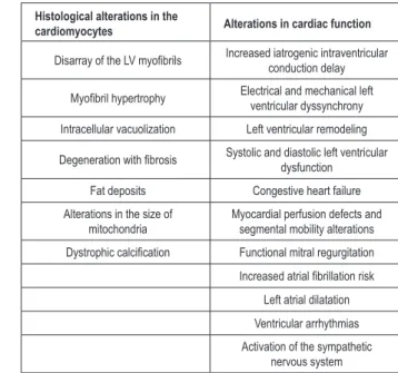
Chart 1 - Deleterious effects of unifocal cardiac stimulation at the right ventricular apex
Histological alterations in the
cardiomyocytes Alterations in cardiac function
Disarray of the LV myoibrils Increased iatrogenic intraventricular
conduction delay
Myoibril hypertrophy Electrical and mechanical left
ventricular dyssynchrony Intracellular vacuolization Left ventricular remodeling
Degeneration with ibrosis Systolic and diastolic left ventricular dysfunction
Fat deposits Congestive heart failure Alterations in the size of
mitochondria
Myocardial perfusion defects and segmental mobility alterations
Dystrophic calciication Functional mitral regurgitation
Increased atrial ibrillation risk
Left atrial dilatation Ventricular arrhythmias Activation of the sympathetic
nervous system
Alternative sites for endocardial cardiac
stimulation
In Latin America, the importance of the Chagasic etiology, associated with the implant of pacemakers has led to the necessity to find an alternative site for the positioning of the ventricular electrode, considering some characteristics of the disease. The right ventricular apex in Chagasic patients can present an endocardial thinning in 20-30% of the patients, in addition to the presence of intracavitary thrombus in a significant number of cases61.
Due to these characteristics, Kormann and Jatene62 described the subtricuspid position (vertebral-costal-diaphragmatic triangle) (Figure 2) to reduce the risks of cardiac perforation and thromboembolic phenomena triggered by the ventricular electrode. This study represented a landmark for a change in conduct by several surgeons in Latin America, who started to use the position of the RV inflow tract as a preferential site for the implant of the RV electrode in Chagasic patients62-64. Among the patients followed at the Pacemaker Laboratory of Hospital de Clinicas of the Federal University of Minas Gerais, in 2006, 77.8% of them had ventricular electrodes fixed in the subtricuspid region63. However, there is no scientific evidence of the functional outcome of this site of stimulation and there have been no comparative studies with other sites for right ventricular electrode implant.
The scientific evidence at the age of cardiac resynchronization, from 1990 on, led to the introduction of selective stimulation. The basic principle of this technique is to try to reproduce the natural sequence of cardiac depolarization, through the positioning of the electrode in the areas that are closest to the conduction system, using the complex natural electrical distribution network54. In this sense, the stimulation of the bundle of His or para-His7-12 and the stimulation of the RV endocardial region closest to the conduction system, along the middle and upper portions of the interventricular septum have been assessed13-26.
In clinical practice, during the implants of definitive pacemakers, the anatomical location of the bundle of His is difficult to reproduce. In spite of these technical questions,
there have been reports on the superiority of the stimulation of the bundle of His in relation to the degree of narrowing of the stimulated QRS complex and the systolic function, when compared to the RV apex9,11
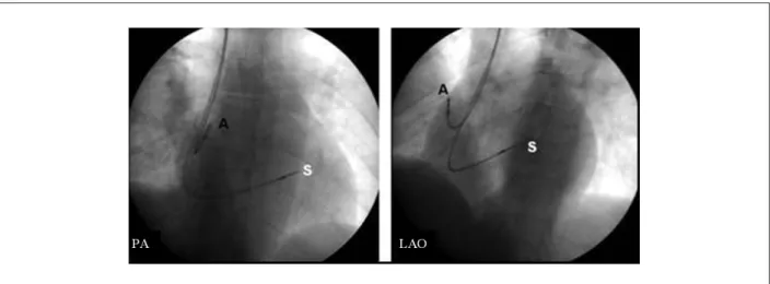
On the other hand, the positioning of the right ventricular electrode in the middle and upper regions of the interventricular septum, with the help of the radiological anatomy and the assessment of the electrocardiographic tracing, is simple and easy to reproduce (Figures 3 and 4). This site of stimulation has been assessed as a very interesting alternative for the implant of the ventricular electrode, as it generates narrower
QRS complexes, with a more physiological activation axis24
(Figures 5 and 6) and because it is feasible at any Service that routinely performs conventional pacemaker implants25-27.
The criteria to obtain the septal position were standardized and described in 2004 by Lieberman et al65. The validation of the septal position depends mainly on the radioscopic incidence at the left anterior oblique (LAO) view, in which the ventricular electrode is turned to the column, in an opposite direction to the free wall of the right ventricle.
There are other radiological parameters used to define the middle and upper32 septal positions (Figures 3 and 4).
In addition to the radiological criteria, some caution must be exercised when using the septal position15,66:
• Mapping of the septum, exhaustively seeking the
narrowest QRS complex (always < 150 ms);
• Obtaining QRS complexes that present an electrical
axis with a variation < 30o from the patient’s basal electrical axis.
• Threshold < 1 volt, to guarantee the stimulation of the septum’s muscular portion.
In spite of the radiological and electrocardiographic criteria to obtain the septal position, there is always the possibility of stimulating the RV free wall, considered one of the main limitations of the technique67. There is also the difficulty to define the upper, middle and lower portions of the septum. Such characteristics contribute to transform the septal position into a heterogeneous group that encompasses
Figure 2 -Radioscopic image in posteroanterior (PA) view, showing the right ventricular electrode in the subtricuspid position. The image on the right shows the delimitation
Figure 5 -Basal 12-lead electrocardiogram (ECG) 12 in patient with deinitive pacemaker, with electrode in the RV apex in the image on the left (duration of the QRS
complex of 200 ms and electrical axis - SÂQRS: -75º). The ECG on the right, resulting from mid-septal stimulation, shows a narrower QRS complex (116 ms) and a more physiological activation axis (SÂQRS: 45º), in comparison with the conventional stimulation.
Figure 3 -Radioscopic image in right anterior oblique (RAO) view at 10º in the image on the left. A division is represented through a 3x3 grid used to deine the different
sites of stimulation in the septal position. The borders are delimited by the vertebral bodies and the cardiac silhouette. Circle A represents the mid-septal position and circle B represents the high septal position. Circle C represents the RV apex. The image on the right shows the radioscopic RAO view of the interventricular septum and the positioning of the electrode in the septal portion of the RVOT (validation of the septal position). Source: Kaye et al32; Lieberman et al65.
Figure 4 -Radioscopic view in PA and LAO views (30º), demonstrating the inal position of the electrodes implanted in the right atrium (A) and mid-septum (S). The septal
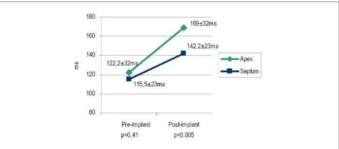
Figure 6 -Comparison of the QRS complex duration resulting from the septal position with the right ventricular apex stimulation (ms). The septal stimulation generates
signiicantly narrower QRS complexes (p = 0.0005). Source: Pachón et al37.
different sites for RV stimulation and translates the need to standardize the method68,69.
Functional outcome assessment of the
alternative sites for RV stimulation (Chart 2)
The first clinical trials comparing the different sites for RV stimulation appeared in the 1990s13,70. Blanc et al70 evaluated the acute hemodynamic effects in 27 individuals with congestive heart failure (CHF). No differences were observed between the stimulation of the RVOT and the apical stimulation through the analysis of the pulmonary capillary pressure and systolic pressure.
Another study of 14 patients in the acute phase of dual-chamber pacemaker implant due to total AV block was carried
out by Schwaab et al66. The septum was carefully mapped
and the electrode was positioned at the site that generated the narrowest QRS complex. The QRS duration decrease was correlated with the homogenization of the left ventricular contraction and improvement in the systolic function. Similar results were obtained by Mera et al15 in 12 patients submitted to a single-chamber pacemaker implant, after ablation of the AV junction. This second study showed left ventricular ejection fraction (LVEF) values at rest that were higher at the septal stimulation, when compared to the RV apical stimulation.
A significant improvement in LVEF was reported by Deshmukh et al8 in 12 patients submitted to a pacemaker implant with the stimulation of the bundle of His. These patients presented narrow QRS complexes, chronic atrial fibrillation (AF) and decreased LVEF (<40%).
Tse et al71 demonstrated that the stimulation of the RVOT region does result in the the same deleterious effects of LVEF decrease or the perfusion defects, in comparison with the stimulation of RV apex.
Victor et al72 published a prospective, randomized study with a crossover every three months, to evaluate the quality
of life and the systolic function in 103 patients submitted to definitive pacemaker implant. These patients presented CHF,
LV systolic dysfunction (LVEF ≤ 40%) and chronic AF. Three
types of stimulation were compared: apical, RVOT and bifocal RV stimulation. The conclusion was that the stimulation of the RVOT and the bifocal RV stimulation promoted a narrowing of the QRS interval, but did not consistently improve the quality of life scores or other clinical outcomes after three months of evolution, when compared to the apical stimulation.
Cai et al73 demonstrated that the stimulation of the RVOT and the bundle of His, in addition to yielding narrower QRS complexes, showed better indices of mechanical synchronization, evaluated by the echocardiogram, when compared to the apical stimulation.
In 2008, Erdoğan et al22 demonstrated that the stimulation
of the RVOT is safe and presents electrophysiological parameters comparable to those obtained with the long-term conventional RV apex stimulation.
Vanerio et al20 retrospectively evaluated 150 patients, with a mean age of 72 ± 7 years, submitted to definitive pacemaker implant. The patients were divided in two groups: apex stimulation (95) and RVOT stimulation (55). The mean follow-up of the patients was 3.4 ± 2 years, between 1999 and 2004. The multivariate analysis showed that the stimulation of the RVOT and the LVEF were independently correlated with survival (p = 0.006 and p = 0.003, respectively). The mortality with the RVOT stimulation was 37.3% lower (long-rank = 0.02). The authors emphasized the need to carry out randomized prospective studies with a larger sample size, to confirm these findings20.
evaluated (apex, RVOT and middle septal region) in a study presented at the meeting of the Heart Rhythm74.
Kypta et al75 carried out a prospective and randomized study to compare the effects of the RV septal and apex stimulation on cardiac function. A total of 98 patients were assessed, with a follow-up of 18 months, through clinical evaluation, brain natriuretic peptide (BNP) levels, functional capacity and LVEF assessment. No significant differences were observed between the two groups regarding the evaluated parameters. The conclusion of the study was that the septal stimulation is not superior to the apical stimulation in individuals with atrioventricular block submitted to definitive
pacemaker implant among a non-selected population75.
Ng et al69 carried out a longitudinal prospective study to evaluate the benefits of long-term septal stimulation. A total of 55 patients were studied and the mean follow-up period was 436 days for the septal position and 2,398 days for the electrode positioned at the RV apex. The echocardiographic parameters of the dimensions of the left chambers and the LVEF were evaluated, as well as the measurements of ventricular dyssynchrony and the QRS duration. The mean QRS duration was shorter in the patients with an electrode positioned at the RV apex (p < 0.001). It was observed that the electrocardiographic and radiological criteria used for the septal implant resulted in a heterogeneous group, with different sites of stimulation. The study concluded that the septal stimulation was associated with a poorer systolic function and a higher degree of dyssynchrony in relation to the RV apex stimulation69.
The four most recent reports21,69,74,75, associated with the description by Victor et al72, suggest that, in spite of the closeness of the artificial electrical stimulation in relation to the bundle of His, the unifocal stimulation is not capable of fully substituting the natural activation through multiple ramifications of the specialized Purkinje fibers. Another interesting aspect is that they represent conflicting evidence regarding the real benefit of the septal position demonstrated in other studies (Chart 2).
Considering the large number of patients that have been successfully followed in the correction of bradyarrhythmias, some centers remain faithful to the RV apex and the subtricuspid region, mainly in patients without structural cardiopathy, with normal systolic function and in cases where it is possible to minimize the stimulation in the ventricular channel76. In this sense, some ongoing studies are using the implant in the RV apex for all patients77,78. This question shows the need to prove the real benefit of septal stimulation in the context of evidence-based Medicine.
Another aspect to considered is that the studies carried out to evaluate alternative sites for RV stimulation encompassed all etiologies related to pacemaker implant and there have been few reports on the septal stimulation in individuals with Chagas disease27,79.
Finally, in an attempt to provide a definitive answer to the question of the superiority of the septal position when compared to the RV apex, three multicenter, prospective, randomized and blind clinical trials are being carried out (OPTIMIZE RV, PROTECT and RASP)32. In total, 58 centers are involved in the study, with an
estimated sample size of 800 patients. The primary outcome is the assessment of the ejection fraction through the echocardiography and radioisotope ventriculography. The secondary objectives include clinical events, 6-minute walking test, BNP measurement and echocardiographic measurements of the left chamber. The time of follow-up of the patients is up to 36 months.
Discussion
During approximately 40 years, since the start of the artificial cardiac stimulation, the RV apex was considered the preferential site for the implant of the ventricular electrode in conventional pacemakers. The concerns regarding the assessment of the damages caused by the iatrogenic left bundle-branch block produced by the stimulation of the RV apex in conventional cardiac pacemakers are relatively recent. In spite of the data obtained with the experimental and clinical studies, the deleterious effects associated with the apical stimulation of the RV depend on the interaction between specific factors of each patient (basal atrial rhythm, inherent atrioventricular and intraventricular conduction, LVEF and underlying cardiomyopathy). Additionally, there are conditions related to the artificial stimulation, such as programming mode, duration of the stimulated QRS complex, percentage and duration of the artificial stimulation in the ventricular channel. Since 1997, alternative sites to the RV apex have been studied, based on the rational search for a more physiological depolarization, caused by the artificial unifocal stimulation57. However, due to the small number of assessed patients, the lack of randomization in some studies, difficulty to standardize the criteria to define the alternative sites and different methods for the assessment of the outcome on cardiac function, there are important limitations in the performed studies.
The stimulation of the bundle of His presents technical difficulties that prevent its practical use68. The electrocardiographic and radiological criteria used for the septal implant are not so accurate and result in a heterogeneous group of different stimulation sites69. To make the question even more complex, the narrowing of the QRS complex in the septal position, when compared to the RV apex stimulation, did not correspond to the improvement in clinical, functional and echocardiographic parameters during the follow-up of the patients in some studies75.
Another aspect to be defined is whether patients with systolic dysfunction should not be treated with cardiac resynchronization, considering the limitations of the RV unifocal stimulation75.
In spite of all the advancements in cardiac electrotherapy in the last 50 years, the RV artificial stimulation is still not capable of fully substituting the natural activation through the specialized fibers of the His-Purkinje system. This limitation can be deleterious to the cardiac function, mainly in patients with systolic dysfunction, and justifies the intensive search for strategies to minimize these deleterious effects.
Conclusions
Chart 2 -Summary of some studies on the functional assessment of alternative sites for right ventricular endocardial stimulation
Authors Study proile N Methods of assessment Results
Blanc and cols., 1997 Observational 27 Systemic arterial and pulmonary capillary pressure
RVOT and apical present similar results regarding hemodynamic parameters*
Giudici and cols., 1997 Observational 89 Cardiac output (ECHO) RVOT results in increased CO in comparison with apical stimulation
de Cock and cols., 1998 Experimental 17 Cardiac output (ECHO) RVOT results in higher cardiac index in comparison with apical stimulation
Mera and cols., 1999 Pilot study 12 ECG and ECHO SS results in narrower QRS complexes and preserves systolic function †
Schwaab and cols., 1999 Observational 14 ECG and ECHO SS results in narrower QRS complexes and acutely increases systolic function
Kolletis and cols., 2000 Observational 20 ECHO RVOT presents better diastolic function parameters in comparison with SS
Bourke and cols., 2002 Prospective 20 Radioisotopic ventriculography RVOT and apical present no differences regarding the duration of the QRS complex and regarding systolic function Tse and cols., 2002 Prospective 24 Myocardial scintigraphy and
radioisotopic ventriculography
RVOT stimulation prevents the long-term deleterious effects on myocardial perfusion and LV systolic function Molina and cols., 2005 Observational 60 Evaluation of cardiac output by
thermodilution
SS was associated with increased CO when compared to apical stimulation
Mazzoca and cols., 2005 Prospective 24 ECG, electrophysiological parameters SS is viable and safe
Victor and cols., 2006 Pilot study 28 functional class, ECG, ECHO, ET RVOT results in narrower QRS complexes, but does not improve quality of life after three months of evolution Pachón and cols., 2006 Prospective 104 Electrophysiological parameters RVOT and apical stimulation present no differences regarding the
electrophysiological parameters
Burri and cols., 2007 Retrospective 362 Electrophysiological parameters and time of luoroscopy SS and apical stimulation present no differences regarding the electrophysiological parameters
Penteado and cols.,2007 Retrospective 21 Electrophysiological parameters SS and apical stimulation are similar regarding technical dificulties and electrical results
Silva Jr. and cols., 2007 Prospective 102 Electrophysiological parameters SS presents excellent electrophysiological parameters in the acute and chronic phases ‡ Alhous and cols., 2008 Experimental 16 Tissue Doppler Apex, SS and RVOT presented mechanical dyssynchrony
Erdoğan and cols., 2008 Prospective 32 Electrophysiological parameters RVOT stimulation is safe in the long term
Cai and cols., 2008 Observational 20 ECG and ECHO Apex and RVOT presented similar CI and CO. RVOT and para-His bundle preserved the mechanical synchronism Ten Cate and cols., 2008 Prospective 14 Tissue Doppler Apex stimulation and RVOT presented signs of mechanical
dyssynchrony § Kypta and cols., 2009 Prospective 98 ECHO, BNP, functional assessment
and clinical follow-up SS is not superior to apical stimulation Ng and cols., 2009 Retrospective 55 ECG, ECHO and tissue Doppler
SS resulted in the long-term worsening of the systolic function and a higher degree of dyssynchrony when compared to apical
stimulation OPTMIZE study Prospective 400 ECHO, BNP, functional assessment
and clinical follow-up Ongoing RASP study Prospective 160 ECHO, BNP, functional assessment
and clinical follow-up Ongoing PROTECT RV study Prospective 238 ECHO, BNP, functional assessment and clinical follow-up Ongoing
RVOT - stimulation of the right ventricular outlow tract; ECHO - echocardiogram; CO - cardiac output; CI - cardiac index; ECG - electrocardiogram; SS - septal stimulation
of the right ventricle; Apex - apical stimulation of the right ventricle; Electrophysiological parameters - command, sensitivity and impedance threshold; ET - ergometric test;
BNP - brain natriuretic peptide measurement. (*) Patients with congestive heart failure; (†) After ablation of the AV junction due to chronic atrial ibrillation in patients with
mild to moderate systolic dysfunction; (‡) Study involving Chagasic patients only; (§) Patients with normal systolic function. Source: adapted from Manolis55.
on the cardiac function. However, the most frequently studied endocardial site, the septal position, encompasses a heterogeneous group of different stimulation sites and there
including all the etiologies associated to the implant of cardiac pacemaker and with a long-term follow-up, to define the selective site of choice for the unifocal stimulation of the right ventricle.
Potential Conflict of Interest
No potential conflict of interest relevant to this article was reported.
Sources of Funding
There were no external funding sources for this study.
Study Association
This article is part of the thesis of doctoral submitted by Otaviano da Silva Júnior, from Universidade Federal do Triângulo Mineiro.
References
1. Pachon MJC, Mosquera JAP, Pachon M Juan C, Vargas RNA, Campos Neto CM. Aspectos epidemiológicos da estimulação cardíaca no Brasil – 12º ano do RBM – Registro Brasileiro de Marcapassos, Desfibriladores e Ressincronizadores Cardíacos. Relampa. 2008; 21 (1): 5-12.
2. Furman S, Schwerdel JB. An intracardiac pacemaker for Stokes-Adams seizures. N Engl J Med. 1959; 261: 943-8.
3. Little WC, Reeves RC, Arciniegas J, Katholl RE, Rogers EW. Mechanisms of abnormal interventricular septal motion during delayed left ventricular activation. Circulation. 1982; 65 (7): 1486-91.
4. Grover M, Glantz SA. Endocardial pacing site affects left ventricular end-diastolic volume and performance in the intact anesthetized dog. Circ Res. 1983; 53: 72-85.
5. Heyndricks GR, Vilaine JP, Knight OR, Vatner SF. Effects of altered site of electrical activation on myocardial performance during inotropic stimulation. Circulation. 1985; 71 (5): 1010-6.
6. Park RC, Little WC, O’Rourke RA. Effect of alteration of left ventricular activation sequence on the left ventricular end systolic pressure–volume relation in closed chest dogs. Circ Res. 1985; 57: 706-17.
7. Scheinman, MM, Saxon LA. Long-term his-bundle pacing and cardiac function. Circulation. 2000; 101 (8): 836-7.
8. Deshmukh P, Casavant DA, Romanyshyn M, Anderson K. Permanent, direct his bundle pacing: a novel approach to cardiac pacing in patients with normal His-Purkinje activation. Circulation. 2000; 101: 869-77.
9. Deshmukh PM, Romanyshyn M. Direct his-bundle pacing: present and future. Pacing Clin Electrophysiol. 2004; 27 (6 Pt 2): 862-70.
10. Catanzariti D, Maines M, Cemin C, Broso G, Marotta T, Vergara GJ. Permanent direct his bundle pacing does not induce ventricular dyssynchrony unlike conventional right ventricular apical pacing: an intrapatient acute comparison study. J Interv Card Electrophysiol. 2006; 16 (2): 81-92.
11. Amitani S, Miyahara K, Sohara H, Kakura H, Koga M, Moriyama Y, et al. Experimental his-bundle pacing: histopathological and electrophysiological examination. Pacing Clin Electrophysiol. 1999; 22 (Pt I): 562-6.
12. Occhetta E, Bortnik M, Marino P. Permanent parahisian pacing. Indian Pacing Electrophysiol J. 2007; 7 (2): 110-25.
13. Giudici MC, Thornburg GA, Buck DL, Coyne EP, Walton MC, Paul DL, et al. Comparison of right ventricular outflow tract and apical lead permanent pacing on cardiac output. Am J Cardiol. 1997; 79 (2): 209-12.
14. De Cock CC, Meyer A, Kamp O, Visser CA. Hemodynamic benefits of right ventricular outflow tract pacing: comparison with right ventricular apex pacing. Pacing Clin Electrophysiol. 1998; 21: 536-41.
15. Mera F, Del Lurgio DB, Patterson RE, Merlino JD, Wade ME, Leon AR. A comparison of ventricular function during high right ventricular septal and apical pacing after his-bundle ablation for refractory atrial fibrillation. Pacing Clin Electrophysiol. 1999; 22: 1234-9.
16. Kolettis TM, Kyriakides ZS, Tsiapras D, Popov T, Paraskevaides IA, Kremastinos DT. Improved left ventricular relaxation during short-term right ventricular outflow tract compared to apical pacing. Chest. 2000; 117: 60-4.
17. Bourke JP, Hawkins T, Keavey P, Tynan M, Jamieson S, Bohulova R, et al. Evolution of ventricular function during permanent pacing from either right ventricular apex or outflow tract following AV-junctional ablation for atrial fibrillation. Europace. 2002; 4: 219-28.
18. Stambler BS, Ellenbogen KA, Zhang X. Right ventricular outflow versus apical pacing in pacemaker patients with congestive heart failure and atrial fibrillation. J Cardiovasc Electrophysiol. 2003; 14: 1180-6.
19. Lewicka-Nowak E, Dabrowska-Kugacka A, Tybura S, Krzyminska-Stasiuk E, Wilczek R, Staniewicz J, et al. Right ventricular apex versus right ventricular outflow tract pacing: prospective, randomised, long-term clinical and echocardiographic evaluation. Kardiol Pol. 2006; 64 (10): 1082-91; discussion 1092-3.
20. Vanerio G, Vidal JL, Fernández Banizi P, Banina Aguerre D, Viana P, Tejada J. Medium- and long-term survival after pacemaker implant: improved survival with right ventricular outflow tract pacing. J Interv Card Electrophysiol. 2008; 21 (3): 195-201.
21. Ten Cate TJ, Scheffer MG, Sutherland GR, Verzijlbergen JF, Van Hemel NM. Right ventricular outflow and apical pacing comparably worsen the echocardioghraphic normal left ventricle. Eur J Echocardiogr. 2008; 9 (5): 672-7.
22. Erdoğan O, Aktöz M, Altun A. Long-term safety and efficacy of right ventricular outflow tract pacing in patients with permanent pacemakers. Anadolu Kardiyol Derg. 2008; 8 (5): 350-3.
23. Medi C, Mond HG. Right ventricular outflow tract septal pacing: long-term follow-up of ventricular lead performance. Pacing Clin Electrophysiol. 2009; 32 (2): 172-6.
24. Assis EG, Pachón MJC, Pachón M Juan C. Reprodutibilidade clínica e comparação da duração do QRS nas estimulações endocárdicas convencional e do septo interventricular.In: 62º Congresso Brasileiro de Arritmias Cardíacas, Porto Alegre (RS); 28/11 a 1/12/2007.
25. Muto C, Ottaviano L, Canciello M, Carreras G, Calvanese R, Ascione L, et al. Effect of pacing the right ventricular mid-septum tract in patients with permanent atrial fibrillation and low ejection fraction. J Cardiovasc Electrophysiol. 2007; 18: 1032-6.
26. Flevari P, Leftheriotis D, Fountoulaki K, Panou F, Rigopoulos AG, Paraskevaidis I, et al. Long-term no outflow septal versus apical right ventricular pacing: relation to left ventricular dyssynchrony. Pacing Clin Electrophysiol. 2009; 32: 354-62.
27. Silva Jr O, Melo CS, Marra M, Tomaz AA, Pachon JC, Pachón Mateos Juan C. Estudo da variação dos parâmetros eletrofisiológicos na estimulação ventricular septal direita em chagásicos. Relampa. 2007; 20 (2): 79-89.
28. Wiggers CJ. The muscular reaction of the mammalian ventricles to artificial surface stimuli. Am J Physiol. 1924; 73: 346-78.
29. Lister JW, Klotz DH, Jomain SL, Stuckey JH, Hoffman BF. Effect of pacemaker site on cardiac output and ventricular activation in dogs with complete heart block. Am J Cardiol. 1964; 14: 494-503.
31. Harris ZI, Gammage MD. Alternative right ventricular pacing sites - where are we going? Europace. 2000; 2: 93-8.
32. Kaye G, Stambler BS, Yee R. Search for the optimal right ventricular pacing site: design and implementation of three randomized multicenter clinical trials. Pacing Clin Electrophysiol. 2009; 32 (4): 426-33.
33. Auricchio A, Salo RW. Acute hemodynamic improvement by pacing in patients with severe congestive heart failure. Pacing Clin Electrophysiol. 1997; 20(2 Pt 1):313-24.
34. Auricchio A, Stellbrink C, Block M, Sack S, Vogt J, Bakker P, et al. Effect of pacing chamber and atrioventricular delay on acute systolic function of paced patients with congestive heart failure. The Pacing Therapies for Congestive Heart Failure Study Group. The Guidant Congestive Heart Failure Research Group. Circulation. 1999; 99 (23): 2993-3001.
35. Xiao HB, Gibson DG. Effects of intermittent left bundle branch block on left ventricular diastolic function: a case report. Int J Cardiol. 1994; 46 (1): 85-8.
36. Xiao HB, Lee CH, Gibson DG. Effect of left bundle branch block on diastolic function in dilated cardiomyopathy. Br Heart J. 1991; 66 (6): 443-7.
37. Pachón M JC, Pachón Mateos JuanC, Vargas RNA, Pachón Mateus EI, Pachón MZC, Lobo TJ, et al. Comparação dos parâmetros eletrofisiológicos das estimulações ventriculares direita e convencional e septal. Reblampa. 2006; 19 (4): 231-7.
38. Karpawich PP, Rabah R, Haas JE. Altered cardiac histology following apical right ventricular pacing in patients with congenital atrioventricular block. Pacing Clin Electrophysiol. 1999; 22: 1372-7.
39. Hanna SR, Chung ES, Aurigemma GP, Meyer TE. Worsening of mitral regurgitation secondary to ventricular pacing. J Heart Valv Dis. 2000; 9: 273-5.
40. Sassone B, De Simone N, Parlangeli R, Tortorici R, Biancoli C, Di Pasquale G. Pacemaker-induced mitral regurgitation: prominent role of abnormal ventricular activation sequence versus altered atrioventricular synchrony. Ital Heart J. 2001; 2: 441-8.
41. Skanes AC, Krahn AD, Yee R and the CTOPP Investigators. Progression to chronic atrial fibrillation after pacing: the Canadian Trial Of Physiologic Pacing. J Am Coll Cardiol. 2001; 38: 167-72.
42. Sharma AD, Rizo-Patron C, Hallstrom AP. and the DAVID Investigators. The DAVID Trial Investigators. Dual-chamber pacing or ventricular backup pacing in patients with an implantable defibrillator: the Dual Chamber and VVI Implantable defibrillator (DAVID) Trial. JAMA. 2002; 288: 3115-23.
43. Prinzen F, Peschar M. Relation between the pacing induced sequence of activation and left ventricular pump function in animals. Pacing Clin Electrophysiol. 2002; 25 (Pt. I): 484-98.
44. Nielsen J, Kristensen L, Andersen H. and the DANISH Trial investigators. A randomized comparison of atrial and dual chamber pacing in 177 consecutive patients with sick sinus syndrome. J Am Coll Cardiol. 2003; 42: 614-23.
45. Sweeney MO, Hellkamp AS, Ellenbogen KA. Adverse effect of ventricular pacing on heart failure and atrial fibrillation among patients with normal baseline QRS duration in a clinical trial of pacemaker therapy for sinus node dysfunction. Circulation. 2003; 23: 2932-7.
46. Thackray SDR, Witte KKA, Nikitin NP, Clark AL, Kaye GC, Cleland JGF. The prevalence of heart failure and asymptomatic left ventricular dysfunction in a typical regional pacemaker population. Eur Heart J. 2003; 24: 1143-52.
47. Thambo JB, Bordachar P, Garrigues A. Detrimental ventricular remodeling in patients with congenital complete heart block and chronic right ventricular apical pacing. Circulation. 2004; 110: 3766-72.
48. Sharma AD, Rizo-Patron C, Hallstrom AP. and the DAVID Investigators. Percent right ventricular pacing predicts outcomes in the DAVID Trial. Heart Rhythm. 2005; 2 (Suppl 2): S75-6.
49. O’keefe JH, Abuissa H, Jones PG. Effect of chronic right ventricular apical pacing on left ventricular function. Am J Cardiol. 2005; 95: 771-3.
50. Steinberg JS, Fisher A, Wang P. The clinical implications of cumulative right ventricular pacing in the Multicenter Automatic Defibrilator Trial II. J Cardiovasc Electrophysiol. 2005; 16: 359-65.
51. Shukla HH, Hellkamp AS, James EA. and the MOST Investigators. Heart
failure hospitalization is more common in pacemaker patients with sinus node dysfunction and a prolonged paced QRS duration. Heart Rhythm. 2005; 2: 245-51.
52. Pachón MJC, Pachón M Juan C, Pachón MEI, Vargas RNS. Síndrome do QRS largo e síndrome ventricular do marcapasso: uma nova fase da estimulação cardíaca artificial. In: Melo CS. Temas de marcapasso. 3ª ed. São Paulo: Casa Editorial Lemos; 2007.
53. Abreu CDG. Análise da dissincronia ventricular e da resposta neuro-humoral em portadores de marcapasso. [dissertação]. Belo Horizonte: Faculdade de Medicina Universidade Federal de Minas Gerais; 2008.
54. Siu CW, Wang M, Zhang XH, Lau CP, Tse HF. Analysis of ventricular performance as a function of pacing site and mode. Prog Cardiovasc Dis. 2008; 51 (2): 171-82.
55. Manolis AS. The deleterious consequences of right ventricular apical pacing: time to seek alternate site pacing. Pacing Clin Electrophysiol. 2006; 29 (3): 298-315.
56. Miyoshi F, Kobayashi Y, Itou H, Onuki T, Matsuyama T, Watanabe N, et al. Prolonged paced QRS duration as a predictor for congestive heart failure in patients with right ventricular apical pacing. Pacing Clin Electrophysiol. 2005; 28 (11): 1182-8.
57. Su Y, Pan W, Gong X, Cui J, Shu X, Ge J. Relationships between paced QRS duration and left cardiac structures and function. Acta Cardiol. 2009; 64: 231-8.
58. Zhang XH, Chen H, Siu CW, Yiu KH, Chan WS, Lee KL, et al. New-onset heart failure after permanent right ventricular apical pacing in patients with acquired high-grade atrioventricular block and normal left ventricular function. J Cardiovasc Electrophysiol. 2008; 19 (2): 136-41.
59. Silva RT, Martinelli Filho M, Oliveira JC, Lima CEB, Martins DGMC, Guirao CI, et al. Remodelamento ventricular na estimulação cardíaca apical do ventrículo direito. Arq Bras Cardiol. 2007; 88 (2): 152-8.
60. Pignalberi C, Ricci RP, Santini M. Deleterious effects of apical right ventricular stimulation. Should we change our method of pacemaker implantation? Ital Heart J Suppl. 2005; 6 (10): 635-48.
61. McGavigan AD, Mond HG. Selective site ventricular pacing. Curr Opin Cardiol. 2006; 21 (1): 7-14.
62. Kormann DS, Jatene AD. Triângulo eletrodo vértebro diafragmático no posicionamento de eletrodo endocavitário para marcapasso cardíaco. Arq Bras Cardiol. 1977; 39 (supl. 2): 380.
63. Gauch PR, Kormann DS, Kormann SJ, Jatene AD. Estudo comparativo do limiar de estimulação agudo e crônico em portadores de doença de Chagas e miocardiosclerose com marcapasso cardíaco. Arq Bras Cardiol. 1980; 35: 359-61.
64. Lieberman R, Grenz D, Mond HG, Gammage MD. Selective site pacing. Pacing Clin Electrophysiol. 2004; 27: 883-6.
65. Rincón LG, Rocha MO, Pires MT, Oliveira BG, Barros VDAC, Barros MV, et al. Perfil clínico de pacientes chagásicos e não-chagásicos portadores de marcapasso cardíaco artificial. Rev Soc Bras Med Trop. 2006; 39 (3): 245-9.
66. Schwaab B, Froehlig G, Alexander C. Influence of right ventricular stimulation site on left ventricular function in atrial synchronous ventricular pacing. J Am Coll Cardiol. 1999; 33: 317-23.
67. Burri H, Sunthorn H, Dorsaz PA, Viera I, Shah D. Thresholds and complications with right ventricular septal pacing compared to apical pacing. Pacing Clin Electrophysiol. 2007; 30 (Suppl 1): S75-8.
68. Giudici MC, Karpawich PP. Alternative site pacing: it’s time to define terms. Pacing Clin Electrophysiol. 1999; 22 (4 Pt 1):551-3.
69. Ng AC, Allman C, Vidaic J, Tie H, Hopkins AP, Leung DY. Long-term impact of right ventricular septal versus apical pacing on left ventricular synchrony and function in patients with second- or third-degree heart block. Am J Cardiol. 2009; 13 (8): 1096-100.
70. Blanc JJ, Etienne Y, Gilard M. Evaluation of different ventricular pacing sites in patients with severe heart failure: results of an acute hemodynamic study. Circulation. 1997; 96: 3273-7.
abnormalities in patients with permanent right ventricular pacing: the effect of sites of electrical stimulation. J Am Coll Cardiol. 2002; 40 (8): 1451-8.
72. Victor F, Mabo P, Mansour H, Pavin D, Kabalu G, De Place C, et al. A randomized comparison of permanent septal versus apical right ventricular pacing: short-term results. J Cardiovasc Electrophysiol. 2006; 17 (3): 238-42.
73. Cai L, Huan DJ, Yan CB, Rao L, Liu J, Liu H. Report on initiating clinical research for electrical and mechanical synchronism of selective region pacing in the right ventricular. Zhonghua Xin Xua Guan Bing Za Zhi. 2007; 35 (2): 147-50.
74. Alhous HA, Hillis G, Small G, Hannah A, Broadhurs TP. The impact of alternate right ventricular pacing sites on indices of left ventricular synchrony: an acute pacing study. In: Heart Rhytm Society meeting; 2008. San Franscisco-CA.USA.
75. Kypta A, Steinwender C, Kammler J, Leisch F, Hofmann R. Long-term outcomes in patients with atrioventricular block undergoing septal ventricular lead implantation compared with standard apical pacing. Europace. 2008; 10: 574–9.
76. Brito Jr HL. Artigo de intercâmbio: a opinião de estimulista sobre o local ideal para o implante de cabos-eletrodos no ventrículo direito e o uso de marcapassos monocamerais atriais exclusivos. Reblampa. 2006; 19 (3): 148-54.
77. Kachboura S, Ben Halima A, Fersi I, Marrakchi S, Zouaoui W, Kammoun I. Assessment of heart failure and left ventricular systolic dysfunction after cardiac pacing in patients with preserved left ventricular systolic function. Ann Cardiol Angeiol (Paris). 2008; 57 (1): 29-36.
78. Liu WH, Chen MC, Chen YL, Guo BF, Pan KL, Yang CH, et al. Right ventricular apical pacing acutely impairs left ventricular function and induces mechanical dyssynchrony in patients with sick sinus syndrome: a real-time three-dimensional echocardiographic study. J Am Soc Echocardiogr. 2008; 21 (3): 224-9.